Journal List > Restor Dent Endod > v.48(3) > 1516084616

Alessi, Jitumori, Bittencourt, Gomes, and Gomes: Effect of irrigation protocols on smear layer removal, bond strength and nanoleakage of fiber posts using a self-adhesive resin cement

Abstract

Objectives

This study aimed to investigate the effect of the application method of 2% chlorhexidine (CHX) and its influence on the adhesion of fiberglass posts cemented with a self-adhesive resin cement.

Materials and Methods

Sixty human mandibular premolars were endodontically treated and divided into 5 groups (n = 12), according to the canal irrigant and its application method: 2 groups with conventional syringe irrigation (CSI)—2.5% sodium hypochlorite (NaOCl) (control) and 2% CHX— and 3 groups with 2% CHX irrigation/activation—by passive ultrasonic irrigation (PUI), Easy Clean file, and XP-Endo Finisher file. Two roots per group were evaluated for smear layer (SL) removal by scanning electron microscopy. For other roots, fiber posts were luted using a self-adhesive resin cement. The roots were sectioned into 6 slices for push-out bond strength (BS) (7/group) and nanoleakage (NL) (3/group). Data from SL removal were submitted to Kruskal-Wallis and Student-Newman-Keuls tests (α = 0.05). Data from BS and NL were evaluated by 2-way analysis of variance and Tukey’s test (α = 0.05).

Results

For SL removal and BS, the CHX irrigation/activation promoted better values than CSI with CHX (p < 0.05), but it was not significantly different from CSI with NaOCl (p > 0.05). For NL, the lowest values were obtained by the chlorhexidine irrigation/activation groups (p < 0.05).

Conclusions

Active 2% CHX irrigation can be used to improve the post space cleaning and adhesion before fiber post cementation with self-adhesive resin cements.

INTRODUCTION

The treatment with fiberglass posts is highly dependent on the adhesive cementation, which should promote adequate bond strength (BS) at the dentin-cement-fiber post interface [1]. For this, conventional resin cements or self-adhesive resin cements may be used; the last one requires no pretreatment (acid conditioning or application of an adhesive system), being applied in a single clinical step, which simplifies clinical procedures, reduces the sensitivity of the technique, and demonstrates adhesive performance similar or superior to conventional cements [234].
However, the root canal preparation to receive fiberglass posts produces a smear layer formed by gutta-percha, endodontic cement, and dentin remnants, directly affecting the BS between dentin and resin cement as they hinder dentin demineralization by the self-adhesive cement, producing a hybrid layer and resinous tags, especially when the smear layer is very thick [56789]. Although the self-adhesive resin cement RelyX U200 (3M ESPE, St. Paul, MN, USA) manufacturer suggests the use of sodium hypochlorite (NaOCl) as an irrigating solution prior to post cementation, neither the criterion used for such indication is clear, nor its concentration, however, some studies demonstrate that this solution negatively affects the results of BS [101112].
Contrasting advantages and disadvantages, the literature is conflicting when chlorhexidine is used prior to fiberglass posts cementation. On one side, some studies show a reduction in BS or even no effect, on the other hand, there are studies that have obtained an increase in adhesion for immediate results and for the long term [69131415161718]. Chlorhexidine has been indicated as an alternative irrigator due to its antimicrobial activity, biocompatibility, and substantivity and it does not interfere with the collagen of the organic matrix of the root dentin, thus maintaining the quality of the substrate [6192021]. However, studies show that chlorhexidine, as NaOCl, is not efficient to remove the smear layer, especially when it is passively used without any type of activation [222324].
Techniques for irrigating solution activation during endodontic treatment have been developed and studied in order to effectively remove microorganisms, organic tissues, and the smear layer, like passive ultrasonic irrigation (PUI), Easy Clean File (Easy, Belo Horizonte, MG, Brazil), and XP-Endo Finisher File (FKG, La Chaux-de-Fonds, Switzerland) [252627]. PUI can contribute to debris removal [2829]. Thus, there is a better exposure of the dentinal tubules, which enhances the action of irrigating solutions in the root canal, and favors the penetration of the resin cement in the dentinal tubules, contributing to higher values in the BS of the fiber posts [102830].
This method consists of activating the irrigating solution in the root canal already prepared by means of an ultrasonic tip coupled to an ultrasound device, which converts electrical energy into ultrasonic waves of a certain frequency (30 kHz), producing an acoustic flow in the solution due its rapid agitation [28]. Easy Clean (Easy) is a pre-sterilized single-use plastic finishing file made of acrylonitrile-butadiene-styrene (ABS) plastic developed to maximize the smear layer removal by scraping root canal walls and agitation of irrigating solution with less or any damage by contact of the file with dentinal walls [2731]. This file is used in reciprocating motion, which means the file makes a 180° clockwise turn followed by a 90° counterclockwise turn, according to the manufacturer. The XP-Endo Finisher file is a nickel-titanium rotary instrument with an ISO 25 diameter and a taper of 0, is flexible, and its use is recommended after instrumentation of the root canals in order to improve the cleaning of the canals and preserve the dentin [2732]. Although these methods have been studied during endodontic treatment, there are few studies using the activation of the irrigating solutions before fiberglass posts cementation.
In this context, the aim of this study was to evaluate the immediate effect of different chlorhexidine irrigation/activation methods (PUI, Easy Clean file, and XP-Endo Finisher file) used after post space preparation in the bond properties between root dentin and a self-adhesive cement. The null hypotheses of this study were that the chlorhexidine activation would not affect 1) the push-out BSs, 2) the smear layer pattern, and 3) the nanoleakage (NL) at the adhesive interface.

MATERIALS AND METHODS

This study was submitted to and approved by the Ponta Grossa State University Research Ethics Committee at number 01595618.6.0000.0105. Sixty sound human mandibular premolars with at least 14 mm of root length without previous endodontic treatment, caries, root fractures, or resorptions were selected. The teeth were radiographed and stored in 0.1% thymol.
The sample size was performed for the push-out test (primary outcome), based on the results of a pilot study (mean 13.90 MPa and standard deviation of 1.8 MPa) and using the following equation:
n = Z2×S2/e2
where Z = 1.96, the acceptable error (e) = 10% of the mean, and s = 1.9 (standard deviation). Thus, the sample size would be at least 6.45, and, therefore, 7 teeth were randomly selected. For the secondary outcomes (smear layer removal and NL), a formal sample size calculation was not performed, but the literature was used as a reference [33343536].
The teeth were decoronated at the cement-enamel junction. The root canals were located and explored using a C+ #10 K-file (Dentsply Maillefer, Ballaigues, Switzerland) and shaped by rotatory instruments (ProTaper Universal; Dentsply Maillefer) in order (S1, S2, F1, F2, and F3) applying the crown-down technique. Irrigation was performed with 2.5% NaOCl throughout the progression of file sizes and final irrigation with 17% ethylenediaminetetraacetic acid (EDTA) and saline solution for 1 minute. The root canals were obturated using a warm vertical condensation technique performed with gutta-percha cones (ProTaper Universal F3; Dentsply Maillefer) and root canal sealer (Sealer 26; Dentsply Sirona, Pirassununga, SP, Brazil) and in sequence, the roots were covered with glass ionomer cement (Vitro Fil; DFL, Rio de Janeiro, RJ, Brazil) in the exposed cervical region.
After one week of storage in 100% relative humidity with distilled water at 37°C ± 1°C, the root canal filling was removed using sizes 1, 2, 3, and 4 Largo drills (Dentsply Maillefer). Periapical radiographs were performed to confirm the removal of the root filling. Next, the root canals were prepared with the corresponding drill to the fiberglass post (White Post DC # 2; FGM, Joinville, SC, Brazil) to a working length of 10 mm. Drills were changed after 5 root canal preparations.
After mechanical preparation of the root canals, the roots were randomly divided by block randomization (www.sealedenvelope.com) into 5 groups (n = 12), by restricted randomization, according to the dentinal treatment prior to the cementation of the fiberglass post (irrigation solution + irrigation method)—NaOCl 2.5% with conventional syringe irrigation (CSI), which is the manufacturer’s suggestion (control group), 2% chlorhexidine with CSI, 2% chlorhexidine irrigation and activation by PUI, 2% chlorhexidine irrigation and activation by Easy Clean file (Easy), and 2% chlorhexidine irrigation and activation by XP-Endo Finisher file (FKG).
The irrigation/activation protocols used were adapted from Van der Sluis et al. [28] and Duque et al. [37]:
  • - 2.5% NaOCl with CSI (NaOCl-CSI; control group): continuous irrigation of 6 mL of 2.5% NaOCl for 90 seconds with a disposable syringe and a 30-gauge needle (NaviTip; Ultradent, South Jordan, UT, USA) was performed.

  • - 2% chlorhexidine with CSI (CHX-CSI): continuous irrigation of 6 mL of 2% chlorhexidine solution for 90 s with a disposable syringe and a 30-gauge needle was performed.

  • - 2% chlorhexidine with activation by PUI (CHX-PUI): irrigation of 2 mL of 2% chlorhexidine for 10 seconds followed by activation with an ultrasonic tip (E1 Irrisonic; Helse Dental Technology, Santa Rosa de Viterbo, SP, Brazil) for 20 seconds in an ultrasound device (Profi Neo, Dabi Atlante, SP, Brazil) at the power of 20%. This procedure was repeated 3 times.

  • - 2% chlorhexidine with activation by Easy Clean file (Easy) (CHX-EC): irrigation of 2 mL of 2% chlorhexidine for 10 seconds followed by activation with Easy Clean file for 20 seconds coupled to a low rotation instrument (20,000 rpm). This procedure was repeated 3 times.

  • - 2% chlorhexidine with activation by XP-Endo Finisher file (FKG) (CHX-XP): irrigation of 2 mL of 2% chlorhexidine for 10 seconds followed by activation with XP-Endo Finisher file (FKG) for 20 seconds coupled to the endodontic device (X-Smart Plus; Dentsply Maillefer) at 800 rpm and torque 1 N. This procedure was repeated 3 times.

After the surface treatments, the root canals were rinsed with 10 mL of distilled water for 1 minute and air-dried for 5 seconds at a distance of 2 cm and 2 # F3 paper points (ProTaper Universal; Dentsply Maillefer).
Two roots per group (n = 2) were randomly selected for open dentinal tubule evaluation. After post space irrigation/agitation procedures, each tooth was split longitudinally in the mesiodistal direction. The samples were fixed to a metallic stub, and stored in colloidal silica in a dry oven at 37°C for 48 hours. Then, they were gold-sputter coated (Balzers SCD 050 Sputter Coater; Bal-Tec, Lübeck, Germany) for scanning electron microscope (SEM) analysis (SSX 550; Shimadzu, Kyoto, Japan) in secondary electron mode. Images at ×2,000 original magnification were obtained at the coronal, middle, and apical third of each hemi-section.
Using the ×2,000 images, 2 previously calibrated and blinded operators classified the proportion of smear layer removal, according to the following scores: 1) no smear layer (SL), when the root canal surface does not have SL and all dentinal tubules are clean and open, 2) a moderate amount of SL, without SL on the root canal surface, however, the tubules contain smear plugs, and 3) heavy SL covering the surface of the root canal and dentinal tubules (Figure 1) [3839]. In case of disagreement between the evaluators, the sample was re-analyzed by both evaluators until a consensus was reached.
Figure 1

Scanning electron microscope micrographs (×2,000) showing: (A) Score 1: no smear layer; (B) Score 2: moderate amount of smear layer; and (C) Score 3: heavy smear layer.

rde-48-e28-g001
In each experimental group, a fiberglass post was cemented in each of the remaining eleven roots using a self-adhesive resin cement (RelyX U200; 3M ESPE). The fiberglass posts (Whitepost DC # 2; FGM) were sectioned in the cervical region with a high-speed diamond bur under water cooling, to obtain 13 mm in post length, where 10 mm corresponded to the post space preparation, and the other 3 mm were used as a guide for standardizing the distance of the light-curing device during photoactivation of specimens. Then, the posts were cleaned with 70% alcohol for 5 seconds prior to cementation.
The self-adhesive resin cement was handled according to the manufacturer’s instructions, according to Table 1. The cement was photoactivated for 40 seconds using an LED light-curing unit with a light intensity of 1,200 mW/cm2 (Radii Cal; SDI, Bela Vista, SP, Brazil) positioned perpendicular to the post. After the cementation procedures, all roots were covered with glass ionomer cement (Vitro Fil; DFL) in the exposed cervical region and stored in 100% relative humidity at 37°C ± 1°C for 24 hours.
Table 1

Cement composition, cement application and batch number of the adhesive cement used in present study

rde-48-e28-i001
Adhesive cement Cement composition Cement application Batch No.
RelyX U200 (3M ESPE, St. Paul, MN, USA) Base: Methacrylate monomers containing phosphoric acid groups, methacrylate monomers, initiators, stabilizers, rheological additives. Application of the adhesive cement into the root canal with a syringe (Centrix, DFL, Rio de Janeiro, RJ, Brazil) and an elongated tip. Application of the adhesive cement on the post. Insertion of post into the root canal. Light cure for 40 seconds. 1816500278
Catalyst: Methacrylate monomers, alkaline stabilizers, pigments, rheological additives.
The roots were perpendicularly sectioned to its long axis into 6 1 mm-thick slices by a low-speed diamond saw (Isomet 1000; Buehler, Lake Buff, IL, USA) under water cooling. The first coronal slice was discarded due to the presence of excess cement, resulting in 6 slices, 2 slices for each third (cervical, middle, and apical). The slice thickness was verified using a 0.01 mm precision digital caliper (Digimatic Caliper, Mitutoyo, Japan) and the cervical side of each slice was identified. Seven roots for the group were randomly selected for the push-out BS testing and 3 roots for the NL evaluation of the interface adhesive between the dentin/resin cement.
Prior to the push-out test, all slices of 7 roots per experimental group (n = 7) were photographed on both sides, with a ×40 magnification, under an optical microscope (BX 51; Olympus, Tokyo, Japan) associated with a digital camera (DP72; Olympus). The measurements of the diameter of the adhesive area (post + cement) in the coronary and apical portion were performed, using software (ImageJ; National Institute of Health, Bethesda, MD, USA) and to determine the adhesive area, the tapered design of the glass-fiber post was considered and the formula of a lateral surface of a truncated cone was used. The push-out test was performed on a universal testing machine (AG-I; Shimadzu Autograph, Kyoto, Japan) at a crosshead speed of 0.5 mm/min. Each specimen was positioned with its more cervical side downwards on a metallic device with a central opening. A compressive force (50-kg load cell) was applied in the apico-coronal direction by a cylindrical metallic tip corresponding to the post diameter until debonding. The load at post dislodgment was recorded in Newton (N), and the BSs (MPa) were obtained by dividing the load by the bonding area in mm2.
The failure modes of all specimens are analyzed using an optical microscope at ×40 magnification and classified as: 1) adhesive between luting cement and dentin, 2) adhesive between luting cement and post, 3) cohesive within luting cement, 4) cohesive within the post, 5) cohesive within dentin, and 6) mixed failure.
For NL analysis, the coronal slice of each third root was used. Each slice was immersed in a 50 wt% ammoniacal silver nitrate solution for 24 hours at 37°C in a dark container, then photo-developed (Carestream Dental, Atlanta, GA, USA) for 8 hours under indirect fluorescent light. The specimens were washed abundantly with running tap water, fixed on metallic stubs and polished wet using 600-, 1,200-, 1,500-, 2,000-, 2,500-, and 3,000-grit silicon carbide papers (3M, Sumare, SP, Brazil) for 30 seconds each. After, the samples were kept at 37°C for 48 hours. Finally, the samples were gold sputter-coated. An SEM image at ×60 magnification of the coronal side of each slice was obtained and analyzed using ImageJ, wherewith it was measured the total interfacial perimeter and later tally the total peripheral length occupied by the silver. NL was calculated as the ratio between the lengths of cement–dentin adhesive interface infiltrated with silver nitrate and the total perimeter of this interface.
Data obtained for the smear layer pattern were analyzed by the Kruskal-Wallis and Student-Newman-Keuls tests (α = 0.05), and the images at ×100 and ×500 were only qualitatively evaluated. For BS and NL, the data were subjected to 2-way analysis of variance (ANOVA) (dentinal treatment vs. root third) and post hoc Tukey’s test (α = 0.05) using statistical software (Statistica 13.2; Dell, Santa Clara, CA, USA). The data from the fracture pattern were only evaluated qualitatively.

RESULTS

A 2% CHX irrigation and activation was found to have higher smear layer removal ability than CHX-CSI group (p = 0.005; Table 2). CHX-CSI group was not able to remove the smear layer (higher median scores) while CHX activation demonstrated partial smear layer removal similar to NaOCl 2.5% (p < 0.05).
Table 2

Median scores and interquartile ranges for smear layer pattern

rde-48-e28-i002
Experimental group Score
NaOCl-CSI 2 (1–3)A
CHX-CSI 3 (3–3)B
CHX-PUI 2,5 (2–3)A
CHX-EC 2,5 (2–3)A
CHX-XP 2 (2–3)A
NaOCl-CSI, 2.5% sodium hypochlorite with conventional syringe irrigation; CHX-CSI, 2% chlorhexidine with conventional syringe irrigation; CHX-PUI, 2% chlorhexidine with activation by passive ultrasonic irrigation; CHX-EC, 2% chlorhexidine with activation by Easy Clean file; CHX-XP, 2% chlorhexidine with activation by XP-Endo Finisher file.
A,BDifferent superscript letters indicate statistically significant differences (Student-Newman-Keuls test, p < 0.05). 1: no smear layer; 2: moderate amount of smear layer; 3: heavy smear layer.
For BS, 2-way ANOVA revealed a significant difference among the groups (p < 0.001; Table 3). In the coronal third, all groups with CHX activation showed higher mean BS values compared to CHX with CSI (p < 0,001), but similar to NaOCl 2.5% (p > 0,05). In the medium third, the PUI and Easy Clean groups demonstrated significant superiority compared to CHX with CSI (p < 0,001), but similar to XP-Endo Finisher and NaOCl groups. CHX with CSI showed similar BS values to XP-Endo Finisher and NaOCl groups. In the apical third, the CHX irrigation/activation groups (CHX-PUI, CHX-EC, and CHX-XP) were similar to the NaOCl group. CHX with CSI showed similar mean BS values with the NaOCl and CHX-PUI groups.
Table 3

Bond strength means and standard deviation (MPa) for the different experimental groups

rde-48-e28-i003
Root region Experimental group (dentinal treatment)
NaOCl-CSI (control) CHX-CSI CHX-PUI CHX-EC CHX-XP
Coronal 13.2 ± 2.1ab 6.4 ± 1.9de 16.2 ± 2.8a 15.4 ± 2.4a 13.9 ± 3.0ab
Medium 8.1 ± 1.5cd 6.2 ± 1.3de 11.3 ± 2.5bc 11.3 ± 1.7bc 9.5 ± 1.9cd
Apical 7.7 ± 1.0de 4.5 ± 0.7e 7.4 ± 1.4de 9.0 ± 1.8cd 8.0 ± 1.8cd
NaOCl-CSI, 2.5% sodium hypochlorite with conventional syringe irrigation; CHX-CSI, 2% chlorhexidine with conventional syringe irrigation; CHX-PUI, 2% chlorhexidine with activation by passive ultrasonic irrigation; CHX-EC, 2% chlorhexidine with activation by Easy Clean file; CHX-XP, 2% chlorhexidine with activation by XP-Endo Finisher file.
a-eDifferent letters indicate statistically significant differences (p < 0.05, Tukey’s test).
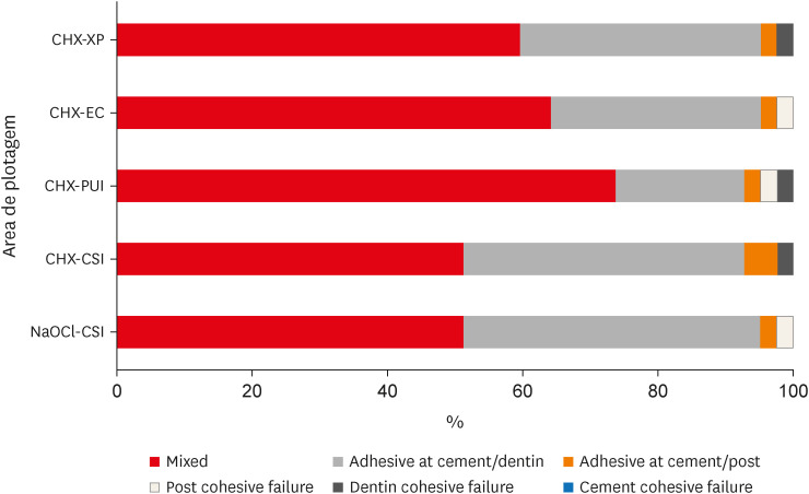
Mixed failure mode was predominant for all groups (Figure 2). Cohesive failures occurred least frequently (Figure 2).
Figure 2

Graph showing the failure mode distribution.

NaOCl-CSI, 2.5% sodium hypochlorite with conventional syringe irrigation; CHX-CSI, 2% chlorhexidine with conventional syringe irrigation; CHX-PUI, 2% chlorhexidine with activation by passive ultrasonic irrigation; CHX-EC, 2% chlorhexidine with activation by Easy Clean file; CHX-XP, 2% chlorhexidine with activation by XP-Endo Finisher file.
rde-48-e28-g002
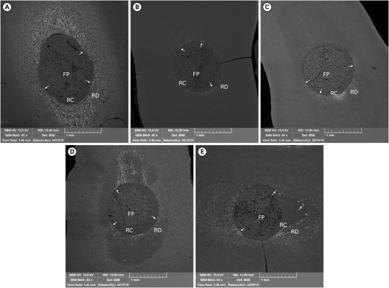
For NL, 2-way ANOVA demonstrated that the cross-product interaction of dentinal treatment vs. root region was not significant (p = 0.193; Table 4), but it was significant for the main factors root region (p < 0.001; Table 4) and dentinal treatment (p < 0.001; Table 4). For the root region, the coronal third showed the lowest NL values, and the apical third the highest. For dentinal treatment, the highest NL values were in the NaOCl group, and the PUI group demonstrated the lowest value similar to the XP-Endo Finisher group (Figure 3).
Table 4

Means and standard deviations of nanoleakage (%) for the different experimental groups

rde-48-e28-i004
Root region Experimental group (dentinal treatment) Main factor
NaOCl-CSI CHX-CSI CHX-PUI CHX-EC CHX-XP
Coronal 56.2 ± 5.3 41.5 ± 3.6 22.0 ± 5.2 37.5 ± 6.3 30.3 ± 6.9 37.5 ± 12.8A
Medium 68.6 ± 7.3 56.6 ± 3.2 32.2 ± 4.3 43.8 ± 4.8 41.7 ± 7.2 48.6 ± 13.9B
Apical 77.9 ± 8.0 73.7 ± 4.4 53.0 ± 2.0 50.5 ± 5.3 57.1 ± 9.7 62.4 ± 12.8C
Main factor 67.5 ± 11.2d 57.2 ± 14.3c 35.7 ± 14.2a 43.9 ± 7.4b 43.0 ± 13.6ab
NaOCl-CSI, 2.5% sodium hypochlorite with conventional syringe irrigation; CHX-CSI, 2% chlorhexidine with conventional syringe irrigation; CHX-PUI, 2% chlorhexidine with activation by passive ultrasonic irrigation; CHX-EC, 2% chlorhexidine with activation by Easy Clean file; CHX-XP, 2% chlorhexidine with activation by XP-Endo Finisher file.
A-CDifferent superscript letters indicate significant differences within each row; and a-cdifferent uppercase letters indicate significant differences within each column (p < 0.05, Tukey’s test).
Figure 3

Representative scanning electron photomicrographs (original magnification ×60) of post cementation interfaces for each dentinal treatment on middle third of root canal. (A) NaOCl-CSI group. (B) CHX-CSI group. (C) CHX-PUI group. (D) CHX-EC group. (E) CHX-XP group. Arrows indicate silver nitrate infiltrated in the adhesive interface. Interfacial marginal gap formation was significantly reduced in groups CHX-PUI, CHX-EC and CHX-XP compared with NaOCl-CSI (control group) and CHX-CSI.

NaOCl-CSI, 2.5% sodium hypochlorite with conventional syringe irrigation; CHX-CSI, 2% chlorhexidine with conventional syringe irrigation; CHX-PUI, 2% chlorhexidine with activation by passive ultrasonic irrigation; CHX-EC, 2% chlorhexidine with activation by Easy Clean file; CHX-XP, 2% chlorhexidine with activation by XP-Endo Finisher file; FP, fiber post; RC, resin cement; RD, root dentin.
rde-48-e28-g003

DISCUSSION

According to the results obtained, the active irrigation with chlorhexidine affected the proportion of open dentinal tubules, BS, and NL of the self-adhesive composite cement tested; thus, the hypotheses must be rejected.
This study demonstrated, through the images obtained by SEM, none of the methods used was efficient for the complete smear layer removal. However, the groups where chlorhexidine was activated (PUI, EC, and XP) promoted a partial removal of the smear layer, similar to the removal promoted by NaOCl 2.5%. As expected, CHX with CSI was not capable to remove the smear layer, which is confirmed by other studies [2340].
Literature provides us with conflicting results regarding the effect of chlorhexidine on fiber posts BS in root dentin when self-adhesive resin cements are used. Kul et al. [6] and Angeloni et al. [9] showed no difference in BS when CHX pretreatment was performed before fiber posts cementation. Di Hipólito et al. [40] found reduced BS when CHX was used as a pretreatment. Durski et al. [15] suggested higher BS values after pretreatment with CHX immediately and in the long term. However, this study differs from others in the use of instruments that are generally used in endodontics to improve the cleaning and disinfection of the root canals prior to endodontic filling, to promote CHX agitation.
CSI has limited ability to remove the smear layer [243141]. Thus, this study performed chlorhexidine agitation using different instruments that are generally used in endodontics to improve the cleaning and disinfection of the root canals prior to endodontic filling. The activation of irrigating solutions promotes better cleaning of the root canals as it increases the flow of the solution and the contact of the irrigating solution with the dentinal walls and creates a flow-reflux mechanism that drives the debris out of the root canal [2931]. However, the difficulty to obtain a uniform cleaning of the root dentinal surface causes controversial results [3142].
A better contact of resin cements with the dentin surface, by removing the smear layer, increases the BS values. This may be explained by the greater exposure of root dentin that provides greater dentin permeability, which in turn, improves resin cement penetration within dentinal tubules [111943].
The lowest results for the BS test were observed in the chlorhexidine with CSI group, which is in agreement with other studies, indicating that chlorhexidine applied without agitation and/or removing the smear layer negatively affects adhesion [1314]. However, in the groups where chlorhexidine was activated, the results were similar to the control group, with 2.5% NaOCl irrigation, especially in the cervical and middle thirds. These results indicate that the activation of the irrigating solution promotes an additional bonding effect between fiberglass posts resin cement and root dentin. Another possible explanation for these results is the high adsorption of chlorhexidine which favors the resin cement infiltration in dentinal tubules, in addition to other characteristics, such as the strong positive ionic charge, fast binding to phosphate groups, a strong affinity for dentin, and ability to promote an increase in surface energy, which favors the adhesion process [15].
Among the root thirds, the BS decreased in the middle and apical thirds in all groups, except for the chlorhexidine with CSI group, which showed similar results for the middle and apical thirds. The variation between the root thirds can be attributed to morphological differences in the dentinal structure, in the density of the dentinal tubules, and in the technical difficulties encountered during cementation, such as narrowing of the root canal towards the apex, which can compromise the resin cement insertion and the limited light transmission to the apical region, hindering adequate polymerization [64344].
Although self-adhesive resin cements present limited demineralization and hybridization of root dentin, we can see in this study that the smear layer removal, through the activation of the irrigating solution, significantly influenced the BS values, which reinforces the fact that the chemical interaction between self-adhesive resin cement and hydroxyapatite is fundamental and more important for proper adhesive cementation in the root canal than the formation of a hybrid layer [45]. This interaction is due to the chelating action of calcium ions promoted by acidic groups present in self-adhesive cement producing a chemical union between cement and hydroxyapatite [46].
The analysis of the results of this study demonstrated that the previous treatment with CHX significantly reduced NL in relation to the control group (NaOCl-CSI), and when chlorhexidine was actively applied, this reduction proved to be even greater. This result is in agreement with another study that demonstrated that chlorhexidine reduced NL when self-adhesive resin cements were used [47]. These findings can be explained by the increase in dentin surface energy promoted by chlorhexidine since the interaction between dentin surface and resin cement is highly dependent on the balance between surface energy and high wettability [4047].
Although self-adhesive resin cements promote neither distinct demineralization, nor a hybrid layer formation, the chemical interaction with hydroxyapatite by these cements, and the partial smear layer removal, that promote better cement contact with the root canal, by activation with instruments during CHX irrigation may have contributed to the reduction of gaps on the adhesive interface and consequently to improve BS results [74849].
Finally, it is worth mentioning the limitations of the present study since only the immediate results (24 hours) after cementation were evaluated and the greatest benefits of the previous use of chlorhexidine in the cementation of fiber posts can be observed over time [15]. Therefore, the better efficacy of smear layer removal through the activation of chlorhexidine (PUI, Easy Clean, and XP-Endo Finisher), and adhesion in relation to passive irrigation, may be indicative of better long-term adhesion stability, preserving the longevity of the BS [1550]. In addition, there are few studies that have evaluated the CHX activation prior to endodontic filling or cementation of fiberglass posts, and their methodological differences prevent the comparison between studies.

CONCLUSIONS

Considering the limitations imposed by the methods used, 2% CHX irrigation/activation, regardless of the protocol, promoted partial smear layer removal, improved the immediate BS and interface permeability of luting self-adhesive cement for glass fiber posts cementation and decreased the NL values. In this way, the previous activation of 2% CHX treatment can be a good alternative when fiberglass posts are cemented with self-adhesive resin cements.

Notes

Conflict of Interest: No potential conflict of interest relevant to this article was reported.

Author Contributions:

  • Conceptualization: Gomes GM, Gomes JC.

  • Formal analysis: Gomes GM, Gomes JC.

  • Funding acquisition: Gomes JC.

  • Investigation: Alessi RS, Jitumori RT.

  • Methodology: Alessi RS, Jitumori RT, Gomes GM.

  • Software: Jitumori RT, Gomes GM.

  • Supervision: Gomes GM, Gomes JC, Bittencourt BF.

  • Validation: Gomes GM, Gomes JC, Bittencourt BF.

  • Writing - original draft: Alessi RS, Jitumori RT.

  • Writing - review & editing: Gomes GM, Gomes JC, Bittencourt BF.

References

1. Tian Y, Mu Y, Setzer FC, Lu H, Qu T, Yu Q. Failure of fiber posts after cementation with different adhesives with or without silanization investigated by pullout tests and scanning electron microscopy. J Endod. 2012; 38:1279–1282. PMID: 22892751.
crossref
2. Faria-e-Silva AL, Menezes MS, Silva FP, Reis GR, Moraes RR. Intra-radicular dentin treatments and retention of fiber posts with self-adhesive resin cements. Braz Oral Res. 2013; 27:14–19. PMID: 23306622.
crossref
3. Ferracane JL, Stansbury JW, Burke FJ. Self-adhesive resin cements - chemistry, properties and clinical considerations. J Oral Rehabil. 2011; 38:295–314. PMID: 21133983.
crossref
4. Gomes GM, Gomes OM, Reis A, Gomes JC, Loguercio AD, Calixto AL. Regional bond strengths to root canal dentin of fiber posts luted with three cementation systems. Braz Dent J. 2011; 22:460–467. PMID: 22189640.
crossref
5. Vichi A, Grandini S, Davidson CL, Ferrari M. An SEM evaluation of several adhesive systems used for bonding fiber posts under clinical conditions. Dent Mater. 2002; 18:495–502. PMID: 12191661.
crossref
6. Kul E, Yeter KY, Aladag LI, Ayrancı LB. Effect of different post space irrigation procedures on the bond strength of a fiber post attached with a self-adhesive resin cement. J Prosthet Dent. 2016; 115:601–605. PMID: 26774312.
crossref
7. Monticelli F, Osorio R, Mazzitelli C, Ferrari M, Toledano M. Limited decalcification/diffusion of self-adhesive cements into dentin. J Dent Res. 2008; 87:974–979. PMID: 18809754.
crossref
8. Hayashi M, Takahashi Y, Hirai M, Iwami Y, Imazato S, Ebisu S. Effect of endodontic irrigation on bonding of resin cement to radicular dentin. Eur J Oral Sci. 2005; 113:70–76. PMID: 15693832.
crossref
9. Angeloni V, Mazzoni A, Marchesi G, Cadenaro M, Comba A, Maravi T, Scotti N, Pashley DH, Tay FR, Breschi L. Role of chlorhexidine on long-term bond strength of self-adhesive composite cements to intraradivular dentin. J Adhes Dent. 2017; 19:341–348. PMID: 28849803.
10. Barreto MS, Rosa RA, Seballos VG, Machado E, Valandro LF, Kaizer OB, Só M, Bier C. Effect of intracanal irrigants on bond strength of fiber posts cemented with a self-adhesive resin cement. Oper Dent. 2016; 41:e159–e167. PMID: 27603176.
crossref
11. Alkhudhairy FI, Yaman P, Dennison J, McDonald N, Herrero A, Bin-Shuwaish MS. The effects of different irrigation solutions on the bond strength of cemented fiber posts. Clin Cosmet Investig Dent. 2018; 10:221–230.
crossref
12. Seballos VG, Barreto MS, Rosa RA, Machado E, Valandro LF, Kaizer OB. Effect of post-space irrigation with NaOCl and CaOCl at different concentrations on the bond strength of posts cemented with a self-adhesive resin cement. Braz Dent J. 2018; 29:446–451. PMID: 30517442.
crossref
13. Lindblad RM, Lassila LV, Salo V, Vallittu PK, Tjäderhane L. Effect of chlorhexidine on initial adhesion of fiber-reinforced post to root canal. J Dent. 2010; 38:796–801. PMID: 20600556.
crossref
14. Lührs AK, Guhr S, Günay H, Geurtsen W. Shear bond strength of self-adhesive resins compared to resin cements with etch and rinse adhesives to enamel and dentin in vitro . Clin Oral Investig. 2010; 14:193–199.
crossref
15. Durski M, Metz M, Crim G, Hass S, Mazur R, Vieira S. Effect of chlorhexidine treatment prior to fiber post cementation on long-term resin cement bond strength. Oper Dent. 2018; 43:E72–E80. PMID: 29504878.
crossref
16. Ghazvehi K, Saffarpour A, Habibzadeh S. Effect of pretreatment with matrix metalloproteinase inhibitors on the durability of bond strength of fiber posts to radicular dentin. Clin Exp Dent Res. 2022; 8:893–899. PMID: 35726182.
crossref
17. Khanum Hm K, Abdulmajeed Barakat A, Qamar Z, Reddy RN, Vempalli S, Ramadan AH, Niazi F, Noushad M. Glass fiber post resistance to dislodgement from radicular dentin after using contemporary and conventional methods of disinfection. Photodiagnosis Photodyn Ther. 2022; 40:103026. PMID: 35872354.
18. Afkhami F, Sadegh M, Sooratgar A, Amirmoezi M. Comparison of the effect of QMix and conventional root canal irrigants on push-out bond strength of fiber post to root dentin. Clin Exp Dent Res. 2022; 8:464–469. PMID: 34664421.
crossref
19. Bitter K, Hambarayan A, Neumann K, Blunck U, Sterzenbach G. Various irrigation protocols for final rinse to improve bond strengths of fiber posts inside the root canal. Eur J Oral Sci. 2013; 121:349–354. PMID: 23841787.
crossref
20. Leitune VC, Collares FM, Werner Samuel SM. Influence of chlorhexidine application at longitudinal push-out bond strength of fiber posts. Oral Surg Oral Med Oral Pathol Oral Radiol Endod. 2010; 110:e77–e81. PMID: 20692186.
crossref
21. Cecchin D, Giacomin M, Farina AP, Bhering CL, Mesquita MF, Ferraz CC. Effect of chlorhexidine and ethanol on push-out bond strength of fiber posts under cyclic loading. J Adhes Dent. 2014; 16:87–92. PMID: 24027772.
22. Violich DR, Chandler NP. The smear layer in endodontics - a review. Int Endod J. 2010; 43:2–15. PMID: 20002799.
crossref
23. Elnaghy AM. Effect of QMix irrigant on bond strength of glass fibre posts to root dentine. Int Endod J. 2014; 47:280–289. PMID: 23829648.
crossref
24. Kuah HG, Lui JN, Tseng PS, Chen NN. The effect of EDTA with and without ultrasonics on removal of the smear layer. J Endod. 2009; 35:393–396. PMID: 19249602.
crossref
25. FundaoğLu KüçükekenciF. Küçükekenci AS. Effect of ultrasonic and Nd: Yag laser activation on irrigants on the push-out bond strength of fiber post to the root canal. J Appl Oral Sci. 2019; 27:e20180420. PMID: 31166549.
crossref
26. Kato AS, Cunha RS, da Silveira Bueno CE, Pelegrine RA, Fontana CE, de Martin AS. Investigation of the efficacy of passive ultrasonic irrigation versus irrigation with reciprocating activation: an environmental scanning electron microscopic study. J Endod. 2016; 42:659–663. PMID: 26906240.
crossref
27. Zand V, Mokhtari H, Reyhani MF, Nahavandizadeh N, Azimi S. Smear layer removal evaluation of different protocol of Bio Race file and XP- endo Finisher file in corporation with EDTA 17% and NaOCl. J Clin Exp Dent. 2017; 9:e1310–e1314. PMID: 29302283.
crossref
28. van der Sluis LW, Vogels MP, Verhaagen B, Macedo R, Wesselink PR. Study on the influence of refreshment/activation cycles and irrigants on mechanical cleaning efficiency during ultrasonic activation of the irrigant. J Endod. 2010; 36:737–740. PMID: 20307755.
crossref
29. Metzger Z, Solomonov M, Kfir A. The role of mechanical instrumentation in the cleaning of root canals. Endod Topics. 2013; 29:87–109.
crossref
30. Guerreiro-Tanomaru JM, Chávez-Andrade GM, de Faria-Júnior NB, Watanabe E, Tanomaru-Filho M. Effect of the passive ultrasonic irrigation on Enterococcus faecalis from root canals: an ex vivo study. Braz Dent J. 2015; 26:342–346. PMID: 26312969.
crossref
31. Prado MC, Leal F, Gusman H, Simão RA, Prado M. Effects of auxiliary device use on smear layer removal. J Oral Sci. 2016; 58:561–567. PMID: 28025441.
crossref
32. Elnaghy AM, Mandorah A, Elsaka SE. Effectiveness of XP-endo Finisher, EndoActivator, and File agitation on debris and smear layer removal in curved root canals: a comparative study. Odontology. 2017; 105:178–183. PMID: 27206916.
crossref
33. Jitumori RT, Bittencourt BF, Reis A, Gomes JC, Gomes GM. Effect of root canal irrigants on fiber post bonding using self-adhesive composite cements. J Adhes Dent. 2019; 21:537–544. PMID: 31802069.
34. Gruber YL, Bakaus TE, Gomes OM, Reis A, Gomes GM. Effect of dentin moisture and application mode of universal adhesives on the adhesion of glass fiber posts to root canal. J Adhes Dent. 2017; 19:385–393. PMID: 28944376.
35. Gruber YL, Jitumori RT, Bakaus TE, Reis A, Gomes JC, Gomes GM. Effect of the application of different concentrations of EDTA on the adhesion of fiber posts using self-adhesive cements. Braz Oral Res. 2020; 35:e012. PMID: 33237242.
crossref
36. Gruber YL, Bakaus TE, Gomes OM, Reis A, Gomes GM. Bonding properties of universal adhesives to root canals prepared with different rotary instruments. J Prosthet Dent. 2019; 12:298–305.
37. Duque JA, Duarte MA, Canali LC, Zancan RF, Vivan RR, Bernardes RA, Bramante CM. Comparative effectiveness of new mechanical irrigant agitating devices for debris removal from the canal and isthmus of mesial roots of mandibular molars. J Endod. 2017; 43:326–331. PMID: 27989584.
crossref
38. Torabinejad M, Khademi AA, Babagoli J, Cho Y, Johnson WB, Bozhilov K, Kim J, Shabahang S. A new solution for the removal of the smear layer. J Endod. 2003; 29:170–175. PMID: 12669874.
crossref
39. Poletto D, Poletto AC, Cavalaro A, Machado R, Cosme-Silva L, Garbelini CC, Hoeppner MG. Smear layer removal by different chemical solutions used with or without ultrasonic activation after post preparation. Restor Dent Endod. 2017; 42:324–331. PMID: 29142881.
crossref
40. Di Hipólito V, Rodrigues FP, Piveta FB, Azevedo LC, Bruschi Alonso RC, Silikas N, Carvalho RM, De Goes MF, Perlatti D’Alpino PH. Effectiveness of self-adhesive luting cements in bonding to chlorhexidine-treated dentin. Dent Mater. 2012; 28:495–501. PMID: 22204915.
crossref
41. Mozo S, Llena C, Chieffi N, Forner L, Ferrari M. Effectiveness of passive ultrasonic irrigation in improving elimination of smear layer and opening dentinal tubules. J Clin Exp Dent. 2014; 6:e47–e52. PMID: 24596635.
crossref
42. Mayer BE, Peters OA, Barbakow F. Effects of rotary instruments and ultrasonic irrigation on debris and smear layer scores: a scanning electron microscopic study. Int Endod J. 2002; 35:582–589. PMID: 12190897.
crossref
43. Ferrari M, Mannocci F, Vichi A, Cagidiaco MC, Mjör IA. Bonding to root canal: structural characteristics of the substrate. Am J Dent. 2000; 13:255–260. PMID: 11764112.
44. Culhaoglu AK, Özcan E, Kilicarslan MA, Seker E. Effect of boric acid versus conventional irrigation solutions on the bond strength between fiber post and root dentin. J Adhes Dent. 2017; 19:137–146. PMID: 28443835.
45. Farina AP, Cecchin D, Garcia LF, Naves LZ, Pires-de-Souza FC. Bond strength of fibre glass and carbon fibre posts to the root canal walls using different resin cements. Aust Endod J. 2011; 37:44–50. PMID: 21771181.
crossref
46. Radovic I, Monticelli F, Goracci C, Vulicevic ZR, Ferrari M. Self-adhesive resin cements: a literature review. J Adhes Dent. 2008; 10:251–258. PMID: 18792695.
47. Pontes DG, Araujo CT, Prieto LT, de Oliveira DC, Coppini EK, Dias CT, Paulillo LA. Nanoleakage of fiber posts luted with different adhesive strategies and the effect of chlorhexidine on the interface of dentin and self-adhesive cements. Gen Dent. 2015; 63:31–37. PMID: 25945761.
48. Temel UB, Van Ende A, Van Meerbeek B, Ermis RB. Bond strength and cement-tooth interfacial characterization of self-adhesive composite cements. Am J Dent. 2017; 30:205–211. PMID: 29178703.
49. Goracci C, Sadek FT, Fabianelli A, Tay FR, Ferrari M. Evaluation of the adhesion of fiber posts to intraradicular dentin. Oper Dent. 2005; 30:627–635. PMID: 16268398.
50. Toman M, Toksavul S, Tamaç E, Sarikanat M, Karagözoğlu I. Effect of chlorhexidine on bond strength between glass-fiber post and root canal dentine after six month of water storage. Eur J Prosthodont Restor Dent. 2014; 22:29–34. PMID: 24922997.
TOOLS
Similar articles