Journal List > Restor Dent Endod > v.47(4) > 1516084582

de Souza, Magalhães, Freitas, Lemus, Barbosa, Silva, and Moura: Calcium-doped zinc oxide nanocrystals as an innovative intracanal medicament: a pilot study

Abstract

Objectives

This study investigated the cytotoxicity, radiopacity, pH, and dentinal tubule penetration of a paste of 1.0% calcium-doped zinc oxide nanocrystals (ZnO:1.0Ca) combined with propylene glycol (PRG) or polyethylene glycol and propylene glycol (PEG-PRG).

Materials and Methods

The pastes were prepared by mixing calcium hydroxide [Ca(OH)2] or ZnO:1.0Ca with PRG or a PEG-PRG mixture. The pH was evaluated after 24 and 96 hours of storage in deionized water. Digital radiographs were acquired for radiopacity analysis and bubble counting of each material. The materials were labeled with 0.1% fluorescein and applied to root canals, and images of their dentinal tubule penetration were obtained using confocal laser scanning microscopy. RAW264.7 macrophages were placed in different dilutions of culture media previously exposed to the materials for 24 and 96 hours and tested for cell viability using the MTT assay. Analysis of variance and the Tukey test (α = 0.05) were performed.

Results

ZnO:1.0Ca materials showed lower viability at 1:1 and 1:2 dilutions than Ca(OH)2 materials (p < 0.0001). Ca(OH)2 had higher pH values than ZnO:1.0Ca at 24 and 96 hours, regardless of the vehicle (p < 0.05). ZnO:1.0Ca pastes showed higher radiopacity than Ca(OH)2 pastes (p < 0.01). No between-material differences were found in bubble counting (p = 0.0902). The ZnO:1.0Ca pastes had a greater penetration depth than Ca(OH)2 in the apical third (p < 0.0001).

Conclusions

ZnO:1.0Ca medicaments presented higher penetrability, cell viability, and radiopacity than Ca(OH)2. Higher values of cell viability and pH were present in Ca(OH)2 than in ZnO:1.0Ca.

INTRODUCTION

The vast majority of dental trauma in permanent teeth has been reported to occur in children and adolescents [1]. The combination of 2 different types of traumatic injuries on the same tooth—luxation and a concomitant crown fracture injury—may significantly increase the risk of pulp necrosis and infection, compromising root development in immature teeth [12]. Among the more recent treatment modalities proposed for necrotic immature permanent teeth are apical plugs using mineral trioxide aggregate (MTA) and revascularization [3]. However, these treatment modalities require a substantial learning curve and are not performed by most dental surgeons, who continue to perform conventional endodontic therapies with multiple visits and routine use of pastes containing calcium hydroxide [Ca(OH)2] for apexification [4]. Additionally, the European Society of Endodontology recommends that in cases of external infection-related resorption, the duration of Ca(OH)2 paste application should be between 4 weeks and several months [3]. In such situations, the use of Ca(OH)2 pastes for inter-visit dressings is justified by their antimicrobial properties and by their ability to stimulate hard tissue formation [4].
Some properties of Ca(OH)2 pastes have been related to the availability of hydroxyl ions, which in turn is associated with the type of vehicle used. Although aqueous vehicles such as water and saline are commonly used, other nonaqueous preparations, such as polyethylene glycol (PEG) and propylene glycol (PRG), have also been recommended due to their ability to create a reservoir for prolonged release of hydroxyl ions [45]. Another advantage of PEG and PRG is that both increase the viscosity of the medication, act as thickeners, and improve the consistency of the product, which makes it easier to insert into the root canals [46]. Regardless of the vehicle used, one of the challenges for apexification using Ca(OH)2 pastes involves the need for periodic changes of the intracanal dressing. To solve this limitation, other long-term medications have been proposed, such as a combination of Ca(OH)2, zinc oxide (ZnO), and chlorhexidine [789]. The formulation of this paste is based on a proportional mixture, in which Ca(OH)2 is dissolved in a few weeks and ZnO acts as an inert sealing material [7].
Previously, the pH and antimicrobial activity of Ca(OH)2 paste combined with nanoparticulate ZnO were evaluated [10]. Instead of mixing substances, such as Ca(OH)2 and ZnO, doping techniques have been used to modify the composition of materials by adding dopants to improve their functionality, resulting in synergistic effects. A previous study synthesized calcium-doped zinc oxide (ZnO:Ca) nanocrystals with different percentages of calcium ion (Ca2+) by weight, evaluating their chemistry, physical properties, and cytotoxicity [1112]. That investigation indicated the potential applicability in dentistry of 1.0% Ca2+-doped ZnO (ZnO:1.0Ca) nanocrystals. However, to enable the use of ZnO:1.0Ca as a temporary medication for traumatized teeth, it is necessary to combine it with vehicles already established in endodontic practice, to achieve an adequate consistency for root canal insertion.
Therefore, the present study examined selected chemical-physical properties of materials with potential clinical implications (radiopacity, pH, dentinal tubule penetration) and the cytotoxicity of a paste of ZnO:1.0Ca combined with PRG or PEG-PRG on a leukemia macrophage murine cell line (RAW 264.7). The null hypothesis tested was that all pastes would present similarities in the evaluated parameters.

MATERIALS AND METHODS

Materials

The pastes were prepared in a laminar flow chamber with 0.28 g of Ca(OH)2 nanocrystals or 0.28 g of ZnO:1.0Ca with 240 µL of PRG or 240 µL of the vehicle mixture (120 µL PEG + 120 µL PRG). This proportion was previously tested to obtain a consistency similar to Coltosol (Coltene-Vigodent S/A Indústria e Comércio, Rio de Janeiro, RJ, Brazil), facilitating its clinical use. The pastes were used for experiments using teeth as the experimental unit and for material characterization.

Cell viability assay

RAW 264.7 cells (Cell Bank of Rio de Janeiro, Rio de Janeiro, RJ, Brazil) were plated at a density of 2 × 10⁴ cells/well in 96-well plates (Corning, New York, NY, USA) containing Dulbecco's modified Eagle’s medium (DMEM) (Vitrocell Embriolife, Campinas, SP, Brazil) supplemented with 10% fetal bovine serum (Vitrocell) and 1% penicillin-streptomycin (Sigma–Aldrich, St. Louis, MO, USA). The cells were incubated at 37°C in 5% CO2 and 95% humidity for 24 hours to allow them to adhere to the plates.
The freshly prepared pastes were inserted into a sterile cylindrical polyethylene tube (diameter: 5 mm; height: 2 mm), kept in an incubator at 37°C for 24 hours, and sterilized by ultraviolet irradiation for 1 hour. Subsequently, the pastes were transferred to a 24-well culture plate and covered with 2.5 mL of DMEM (Vitrocell) for 24 hours at 37°C. The undiluted extraction media were filtered with a 0.22 µm filter (Kasvi, São José dos Pinhais, PR, Brazil) and used to prepare serially diluted extracts until a dilution of 1:16. The culture medium was then removed, and the cells were incubated with 200 µL of the extracts at predetermined dilutions (1:1, 1:2, 1:4, 1:8, and 1:16). The positive control group contained cells maintained in 10% DMEM. Three independent experiments were performed in triplicate.
Cell viability analysis was performed with incubation periods of 24 hours and 96 hours using the MTT method as previously described [12]. After exposure to each conditioned medium, 1 mg/mL MTT solution (Sigma–Aldrich) was added and incubated for an additional 4 hours. To solubilize the formazan crystals, the supernatant was removed, 10% dimethyl sulfoxide solution (LGC Biotecnologia, Cotia, SP, Brazil) was added to each well, and the plates were incubated at 37°C for 10 minutes. The optical densities were measured in a 570-nm wavelength filter (Biochrom, Cambridge, UK). The absorbance readings were normalized in proportion to the absorbance of the positive control, which represented succinate dehydrogenase activity (cell metabolism).

pH

To determine the pH, polyethylene tubes that were 10 mm long with a 1.6 mm internal diameter (Embramed, Sao Paulo, SP, Brazil) were filled with each material (n = 5). To insert the materials inside the tubes, a #25 K-file (Dentsply Maillefer, Ballaigues, Switzerland) was used, followed by condensation with a 1-2 plugger (Odous de Deus, Belo Horizonte, MG, Brazil) under ×3.0 magnification (D.F. Vasconcellos, Valença, RJ, Brazil) to confirm complete filling. Next, the samples were immersed in flasks containing 10 mL of deionized water (DW) and stored at 37°C. DW was used as a control. The pH of the solutions was measured using a previously calibrated digital pH meter (Digimed Analítica Ltda., Sao Paulo, Brazil) after 24 hours and 96 hours of immersion. The mean pH values of each group were calculated after each measurement in triplicate.

Radiopacity and bubbles

Using a silicone mold, 4 specimens (ZnO:1.0Ca+PRG; ZnO:1.0Ca+PEG-PRG; Ca(OH)2+PRG; Ca(OH)2+PEG-PRG), 6 mm in thickness and 5.4 mm in diameter, were prepared from each material. All specimens were radiographed once on the same day of preparation. A VistaScan Mini Plus® photostimulable phosphor (PSP) system (Dürr Dental, Bietigheim-Bissingen, Germany) was used, and the samples were imaged by a size 2 (3 × 4 cm) PSP plate using the settings of 70 kV, 7.0 mA, 0.14 seconds exposure time, and a focus-film distance of 30 cm (Timex 70E X-ray unit, Gnatus, Ribeirão Preto, SP). Subsequently, PSP plate scanning was performed, and the images were exported to ImageJ software (National Institutes of Health, Bethesda, MD, USA) for radiopacity analysis and bubble counting.
To determine the gray values, 3 points were randomly selected on each image in the region corresponding to the restorative material: one positioned to the right, one centrally, and one to the left. The gray value of each point was determined by the software, and it ranged from 0 to 255, as the radiographs were 8-bit images. The value 0 corresponded to black and 255 indicated white, with 254 different shades of gray between them, representing the variations between the most radiolucent to the most radiopaque tone.
The same radiographic images were used to count the number of bubbles on each material. For this analysis, a round region of interest of 250 pixels was selected, encompassing all of the specimen areas. Subsequently, the image was duplicated and made binary. A sequence of imaging tools was applied to remove outliers and perform despeckling. The resultant image was eroded and dilated once to reduce noise. To obtain a more homogeneous image and perform a proper bubble count, a closing tool was applied. The image was inverted, and a software tool to analyze particles was used to determine the number of bubbles.

Dentinal tubule penetration

Twenty bovine central incisors (30-38 months) with similar dimensions and pulp spaces were selected according to Oliveira et al. [13] and stored in distilled water at 4°C until use. All teeth were standardized in a cut machine (Isomet 5000; Buehler, Lake Bluff, IL, USA) to remove the dental crown, leaving a remaining height of 12 mm. The root canals were enlarged with Peeso reamers #1 to #6 (1.50 mm). Then, each root was flushed with 20 mL of 2.5% sodium hypochlorite followed by 20 mL of distilled water. The root canals were dried with paper points, and the external surfaces were dried by wipers (Kimberly Clark, Irving, TX, USA). The external surface was covered with a double layer of nail polish (Colorama, L’Oréal Brasil LTDA., Rio de Janeiro, RJ, Brazil), and each root was embedded in a polystyrene resin (Cristal, Piracicaba, SP, Brazil) and polyether impression material (Impregum F; 3M ESPE, Seefeld, Germany) to simulate periapical tissues. The prepared roots were randomly divided into 6 experimental groups (n = 5) based on the material used as follows: ZnO:1.0Ca+PRG; ZnO:1.0Ca+PEG-PRG; Ca(OH)2+PRG; Ca(OH)2+PEG-PRG. The pastes were mixed with 0.1% fluorescein (Sigma–Aldrich) as a fluorescence tracer for confocal laser scanning microscopic analysis. The materials were then incrementally placed in an orthograde direction into the root canals until they appeared from the apex and up to the cervical line with a medium-sized stainless steel endodontic plugger (Easy, Belo Horizonte, MG, Brazil). Then, the access cavities were sealed with temporary filling material (Cavit W; 3M ESPE), and the samples were incubated at 37°C for 24 hours before analysis. Three 1-mm-thick slices perpendicular to the long axis of the root canal, corresponding to the cervical, middle, and apical thirds, were obtained from each specimen using a slow-speed, water-cooled 0.3-mm microtome saw (Isomet 5000). All specimens were mounted onto glass slides and scanned under a confocal laser scanning microscope (LSM Pascal; Carl Zeiss, Jena, Germany) using the 543-nm wavelength of a helium laser under ×10 magnification (numeric aperture = 0.12). The depth at which the material penetrated the dentin tubules was measured. This measurement was performed in 4 quadrants: upper left (a), upper right (b), lower left (c), and lower right (d) (Figure 1). The measurement started from the wall of the root canal, following the trajectory of the dentinal canaliculus to the maximum point of penetration of the intracanal medicament.
Figure 1

Schematic diagram showing dentin penetration in a tooth sample. (A) Delimitation of the section region; (B) after preparation and insertion of the medications, the roots were sectioned in the cervical, middle, and apical thirds to evaluate dentinal penetration under fluorescence microscopy; (C) penetration measurements were made in quadrants: upper left (a), upper right (b), lower left (c), and lower right (d).

rde-47-e38-g001

Statistical analysis

The cell viability, pH, radiopacity, bubble count and dentinal tubule penetration data were analyzed for normality and homoscedasticity using the Shapiro–Wilk and Levene tests. For cell viability, pH and dentinal tubule penetration, 2-way analysis of variance (ANOVA) and the Tukey test were used to compare the data between the treated groups. One-way ANOVA and the Tukey test were used to compare the radiopacity of the materials. The Dunnett test was used to compare the experimental groups with the control group. The statistical analysis was performed using GraphPad Prism, a statistical software package (GraphPad LLC, San Diego, CA, USA). The significance level was set at 95% for all analyses.

RESULTS

Cell viability assays

At 24 (Figure 2A) and 96 hours (Figure 2B), it was observed that all materials were more cytotoxic at the 1:1 dilution than at the other dilutions (p < 0.0001). At dilutions of 1:4 and higher, the cytotoxic effects stabilized, with similar cell viability values between the dilutions for each material (p > 0.05). At 24 hours and 96 hours, the ZnO:1.0Ca+PRG and ZnO:1.0Ca+PEG-PRG groups at 1:1 and 1:2 dilutions presented the lowest percentages of viability (p < 0.0001). Within 24 hours, the cells in contact with extracts of ZnO:1.0Ca+PRG and ZnO:1.0Ca+PEG-PRG at 1:4, 1:8, and 1:16 showed the highest levels of viability (p < 0.0001), while those in contact with extracts of Ca(OH)2+PEG-PRG had the lowest values (p < 0.0001) (Figure 2A). After 96 hours, all groups showed similar viability values at 1:8 and 1:16 (p > 0.05) (Figure 2B). For both periods evaluated, the experimental groups had lower viability than the control group (p < 0.0001).
Figure 2

Cell viability percentage of RAW264.7 cells after (A) 24 hours and (B) 96 hours of exposure to extracts according to the material tested and dilution by the MTT formazan method. Capital letters indicate comparisons among different dilutions of extracts and the control group for each material. Lowercase letters indicate comparisons of the same material among the different dilutions. Two-way analysis of variance and the Tukey test (p < 0.05).

ZnO:1.0Ca, 1.0% Ca2+-doped zinc oxide nanocrystals; Ca(OH)2, calcium hydroxide; PRG, propylene glycol; PEG-PRG, polyethylene glycol and propylene glycol.
rde-47-e38-g002

pH

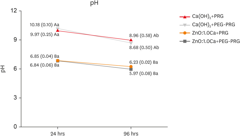
Ca(OH)2 had higher pH values than ZnO:1.0Ca at 24 and 96 hours, regardless of the vehicle used (p < 0.05). The pH of the Ca(OH)2 mixtures decreased over time (p < 0.05). ZnO:1.0Ca did not show pH variations between the evaluated periods, regardless of the vehicle used (p > 0.05) (Figure 3).
Figure 3

pH values after 24 hours and 96 hours of immersion in distilled water. Capital letters indicate comparisons among materials. Lowercase letters indicate comparisons of the same material among the different times of exposure. Two-way analysis of variance and the Tukey test (p < 0.05).

ZnO:1.0Ca, 1.0% Ca2+-doped zinc oxide nanocrystals; Ca(OH)2, calcium hydroxide; PRG, propylene glycol; PEG-PRG, polyethylene glycol and propylene glycol.
rde-47-e38-g003

Radiopacity

Table 1 presents the mean and standard deviation values for radiopacity, expressed as gray values immediately after material insertion. The ZnO:1.0Ca pastes showed higher radiopacity than the Ca(OH)2 pastes (p < 0.01).
Table 1

Mean radiopacity (gray values) in the different groups evaluated

rde-47-e38-i001
Groups Gray value
Ca(OH)2+PRG 56.83 ± 6.83 B
Ca(OH)2+PEG-PRG 59.92 ± 5.28 B
ZnO:1.0Ca+PRG 168.92 ± 14.46 A
ZnO:1.0Ca+PEG-PRG 177.83 ± 8.13 A
The values are mean ± standard deviation.
ZnO:1.0Ca, 1.0% Ca2+-doped zinc oxide nanocrystals; Ca(OH)2, calcium hydroxide; PRG, propylene glycol; PEG-PRG, polyethylene glycol and propylene glycol.
*Different capital letters represent statistically significant differences between groups by the Tukey test (p < 0.05).

Bubble count

Table 2 presents the mean bubble count and standard deviation. There were no statistically significant differences between the materials evaluated (p = 0.0902).
Table 2

Mean bubble count in the different groups evaluated

rde-47-e38-i002
Groups Number of bubbles
Ca(OH)2+PRG 13.5 ± 3.87 A
Ca(OH)2+PEG-PRG 11 ± 2.45 A
ZnO:1.0Ca+PRG 8.5 ± 3.42 A
ZnO:1.0Ca+PEG-PRG 8.25 ± 1.71 A
The values are mean ± standard deviation.
ZnO:1.0Ca, 1.0% Ca2+-doped zinc oxide nanocrystals; Ca(OH)2, calcium hydroxide; PRG, propylene glycol; PEG-PRG, polyethylene glycol and propylene glycol.
*Different capital letters represent statistically significant differences between groups by the Tukey test (p < 0.05).

Dentinal tubule penetration

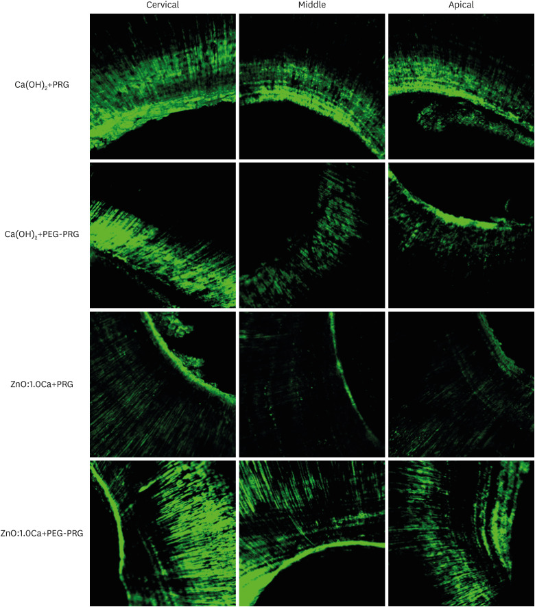
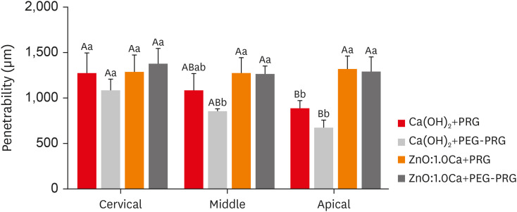
The levels of penetration of the materials into the different radicular thirds were observed (Figure 4). The experimental materials ZnO:1.0Ca+PRG and ZnO:1.0Ca+PEG-PRG exhibited similar penetration depths in all radicular thirds (p > 0.05). CaOH2+PRG, and Ca(OH)2+PEG-PRG had higher penetration in the cervical third than in the apical third (p = 0.01). All materials had similar depths of penetration in the dentinal tubules in the cervical third (p > 0.05). In the middle third, Ca(OH)2+PRG, ZnO1.0Ca+PRG, and ZnO:1.0Ca+PEG-PRG had similar penetration depths (p > 0.05), and Ca(OH)2+PEG-PRG presented the lowest values. ZnO:1.0Ca pastes had a greater penetration depth than Ca(OH)2 pastes in the apical third (p < 0.0001) (Figure 5).
Figure 4

Representative confocal laser scanning microscopy images from each experimental group at ×10 magnification.

ZnO:1.0Ca, 1.0% Ca2+-doped zinc oxide nanocrystals; Ca(OH)2, calcium hydroxide; PRG, propylene glycol; PEG-PRG, polyethylene glycol and propylene glycol.
rde-47-e38-g004
Figure 5

Dentinal tubule penetration depth (µm) of the experimental groups (n =15). Capital letters indicate comparisons of the same material among the different root thirds. Lowercase letters indicate comparisons among the different materials. Two-way analysis of variance and the Tukey test (p < 0.05).

ZnO:1.0Ca, 1.0% Ca2+-doped zinc oxide nanocrystals; Ca(OH)2, calcium hydroxide; PRG, propylene glycol; PEG-PRG, polyethylene glycol and propylene glycol.
rde-47-e38-g005

DISCUSSION

The null hypothesis was rejected, as the paste formulations affected the cell viability, radiopacity, and penetrability. The cytotoxicity of materials can vary depending on the cell model evaluated, the methodology of analysis, and the type of vehicle used for intracanal dressing [61214]. The RAW 264.7 macrophage lineage was chosen for this study because monocytes and macrophages play a pivotal role in periapical repair, releasing mediators related to the recruitment and differentiation of other cells involved in the formation of the mineralized apical barrier [15]. Previously, the same doped materials (ZnO:xCa) with different percentages of Ca2+ by weight (x = 0.7%; 1.0%; 5.0%; 9.0%) were tested against RAW 264.7, SAOS-2, and human dental pulp cells, supporting the use of ZnO:0.7Ca nanocrystals as pulp protection material [12]. However, in that study, the assessments were performed with materials in powder form, which does not represent clinical use, as the combination of Ca(OH)2 [4616] or ZnO with different vehicles is necessary to obtain an adequate consistency of the material [1017]. Specifically, in this study, the paste consistency was chosen to facilitate insertion in immature teeth with large canals, minimize the dispersion of the materials into the tissues, and retain the material in the desired area for a longer time [18]. Thus, an initial test was performed in order to verify the consistency of the material after manipulation with the proposed vehicle, demonstrating that the ZnO:0.7Ca nanocrystals did not have the desired consistency after manipulation. Since ZnO:1.0Ca also exhibited favorable cell viability and low production of reactive oxygen species and nitric oxide, such nanocrystals were selected for conducting the present study [12]. To assess the cytotoxicity of ZnO:1.0Ca and Ca(OH)2, extracts obtained from freshly prepared materials were used [1319]. This is because, in the clinical setting, the cells present in periapical tissues come into contact with progressively lower concentrations of leachable compounds that are continually cleared by extracellular fluids [13]. The MTT assay was chosen for cytotoxicity screening of these materials because it is often used for determining the cytotoxicity of endodontic materials without resulting in overestimation or underestimation [14].
The results revealed that the ZnO:1.0Ca and Ca(OH)2 formulations both presented greater cytotoxic effects at higher concentrations (1:1 and 1:2) regardless of the vehicle or contact time, which is in accordance with other studies using different materials [1319]. However, the behavior of RAW 264.7 against these same formulations at 24 and 96 hours was the opposite at 1:4 extracts, which is considered an intermediate dilution. At a 1:4 dilution, the lower cytotoxicity for ZnO:1.0Ca formulations compared to Ca(OH)2 at 24 hours suggested that the nano-doped ZnO:1.0Ca formulations initially promoted less damage to cell metabolism and consequently affected cell viability to a reduced degree. The alteration of this scenario at 96 hours, with Ca(OH)2 samples showing less cytotoxicity than ZnO:1.0Ca, may indicate that the leachate released by the doped material was altered rapidly, resulting in differences in their chemical composition. A possible explanation for the results at 24 hours may be related to the nanocrystal morphology, which is one of the most important characteristics affecting its cytotoxicity [20]. While the Ca(OH)2 nanocrystals used in the present study exhibit a hexagonal structure, the ZnO:1.0 Ca nanocrystals have a rod shape [1221]. Hexagonally shaped particles possess edge surface atoms, which makes it easier for them to enter into cells without disrupting the cells, while rod-shaped nanoparticles bind to the positively charged cell surface and disrupt them, resulting in cell death [20]. The results observed at 96 hours at the same dilution (1:4) may also be related to the zinc ion (Zn2+) release by doped ZnO:1.0Ca [22].
ZnO nanocrystals have the potential to be a potent means for Zn2+ delivery into intracellular compartments [22]. A previous study demonstrated that the toxicity of ZnO nanoparticles is caused by the dissolution and subsequent release of toxic Zn2+ ions into the culture medium [2223]. The dissolution of Zn2+ ions increases with the size of the particles [23]. The presence of Zn2+ may affect several cellular mechanisms, such as the cell cycle, differentiation, and death, via different pathways [2224]. In models using neural cells, the entrance of Zn2+ into the cells was observed after several days, and it is possible to speculate that the stronger cytotoxic effects of ZnO:1.0Ca at 96 hours are related to the same phenomena. However, despite the possible toxic effect of Zn2+ ions, ZnO has been used for decades in dentistry, including its use in pediatric dentistry in pulpotomies. In addition, ZnO nanoparticles have been shown to be effective against Gram-negative and Gram-positive bacteria [25262728].
The methodology followed in the present study for pH assessment is outlined in ISO 6876, which defines the standard for evaluating the physicochemical properties of root canal sealing materials [29]. Although the materials tested are intracanal medications and are not intended to permanently obturate the canals, we chose to follow the ISO standard due to the “paste-like” consistency of the materials. Previous studies have also utilized this methodology, immersing the plastic tubes containing medications in distilled water [1330]. Oliveira et al. [13] used tubes measuring 10 mm in length and 1.6 mm in internal diameter to evaluate the pH of BioC Temp, BioC Repair, and Biodentine. Villa et al. [30] evaluated the pH using BioC Temp and UltraCal in tubes measuring 10 mm in length and 1 mm in diameter.
The low pH values of doped ZnO:1.0Ca formulations indicate that the presence of Ca2+, a high-valence metal cation, was not able to modify the acid pH characteristic of ZnO nanoparticles produced via the coprecipitation technique, despite the vehicle used [12]. Previously, Guerreiro-Tanomaru et al. [10] evaluated the pH of nanoparticulate ZnO+PEG and compared it to nanoparticulate Ca(OH)2+ZnO+PEG and observed slightly higher pH values than the present study. The method of producing the ZnO nanocrystals, which is a determining factor in the material's characteristics, was different in that study from the method used here [24]. In addition, the doping of materials modifies the composition and properties of nanocrystals through the intentional introduction of dopants [11]. In this regard, the synthetic strategies of doped nanocrystals certainly influence the biological and physicochemical properties of nanocrystalline materials.
Regarding Ca(OH)2 formulations, pH values between 11 and 12 were expected, which were higher than the findings observed in the present study [4]. The reason for this may be attributed to the time of analysis, the vehicles used, differences in methodology and differences regarding the bulk Ca(OH)2 and nanocrystals [41626313233]. No previous studies have compared the dissociation of Ca(OH)2 formulations into OH- and Ca2+ between bulk Ca(OH)2 and nanocrystals or pH values. In this sense, it is not possible to directly compare the extant literature and the present findings. Considering that a pH of approximately 8.6 to 10.3 is required for the biological activity of Ca(OH)2 to promote the activation of alkaline phosphatase enzymes, the pH values obtained by Ca(OH)2+PRG and Ca(OH)2+PEG-PRG are within an acceptable range [32].
Interestingly, the vehicle did not affect the evaluated parameters of the ZnO:1.0Ca and Ca(OH2) nanocrystal formulations. The reason for this may be that both PEG and PRG are viscous vehicles with a high molecular weight [3435]. The characteristics of PEG and PRG were previously evaluated in a study of antibiotic pastes in a 1:1 ratio, demonstrating favorable results regarding cytotoxicity [6]. According to Mohammadi and Dummer [35], viscous vehicles promote the slow and sustained release of Ca2+ and OH- ions from Ca(OH)2 pastes compared with aqueous vehicles. In addition, the use of viscous vehicles has been suggested to optimize the intratubular penetration of intracanal medications [18].
The antimicrobial and penetrability effects of intracanal medications can be affected by the size and shape of the particles and the type of vehicle used [173637]. A previous study demonstrated that as the particle size increased, the particle shape became more rectangular, but as the size decreased, the particle shape became rounder, which was more desirable for deep penetration [37]. Considering that ZnO:1.0Ca nanocrystals used in the present study have a rod shape and are approximately 20 nm in diameter, while the Ca(OH)2 nanocrystals have a hexagonal structure and are approximately 80 nm in diameter, the greater ability of the ZnO:1.0Ca paste to penetrate into the middle and apical thirds could be attributed to these properties [1221]. However, the Ca(OH)2 paste presented a higher fluorescence intensity than ZnO:1.0Ca. Considering that the fluorescence intensity of sodium fluorescein is related to the presence of Ca+2 ions and the opposite effect occurs in the absence of these ions, it is possible to suggest that ZnO:1.0Ca+PRG released a smaller amount of Ca+2 ions than the other evaluated pastes [38]. However, additional studies need to be performed to confirm this speculation.
Another parameter evaluated was the radiopacity of the pastes, an important factor that enables the visualization of the material radiographically, as well as the presence of bubbles or an absence of material in the desired region [39], which facilitates clinical follow-up [30]. Among several radiopacifiers used in endodontic materials, ZnO has often been evaluated [4041]. Ca(OH)2 pastes, regardless of the vehicle used, do not present adequate radiopacity unless radiopacifying substances are added to the mixture [34]. In this sense, the higher radiopacity of the ZnO:1.0Ca formulations was in agreement with the current literature.
To maximize the benefit of dressing materials, the pastes must be in direct contact with the root canal walls and fill the total extension of the main canal space [42]. In the present study, all groups presented bubbles, without significant differences among them, which may be related to the similar consistency of the materials. However, the method used for bubble count based on binarized radiographic images has limitations, especially regarding the use of molds to obtain the images and a lack of volumetric analysis. Micro-computed tomography (µCT) would be a better alternative since it is the method of choice to calculate the volume of filling materials, providing high-resolution 3-dimensional assessment [4344]. Despite the advantages of µCT, it does not represent a tool that is available for clinicians to assess the filling of a given material, which is why radiographic images were chosen to assess the presence of bubbles.
One of the concerns regarding the materials tested as long-term medications is the difficulty of root canal removal, as the remnants of dressing material may prevent the adaptation of endodontic sealers to the root canal wall and the penetration of sealers into dentinal tubules [45]. Considering that intracanal medication remnants can be found in roots after irrigation, regardless of the vehicle used for their manipulation, the combination of chemical removal and mechanical activation of the irrigant solutions should be proposed in order to obtain more favorable results [464748]. Further studies need to be conducted to assess the difficulty of removing the medications proposed in this study.

CONCLUSIONS

In conclusion, ZnO:1.0Ca nanocrystals associated with low-surface-tension vehicles presented higher penetrability, cell viability, and radiopacity than Ca(OH)2 nanocrystals. In contrast, higher values of cell viability and pH were present in Ca(OH)2 nanocrystals than in ZnO:1.0Ca nanocrystals. None of the parameters were influenced by the vehicle (PRG or PEG-PRG).

ACKNOWLEDGEMENTS

This project was developed at CPBio ??Biomechanics, Biomaterials and Cell Biology Research Center of the Federal University of Uberlândia. The research was financed in part by Fundação de Amparo à Pesquisa do Estado de Minas Gerais (Finance code: APQ-02660-21); National Council for Scientific and Technological Development (CNPq) and Coordination for the Improvement of Higher Education Personnel – Brazil (CAPES) (Finance code 001).

Notes

Conflict of Interest: No potential conflict of interest relevant to this article was reported.

Author Contributions:

  • Conceptualization: Souza GL, Moura CCG, de Rezende Barbosa GL, Silva ACA.

  • Data curation: Souza GL, de Rezende Barbosa GL.

  • Formal analysis: Souza GL, Moura CCG, de Rezende Barbosa GL, Silva ACA.

  • Funding acquisition: Moura CCG, Silva ACA.

  • Investigation: Souza GL, Magalhães TEA, Freitas GAN, Lemus NXA.

  • Methodology: Souza GL, Magalhães TEA, Freitas GAN, Lemus NXA.

  • Project administration: Moura CCG.

  • Resources: Moura CCG, de Rezende Barbosa GL, Silva ACA.

  • Software: Souza GL, Magalhães TEA, Freitas GAN, Lemus NXA, de Rezende Barbosa GL.

  • Supervision: Moura CCG.

  • Validation: Souza GL, Moura CCG, de Rezende Barbosa GL.

  • Visualization: Souza GL, Moura CCG.

  • Writing - original draft: Souza GL, Magalhães TEA, Freitas GAN, Lemus NXA, de Rezende Barbosa GL, Silva ACA, Moura CCG.

  • Writing - review & editing: Souza GL, Moura CCG.

References

1. Bourguignon C, Cohenca N, Lauridsen E, Flores MT, O’Connell AC, Day PF, Tsilingaridis G, Abbott PV, Fouad AF, Hicks L, Andreasen JO, Cehreli ZC, Harlamb S, Kahler B, Oginni A, Semper M, Levin L. International Association of Dental Traumatology guidelines for the management of traumatic dental injuries: 1. Fractures and luxations. Dent Traumatol. 2020; 36:314–330. PMID: 32475015.
crossref
2. Lauridsen E, Hermann NV, Gerds TA, Ahrensburg SS, Kreiborg S, Andreasen JO. Combination injuries 1. The risk of pulp necrosis in permanent teeth with concussion injuries and concomitant crown fractures. Dent Traumatol. 2012; 28:364–370. PMID: 22221538.
crossref
3. Krastl G, Weiger R, Filippi A, Van Waes H, Ebeleseder K, Ree M, Connert T, Widbiller M, Tjäderhane L, Dummer PM, Galler K. Endodontic management of traumatized permanent teeth: a comprehensive review. Int Endod J. 2021; 54:1221–1245. PMID: 33683731.
crossref
4. Athanassiadis B, Walsh LJ. Aspects of solvent chemistry for calcium hydroxide medicaments. Materials (Basel). 2017; 10:1219. PMID: 29065542.
crossref
5. Deniz Sungur D, Aksel H, Purali N. Effect of a low surface tension vehicle on the dentinal tubule penetration of calcium hydroxide and triple antibiotic paste. J Endod. 2017; 43:452–455. PMID: 28131413.
crossref
6. Faria G, Rodrigues EM, Coaguila-Llerena H, Gomes-Cornélio AL, Neto Angéloco RR, Swerts Pereira MS, Tanomaru Filho M. Influence of the vehicle and antibiotic formulation on cytotoxicity of triple antibiotic paste. J Endod. 2018; 44:1812–1816. PMID: 30477667.
crossref
7. de Jesus Soares A, Yuri Nagata J, Casarin RC, Flávio Affonso de Almeida J, Gomes BP, Augusto Zaia A, Randi Ferraz CC, José de Souza-Filho F. Apexification with a new intra-canal medicament: a multidisciplinary case report. Iran Endod J. 2012; 7:165–170. PMID: 23056138.
8. Herrera DR, Herrera CM, Lima AR, Nagata JY, Pereira AC, Silva EJ, Soares AJ, Gomes BP. Repair of apical root resorption associated with periodontitis using a new intracanal medicament protocol. J Oral Sci. 2014; 56:311–314. PMID: 25500930.
crossref
9. Lima TF, Ascendino JF, Cavalcante IO, D Assunção FL, Salazar-Silva JR, Silva EJ, Lemos SG, Soares AJ. Influence of chlorhexidine and zinc oxide in calcium hydroxide pastes on pH changes in external root surface. Braz Oral Res. 2019; 33:e005. PMID: 30758405.
crossref
10. Guerreiro-Tanomaru JM, Pereira KF, Nascimento CA, Bernardi MI, Tanomaru-Filho M. Use of nanoparticulate zinc oxide as intracanal medication in endodontics: pH and antimicrobial activity. Acta Odontol Latinoam. 2013; 26:144–148. PMID: 25335366.
11. Silva AC, Zóia MA, Correia LI, Azevedo FV, Paula AT, Maia LP, Carvalho LS, Carvalho LN, Costa MP, Giaretta LC, Rodrigues RS, Ávila VM, Goulart LR, Dantas NO. Biocompatibility of doped semiconductors nanocrystals and nanocomposites. Askin Celik T, editor. Cytotoxicity. London: IntechOpen;2018. p. 149–161.
12. de Souza GL, Moura CC, Silva AC, Marinho JZ, Silva TR, Dantas NO, Bonvicini JF, Turrioni AP. Effects of zinc oxide and calcium-doped zinc oxide nanocrystals on cytotoxicity and reactive oxygen species production in different cell culture models. Restor Dent Endod. 2020; 45:e54. PMID: 33294419.
crossref
13. Oliveira LV, de Souza GL, da Silva GR, Magalhães TE, Freitas GA, Turrioni AP, de Rezende Barbosa GL, Moura CC. Biological parameters, discolouration and radiopacity of calcium silicate-based materials in a simulated model of partial pulpotomy. Int Endod J. 2021; 54:2133–2144. Epub ahead of print. PMID: 34418112.
crossref
14. Pintor AV, Queiroz LD, Barcelos R, Primo LS, Maia LC, Alves GG. MTT versus other cell viability assays to evaluate the biocompatibility of root canal filling materials: a systematic review. Int Endod J. 2020; 53:1348–1373. PMID: 32602945.
crossref
15. Lin LM, Rosenberg PA. Repair and regeneration in endodontics. Int Endod J. 2011; 44:889–906. PMID: 21718337.
crossref
16. Guerreiro-Tanomaru JM, Chula DG, de Pontes Lima RK, Berbert FL, Tanomaru-Filho M. Release and diffusion of hydroxyl ion from calcium hydroxide-based medicaments. Dent Traumatol. 2012; 28:320–323. PMID: 22273011.
crossref
17. Samiei M, Torab A, Hosseini O, Abbasi T, Abdollahi AA, Divband B. antibacterial effect of two nano zinc oxide gel preparations compared to calcium hydroxide and chlorhexidine mixture. Iran Endod J. 2018; 13:305–311. PMID: 30083198.
18. de Almeida Barbosa M, de Oliveira KV, Dos Santos VR, da Silva WJ, Tomazinho FS, Baratto-Filho F, Gabardo MC. Effect of vehicle and agitation methods on the penetration of calcium hydroxide paste in the dentinal tubules. J Endod. 2020; 46:980–986. PMID: 32439224.
crossref
19. Oliveira LV, da Silva GR, Souza GL, Magalhães TE, Barbosa GL, Turrioni AP, Moura CC. A laboratory evaluation of cell viability, radiopacity and tooth discoloration induced by regenerative endodontic materials. Int Endod J. 2020; 53:1140–1152. PMID: 32299123.
crossref
20. Souza GL, Almeida Silva AC, Dantas NO, Silveira Turrioni AP, Moura CC. Cytotoxicity and effects of a new calcium hydroxide nanoparticle material on production of reactive oxygen species by LPS-stimulated dental pulp cells. Iran Endod J. 2020; 15:227–235.
21. Jeevanandam J, Chan YS, Danquah MK, Law MC. Cytotoxicity analysis of morphologically different sol-gel-synthesized MgO nanoparticles and their in vitro insulin resistance reversal ability in adipose cells. Appl Biochem Biotechnol. 2020; 190:1385–1410. PMID: 31776944.
crossref
22. Pan C, Hu Y, Gong Z, Yang Y, Liu S, Quan L, Yang Z, Wei Y, Ye W. Improved blood compatibility and endothelialization of titanium oxide nanotube arrays on titanium surface by zinc doping. ACS Biomater Sci Eng. 2020; 6:2072–2083. PMID: 33455341.
crossref
23. Vasile BŞ, Vasile OR, GhiŢulică DC, Ilie FC, Nicoară IF, Truşcă R, Oprea OC, Surdu VA, Neacşu IA. Eu3+-doped ZnO nanostructures: advanced characterizations, photoluminescence and cytotoxic effect. Rom J Morphol Embryol. 2017; 58:941–952. PMID: 29250673.
24. Mishra PK, Mishra H, Ekielski A, Talegaonkar S, Vaidya B. Zinc oxide nanoparticles: a promising nanomaterial for biomedical applications. Drug Discov Today. 2017; 22:1825–1834. PMID: 28847758.
crossref
25. Grabrucker AM, Garner CC, Boeckers TM, Bondioli L, Ruozi B, Forni F, Vandelli MA, Tosi G. Development of novel Zn2+ loaded nanoparticles designed for cell-type targeted drug release in CNS neurons: in vitro evidences. PLoS One. 2011; 6:e17851. PMID: 21448455.
crossref
26. Yamamoto O. Influence of particle size on the antibacterial activity of zinc oxide. Int J Inorg Mater. 2001; 3:643–646.
crossref
27. Jones N, Ray B, Ranjit KT, Manna AC. Antibacterial activity of ZnO nanoparticle suspensions on a broad spectrum of microorganisms. FEMS Microbiol Lett. 2008; 279:71–76. PMID: 18081843.
crossref
28. Kishen A, Shi Z, Shrestha A, Neoh KG. An investigation on the antibacterial and antibiofilm efficacy of cationic nanoparticulates for root canal disinfection. J Endod. 2008; 34:1515–1520. PMID: 19026885.
crossref
29. International Organization for Standardization. ISO 6876: dental root canal sealing materials. Geneva: International Organization for Standardization;2012. p. 3–4.
30. Villa N, Santos VV, Costa UM, Mendes AT, Duarte PH, Rosa RA, Pereira JR, Só MV. A new calcium silicate-based root canal dressing: physical and chemical properties, cytotoxicity and dentinal tubule penetration. Braz Dent J. 2020; 31:598–604. PMID: 33237230.
crossref
31. Shetty S, Manjunath MK, Tejaswi S. An in-vitro evaluation of the pH change through root dentin using different calcium hydroxide preparations as an intracanal medicament. J Clin Diagn Res. 2014; 8:ZC13–ZC16.
32. Zancan RF, Vivan RR, Milanda Lopes MR, Weckwerth PH, de Andrade FB, Ponce JB, Duarte MA. Antimicrobial activity and physicochemical properties of calcium hydroxide pastes used as intracanal medication. J Endod. 2016; 42:1822–1828. PMID: 27776884.
crossref
33. Louwakul P, Saelo A, Khemaleelakul S. Efficacy of calcium oxide and calcium hydroxide nanoparticles on the elimination of Enterococcus faecalis in human root dentin. Clin Oral Investig. 2017; 21:865–871.
crossref
34. Fava LR, Saunders WP. Calcium hydroxide pastes: classification and clinical indications. Int Endod J. 1999; 32:257–282. PMID: 10551118.
crossref
35. Mohammadi Z, Dummer PM. Properties and applications of calcium hydroxide in endodontics and dental traumatology. Int Endod J. 2011; 44:697–730. PMID: 21535021.
crossref
36. Zand V, Mokhtari H, Hasani A, Jabbari G. Comparison of the penetration depth of conventional and nano-particle calcium hydroxide into dentinal tubules. Iran Endod J. 2017; 12:366–370. PMID: 28808467.
37. Komabayashi T, D’souza RN, Dechow PC, Safavi KE, Spångberg LS. Particle size and shape of calcium hydroxide. J Endod. 2009; 35:284–287. PMID: 19166791.
crossref
38. Paredes RM, Etzler JC, Watts LT, Zheng W, Lechleiter JD. Chemical calcium indicators. Methods. 2008; 46:143–151. PMID: 18929663.
crossref
39. Xuereb M, Sorrentino F, Damidot D, Camilleri J. Development of novel tricalcium silicate-based endodontic cements with sintered radiopacifier phase. Clin Oral Investig. 2016; 20:967–982.
crossref
40. Ordinola-Zapata R, Bramante CM, García-Godoy F, Moldauer BI, Gagliardi Minotti P, Tercília Grizzo L, Duarte MA. The effect of radiopacifiers agents on pH, calcium release, radiopacity, and antimicrobial properties of different calcium hydroxide dressings. Microsc Res Tech. 2015; 78:620–625. PMID: 25990864.
41. Pelepenko LE, Saavedra F, Bombarda GF, Gomes BP, DE-Jesus-Soares A, Zaia AA, Duarte MA, Tanomaru-Filho M, Marciano MA. Dental discoloration caused by Grey-MTAFlow cement: analysis of its physicochemical, biological and antimicrobial properties. J Appl Oral Sci. 2020; 28:e20200269. PMID: 32785521.
crossref
42. Galvão T, Camargo B, Armada L, Alves F. Efficacy of three methods for inserting calcium hydroxide-based paste in root canals. J Clin Exp Dent. 2017; 9:e762–e766. PMID: 28638552.
crossref
43. Zare S, Shen I, Zhu Q, Ahn C, Primus C, Komabayashi T. Micro-computed tomographic evaluation of single-cone obturation with three sealers. Restor Dent Endod. 2021; 46:e25. PMID: 34123761.
crossref
44. Bhandi S, Mashyakhy M, Abumelha AS, Alkahtany MF, Jamal M, Chohan H, Raj AT, Testarelli L, Reda R, Patil S. Complete obturation-cold lateral condensation vs. thermoplastic techniques: a systematic review of micro-CT studies. Materials (Basel). 2021; 14:4013. PMID: 34300930.
crossref
45. Donnermeyer D, Wyrsch H, Bürklein S, Schäfer E. Removal of calcium hydroxide from artificial grooves in straight root canals: sonic activation using EDDY versus passive ultrasonic irrigation and XP endo Finisher. J Endod. 2019; 45:322–326. PMID: 30803540.
crossref
46. De Faria-Júnior NB, Keine KC, Só MV, Weckwerth PH, Guerreiro-Tanomaru JM, Kuga MC. Residues of calcium hydroxide-based intracanal medication associated with different vehicles: a scanning electron microscopy evaluation. Microsc Res Tech. 2012; 75:898–902. PMID: 22279037.
crossref
47. Balvedi RP, Versiani MA, Manna FF, Biffi JC. A comparison of two techniques for the removal of calcium hydroxide from root canals. Int Endod J. 2010; 43:763–768. PMID: 20546045.
crossref
48. da Silva JM, Andrade Junior CV, Zaia AA, Pessoa OF. Microscopic cleanliness evaluation of the apical root canal after using calcium hydroxide mixed with chlorhexidine, propylene glycol, or antibiotic paste. Oral Surg Oral Med Oral Pathol Oral Radiol Endod. 2011; 111:260–264. PMID: 21168349.
crossref
TOOLS
Similar articles