Journal List > Restor Dent Endod > v.47(3) > 1516084483

Ziotti, Paschoini, Corona, and Souza-Gabriel: Chitosan-induced biomodification on demineralized dentin to improve the adhesive interface

Abstract

Objectives

Metalloproteinase-inhibiting agents, such as chitosan, can prevent collagen degradation in demineralized dental substrates, thereby improving the adhesive interface. This study evaluated the bond strength (BS) and chemical and morphological characterization of the adhesive interface after applying chitosan solution to demineralized dentin.

Materials and Methods

The 80 third molars were selected. Forty teeth underwent caries induction using the pH cycling method. The teeth were divided according to the treatment: distilled water (control) and 2.5% chitosan solution. The surfaces were restored using adhesive and composite resins. Half of the specimens in each group were aged, and the other half underwent immediate analyses. The teeth were sectioned and underwent the microtensile bond strength test (µTBS), and chemical and morphological analyses using energy-dispersive spectroscopy and scanning electron microscopy, respectively. Data analysis was performed using 3-way analysis of variance.

Results

For µTBS, sound dentin was superior to demineralized dentin (p < 0.001), chitosan-treated specimens had higher bond strength than the untreated ones (p < 0.001), and those that underwent immediate analysis had higher values than the aged specimens (p = 0.019). No significant differences were observed in the chemical or morphological compositions.

Conclusions

Chitosan treatment improved bond strength both immediately and after aging, even in demineralized dentin.

INTRODUCTION

Minimally invasive dentistry emphasizes prevention and early intervention in caries disease with preservation and minimal restoration [1]. The success of this therapy is based on the concept of selective removal of caries tissue, in which only necrotic dentin needs to be removed, and the affected or demineralized dentin from the bottom of the cavity can be maintained, as it is capable of remineralization [12].
However, achieving a bioadhesive interface in a partially demineralized substrate requires attention because the dentin affected by caries has a disorganized organic matrix and different morphological characteristics from that of sound dentin [3]. Bacterial by-products of the carious process activate dentin metalloproteinases and, combined with hydrolytic absorption, degrade the adhesive interface more quickly [45].
The use of chitosan extracts has been highlighted in several areas of medicine and dentistry [678]. Chitosan is a biopolymer obtained from the deacetylation of chitin and naturally occurs in the cell walls of fungi, yeasts, insects, and mainly in crustaceans’ shells [9]. It is a promising material, biocompatible, biodegradable, non-toxic and bioadhesive, in addition to furthering teeth remineralization through calcium and phosphate deposition. Chemically, the chitosan molecule allows substitution reactions because of its amino groups and forms cross-links with dentin collagen [910111213]. Its adhesiveness occurs as a result of electrostatic bonding, in which the chitosan amine group (NH3 +) is attracted by the collagen carboxyl group (COO) [9]. This process increases the stability of the organic matrix mechanically and chemically and protects it against degradation, reducing the action of metalloproteinases [1415]. For all these reasons combined, the use of chitosan in restorative dentistry is of particular interest, especially with minimally invasive cavities and selective removal of caries lesions [1617].
The proposed treatment of dentin with a metalloproteinase-inhibiting agent has been studied previously; however, the results remain controversial [161718]. Adhesion to the demineralized and dried dentin surface causes fiber collapse, exposes dentin collagen, and harms the infiltration of resin monomers [19].
Considering the selective removal of caries tissue in clinical practice, it is important to improve the structural stability of the dentin collagen matrix using dentin biomodification. Incorporating a chitosan biopolymer into the demineralized dentin could increase the mechanical strength and degradation time of the adhesive interface. The null hypothesis of the study was that the chitosan solution applied to demineralized dentin would lead to no significant difference in: 1) the bond strength of the adhesive material, 2) the chemical composition of the dentin, and 3) the morphology of the adhesive interface.

MATERIALS AND METHODS

Sample preparing

This study was approved by the local research ethics committee (#90731618.2.0000.5419). Sound human molars were selected from the local teeth biobank, and 80 teeth were placed in a 0.1% thymol solution at 4°C. Before use, they were washed in running water for 24 hours to remove residues of thymol solution; to ensure the absence of structural defects, teeth were inspected using a stereoscopic magnifying glass (Nikon, Melville, NY, USA).
The occlusal enamel was removed, and the roots were sectioned 1 mm below the cemento-enamel junction using a precision cutter-coupled diamond disc (IsoMet 1000; Buehler, Lake Bluff, IL, USA). The coronary dentin surface was analyzed using light microscopy to ensure that all enamel was removed, and polished with #600 sandpaper (Hermes Abrasives Ltda., Virginia Beach, VA, USA) for 30 seconds using a refrigerated polishing machine (Arotec, Cotia, SP, Brazil) to standardize the smear layer of the specimens.
Forty teeth underwent artificial caries induction using the pH cycling method, which consists of isolating the enamel surfaces with wax so that only the dentin of the occlusal surface remains exposed. Each tooth was immersed in 10 mL of demineralizing solution for 8 hours, followed by 10 mL of remineralizing solution for 16 hours for 14 days at 25°C [20]. The selective removal of decayed tissue was performed using carbide drills (KG Sorensen, Sao Paulo, SP, Brazil) in a low-speed handpiece (Dabi Atlante, Ribeirão Preto, SP, Brazil).

Division of experimental groups

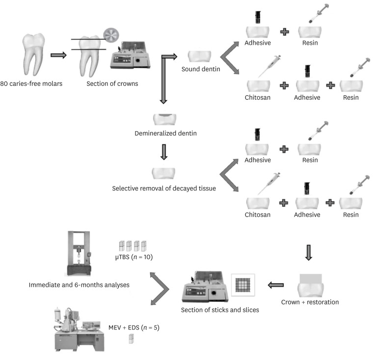
The sound and decayed specimens were randomly assigned into two groups according to dentin treatment: distilled water (control) and 2.5% chitosan solution. Each of these two groups was subdivided into 2 subgroups (10 teeth for each subgroup) and subjected to analysis immediately (24 hours) or after aging (6 months of storage in water + 12,000 thermal cycles + enzymatic degradation). Figure 1 shows the schematic diagram of the experimental workflow.
Figure 1

Schematic diagram of the experimental workflow.

rde-47-e28-g001

Preparation of chitosan solution

To prepare the chitosan solution, 2.5 g of low molecular weight (75%–85% deacetylation) chitosan (Sigma-Aldrich, Saint Louis, MO, USA) was slowly added to 100 mL of 1% acetic acid solution under magnetic stirring (Marconi Equip. Lab. Ltda., Piracicaba, SP, Brazil) for 20 minutes. To avoid particle aggregation and neutralize the pH solution, 1 mol/L NaOH solution was added [1821].

Sample restoration

For the specimens in each group that were subjected to the dentin treatment, chitosan solution was actively applied on the surface for 1 minute, followed by drying with absorbent paper. According to the manufacturer’s instructions, an adhesive layer (Single Bond Universal; 3M ESPE, St. Paul, MN, USA) was actively applied with subsequent solvent evaporation and polymerization using a light-emitting diode (LED) source (Gnatus, Ribeirão Preto, SP, Brazil) for 10 seconds [12]. Two increments of the composite resin (Filtek Z250/3M ESPE, St. Paul, MN, USA) were included and polymerized for 20 seconds in each increment using the LED source to restore the dentin surfaces. The LED source had maximum polymerization power at 1200 mW/cm2, which was measured using a radiometer (RD7; Ecel Indústria e Comércio Ltda., Ribeirão Preto, SP, Brazil). Restored specimens destined for immediate analysis were stored in distilled water at 37°C for 24 hours before the sectioning of the sticks. Table 1 present the material’s particulars used in this study.
Table 1

Material’s particulars used in this study

rde-47-e28-i001
Material Composition Manufacturer
Chitosan solution Low molecular weight chitosan, acetic acid, sodium hydroxide Sigma-Aldrich, Saint Louis, MO, USA
Adhesive System – Single Bond Universal MDP phosphate monomer, dimethacrylate resins, HEMA, Vitrebond Copolymer, filler, ethanol, water, initiators, silane 3M ESPE, St. Paul, MN, USA
Composite resin – Filtek Z250 Bis-GMA, UDMA, Bis-EMA, camphorquinone, zirconia/silica 3M ESPE, St. Paul, MN, USA
MDP, methacryloyloxydecyl dihydrogen phosphate; HEMA, 2-hydroxyethyl methacrylate; Bis-GMA, bisphenol A-glycidyl methacrylate; UDMA, urethane dimethacrylate; Bis-EMA, bisphenol-A glycidyl dimethacrylate.

Aging process

The other restored specimens from each group, destined for the aging process, were aged using a combination of hydrolytic, thermal, and enzymatic degradation protocols [52223].
Specimens were hydrolytically degraded in 20 mL distilled water at 37°C that was exchanged weekly for 6 months [22]. Subsequently, specimens were then subjected to thermal aging in water baths for 30 seconds, at temperatures ranging from 5°C to 55°C in a thermocycling machine (Ética Equip. Científicos S/A, São Paulo, SP, Brazil), with a transfer time of 3 seconds between each bath. There were 500 thermal cycles per week, totaling 12,000 cycles [23].
The specimens were then subjected to enzymatic degradation at the interface. For this purpose, they were immersed in artificial saliva, containing 100 U/mL EDTA-free protease inhibitor cocktail (Roche, Basel, Switzerland) in addition to 100 U/mL Clostridium histolyticum collagenase (Sigma-Aldrich) for 5 days at 37°C [24]. After this period, they were sectioned, as described below.

Sample sectioning

Restored specimens were sectioned into sticks of 1.0 ± 0.2 mm2 cross-sectional area using a precision cutter (IsoMet 1000; Buehler) under water irrigation [15]. The sticks’ thickness was certified using a digital caliper (Mitutoyo, Tokyo, Japan). At least four sticks were extracted from the central portion of the specimen, and one slice from the margin restoration [22].

Microtensile bond strength test

The sticks were attached to a stainless-steel device using cyanoacrylate adhesive (Super Bonder; Henkel Ltda., São Paulo, SP, Brazil) and vertically positioned at a universal testing machine (Instron Corporation, Canton, MA, USA). Then, the specimens were stressed by the device’s extremities until failure, at a load cell of 50 kg/f and a crosshead speed of 0.5 mm/min.
Both halves of each fractured specimen were analyzed under a stereomicroscope (Nikon, Melville, NY, USA) to categorize the failure pattern as adhesive when the failure occurred at the resin-dentin interface, cohesive of material when the surface was entirely covered by composite resin, cohesive of substrate when the failure occurred in dentin, and mixed when both adhesive and cohesive failure types were found in the dentin-resin interface.

Energy dispersive spectrometry (EDS) and scanning electron microscopic (SEM) analyses

EDS analysis identified and quantified the chemical elements in the sample using energy emission from excited atoms and ions. The slices for analyses in EDS and SEM were fixed in acrylic resin and polished with decreasing granulations of sandpaper (#600 and #1,200) and wet polishing cloth in synthetic fiber (Buehler Brazil, São Paulo, SP, Brazil) with 0.3-μm and 0.05-μm alumina slurries (Buehler Brazil) [12]. The surface was then analyzed using SEM with EDS coupled detector (EVO 50; Carl Zeiss, Cambridge, England) under an interface magnification of ×1,000.
Dehydration was performed by immersing specimens in ethanol at concentrations of 25%, 50%, 75%, and 95% for 20 minutes in each solution and 60 minutes in 100% ethanol. The specimens were fixed again in metallic stubs and covered with a thin layer of gold-palladium alloy (Bal-Tec SCD 005 Sputter Coater, Balzers, Liechtenstein) [12]. The adhesive interface was scanned, and the most representative area of each group was photographed at different magnifications.

Data analysis

IBM SPSS Statistics version 25 for Windows (IBM Corporation, Armonk, NY, USA), and a significance level of 5% was used. The Shapiro–Wilk and Levene’s test verified the normal and homogeneous distribution of the sample, and 3-way analysis of variance (ANOVA) was performed with substrate, dentin treatment, and aging as independent factors. The mean bond strength from the four sticks of each tooth was used, providing 40 values per subgroup for analysis. For the failure pattern analysis, the Kruskal–Wallis non-parametric test and Dunn’s post-test were used. The chemical element concentration is expressed as a percentage (%), and photomicrograph examination was performed by 2 blinded examiners (kappa intra-examiner A = 0.92, kappa intra-examiner B = 0.98, and kappa inter-examiner AB = 0.87).

RESULTS

µTBS test

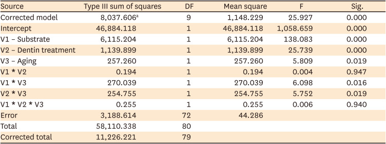
The ANOVA revealed that substrate, dentin treatment, and aging were statistically significant (p < 0.05). Regarding the substrate, specimens of sound dentin had significantly higher values than those of demineralized dentin (p < 0.001). The specimens treated with 2.5% chitosan solution had significantly higher bond strength than that of untreated control specimens (p < 0.001). The immediately tested specimens had higher bond strength values than those of the aged specimens (p = 0.019).
Regarding the interactions, substrate × dentin treatment (p = 0.947) and substrate × dentin treatment × aging (p = 0.940) interactions were not significant. When comparing the substrate with aging, the sound dentin had the highest bond strength compared to the demineralized dentin immediately and after the aging process (p = 0.016). Analyzing the interaction of dentin treatment with aging, 2.5% chitosan solution treatment of dentin increased (p = 0.019) the resin bond strength values compared to those of non-treated dentin for both periods (immediate and after aging). The µTBS values are presented in Table 2, and 3-way ANOVA interaction is displayed in Table 3.
Table 2

Microtensile bond strength mean values and standard deviations of dentin with water (control) and chitosan solution

rde-47-e28-i002
Dental substrate Distilled water (control) Chitosan solution
Immediate Aged* Immediate Aged*
Sound dentin 27.34 ± 4.33Ab 32.20 ± 5.85Ab 38.47 ± 8.21Aa 33.39 ± 9.92Aa
Demineralized dentin 13.54 ± 4.28Bb 9.74 ± 4.90Bb 24.65 ± 9.54Ba 13.93 ± 5.34Ba
*Six months of water storage + 12,000 thermal cycles + enzymatic degradation.
Same capital letters denote groups that are not statistically different in the comparison within columns (p < 0.05); Same lowercase letters denote groups that are not statistically different in the comparison within lines (p < 0.05); Non-bolded letters correspond to immediate results and bolded ones to aged.
Table 3

3-way analysis of variance interaction regarding the substrate, dentin treatment, and aging

rde-47-e28-i003
Source Type III sum of squares DF Mean square F Sig.
Corrected model 8,037.606a 9 1,148.229 25.927 0.000
Intercept 46,884.118 1 46,884.118 1,058.659 0.000
V1 – Substrate 6,115.204 1 6,115.204 138.083 0.000
V2 – Dentin treatment 1,139.899 1 1,139.899 25.739 0.000
V3 – Aging 257.260 1 257.260 5.809 0.019
V1 * V2 0.194 1 0.194 0.004 0.947
V1 * V3 270.039 1 270.039 6.098 0.016
V2 * V3 254.755 1 254.755 5.752 0.019
V1 * V2 * V3 0.255 1 0.255 0.006 0.940
Error 3,188.614 72 44.286
Total 58,110.338 80
Corrected total 11,226.221 79
DF, degrees of freedom.
aR squared = 0.716 (adjusted R squared = 0.688).

Failure pattern analysis

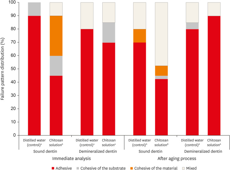
The failure pattern of each experimental group is expressed as the frequency of distribution (Figure 2) and was analyzed using non-parametric tests (p > 0.05). There was a predominance of adhesive failures. A significant difference (p < 0.05) was observed for the sound dentin treated with 2.5% chitosan due to the higher incidence of cohesive material and mixed-type failures in the immediate and after aging analyses, respectively.
Figure 2

Frequency distribution of the failure patterns (%) after microtensile bond strength (µTBS) test. Different letters indicate significant difference among groups. Capital letters correspond to immediate analysis and lowercase letters to after aging process (χ2 test, p < 0.05).

rde-47-e28-g002

EDS and SEM of the adhesive interface

Table 4 presents the data obtained from the EDS analysis of immediately tested sound specimens. In this analysis, it was possible to quantify the concentrations of carbon (C), oxygen (O), phosphorus (P), and calcium (Ca). There were no significant differences in the concentration of these elements in the adhesive interface of the studied groups.
Table 4

Mean and standard deviations of the atomic percentage (wt%) of chemical elements in the specimens

rde-47-e28-i004
Chemical element Sound dentin Demineralized dentin
Distilled water (control) Chitosan solution Distilled water (control) Chitosan solution
24 hours 6 months 24 hours 6 months 24 hours 6 months 24 hours 6 months
C 37.93 ± 0.82 39.97 ± 2.86 37.97 ± 2.79 39.73 ± 3.47 38.28 ± 0.28 39.65 ± 0.74 37.00 ± 2.36 38.60 ± 2.00
O 33.40 ± 0.50 42.17 ± 2.73 42.98 ± 10.20 37.52 ± 3.04 42.43 ± 7.00 41.55 ± 4.18 43.21 ± 1.29 40.62 ± 6.68
P 4.89 ± 0.66 5.09 ± 0.00 5.73 ± 1.94 6.80 ± 0.30 5.94 ± 2.10 5.80 ± 1.12 5.98 ± 0.38 6.33 ± 1.41
Ca 10.87 ± 0.97 11.36 ± 0.12 12.85 ± 5.45 15.65 ± 0.02 13.07 ± 4.45 12.57 ± 2.51 13.43 ± 0.48 13.92 ± 3.31
C, carbon; O, oxygen; P, phosphorus; Ca, calcium.
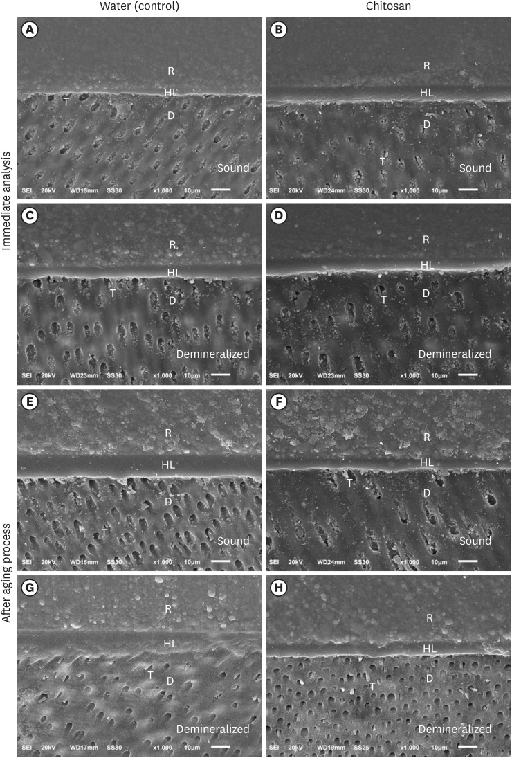
The SEM analysis of the sections restored with an adhesive system/composite resin immediately after the adhesive was applied allowed verification of the presence of resin tags, hybrid layer, and a good adhesive interface, regardless of the application of the chitosan solution. The same morphological pattern with tags, hybrid layer, and good adhesive interface was found in groups after the aging process (Figure 3).
Figure 3

Photomicrographs (×1,000) of the adhesive interface - Immediately after the adhesive procedure: (A) Sound dentin with water (control); (B) Sound dentin with chitosan; (C) Demineralized dentin with water and (D) Demineralized dentin with chitosan. After aging of the adhesive interface: (E) Sound dentin with water (control); F) Sound dentin with chitosan; (G) Demineralized dentin with water; and (H) Demineralized dentin with chitosan. R, resin; HL, hybrid layer; D, dentin; T, resin tags.

rde-47-e28-g003

DISCUSSION

Chitosan has been reported as an important biomaterial that can stabilize the adhesive interface, avoiding the degradation of the dentin organic matrix by metalloproteinases by forming crosslinks with collagen fibrils [13151720]. The present study evaluated the use of chitosan as a strategy to preserve the hybrid layer to allow infiltration of resin monomers inside the interfibrillar spaces of the dentin collagen matrix, which is responsible for the micromechanical retention of the restorative material on the substrate [25].
The choice of 2.5% chitosan solution concentration was based on previous studies because it forms a calcium phosphate layer on dentin and does not influence the wettability of the adhesive on the substrate [1226]. The dissolution of chitosan was carried out in acetic acid due to its insolubility in water, affirming that extra-fibrillar dentin demineralization by chitosan is attributed exclusively to its chelating capacity and not to the effect of the solvent [27].
Bond strength analysis revealed higher values in the sound specimens than in the demineralized specimens. The precipitates inside dentinal tubules differ the structure of caries-affected dentin from the sound dentin, and bond strength is inversely proportional to the degree of dentin involvement [2829].
In our results, the first null hypothesis was rejected. Chitosan was related to higher bond strength values in specimens, which demonstrates its ability to interact with the dental structure [17]. The chitosan cross-links with dental collagen produce a mechanically strong fibril chain and can explain the higher mechanical performance of chitosan-treated groups in this study, corroborating findings from other studies [13161827]. In contrast, Stenhagen et al. [30] evaluated the effect of methacrylate chitosan incorporated in experimental adhesives, and demonstrated no bond strength modifications in dentin. We could infer that this difference may be due to the methacrylation performed in the aforementioned study [30].
The main factor that decreases bond strength over time is the degradation of the dentin collagen matrix after the activation of metalloproteinases present in this matrix, which have gelatinolytic activity [4]. The present study used C. histolyticum collagenase to simulate collagen degradation [5]. The specimens were aged before sectioning of the sticks to reproduce the oral condition, in which the outer part of the restorations is more affected than the inside one. After hydrolytic and enzymatic aging of the specimens, the specimens treated with chitosan solution obtained higher values of bond strength. This result highlights the potential of the chitosan solution to guarantee the durability of the adhesive interface. A previous study also suggested that chitosan could inhibit the enzymatic activity of collagenase, increasing the resistance to collagen degradation and improving the bond strength of the resin-dentin surface [13].
Despite some divergences in applied methodology, mainly due to the manner in which chitosan was used, other authors have also observed favorable results regarding its use to improve the longevity of dental restorations [1831]. When using chitosan dentin treatment, Fawzy et al. [18] found better mechanical properties and a stabilized demineralized dentin substrate when challenged by hydrolytic and/or collagenolytic degradation. Diolosà et al. [31] also demonstrated mechanical advantages in a 5-year simulation of restoration in the oral cavity. Therefore, it is clear that chitosan can improve the adhesive interface and increase the durability of composite resin restorations.
In the failure pattern analysis, adhesive failures were predominant because it is the most common failure type found in µTBS tests. However, the statistical analysis revealed a difference in the group with sound dentine treated with chitosan solution, both in the immediate and aged specimens. In this group, there was an increase in the cohesion of material and mixed failures. A higher percentage of mixed failures suggests an improvement in the adhesive interface and consequently in the bond strength of such groups [32].
The second and third null hypotheses were accepted. EDS analysis revealed no differences in the concentrations of chemical elements at the adhesive interface. Chitosan treatment maintained the sample’s organic and inorganic composition and its adhesive interaction was preserved since changes in the calcium/phosphate ratio can negatively affect the permeability of adhesives in dental substrates [33]. SEM analysis of this study allowed verification of the formation of a hybrid layer and the presence of resinous tags in the dentinal tubules, suggesting a strong adhesive interface and corroborating previous study [8]. Beltrame et al. [34] also affirmed that their experimental chitosan solution had a favorable effect on maintaining the integrity of collagen fibrils; therefore, we can speculate that this biopolymer forms crosslinks with the organic dentin matrix. Despite the limitations of the in vitro study, this research may inspire further analyses to generate new adhesive protocols using chitosan biopolymers. Future studies are necessary to assess and confirm its effects on demineralized dentin as a crosslinking agent, since the inhibition of dentin metalloproteinases is one of the main factors contributing to the improvement of durability bond strength.

CONCLUSIONS

The 2.5% chitosan solution improved the bond strength of the resin to dentin and did not negatively affect the chemical composition and morphology of the adhesive interface. Chitosan increased the restoration’s mechanical resistance even after 6-month of degradation.

ACKNOWLEDGEMENTS

The authors would like to thank the National Council for Scientific and Technological Development (CNPq) for the financial support (425631/2016-8).

Notes

Conflict of Interest: No potential conflict of interest relevant to this article was reported.

Author Contributions:

  • Conceptualization: Ziotti IR.

  • Souza-Gabriel AE. Data curation: Ziotti IR.

  • Souza-Gabriel AE. Formal analysis: Souza-Gabriel AE. Funding acquisition.

  • Souza-Gabriel AE. Investigation: Ziotti IR. Methodology.

  • Ziotti IR. Project administration and Supervision: Souza-Gabriel AE. Resources.

  • Souza-Gabriel AE. Software: Ziotti IR.

  • Souza-Gabriel AE. Writing - original draft: Ziotti IR.

  • Souza-Gabriel AE. Writing - review & editing: Paschoini VL.

References

1. Brostek AM, Walsh LJ. Minimal intervention dentistry in general practice. Oral Health Dent Manag. 2014; 13:285–294. PMID: 24984635.
2. Comert S, Oz AA. Clinical effect of a fluoride-releasing and rechargeable primer in reducing white spot lesions during orthodontic treatment. Am J Orthod Dentofacial Orthop. 2020; 157:67–72. PMID: 31901283.
crossref
3. Perdigão J. Dentin bonding-variables related to the clinical situation and the substrate treatment. Dent Mater. 2010; 26:e24–e37. PMID: 20005565.
crossref
4. Chaussain-Miller C, Fioretti F, Goldberg M, Menashi S. The role of matrix metalloproteinases (MMPs) in human caries. J Dent Res. 2006; 85:22–32. PMID: 16373676.
5. Kato MT, Leite AL, Hannas AR, Calabria MP, Magalhães AC, Pereira JC, Buzalaf MA. Impact of protease inhibitors on dentin matrix degradation by collagenase. J Dent Res. 2012; 91:1119–1123. PMID: 23023765.
crossref
6. Park KM, Lee HJ, Koo KT, Ben Amara H, Leesungbok R, Noh K, Lee SC, Lee SW. Oral soft tissue regeneration using nano controlled system inducing sequential release of trichloroacetic acid and epidermal growth factor. Tissue Eng Regen Med. 2020; 17:91–103. PMID: 31970697.
crossref
7. Mazzi-Chaves JF, Martins CV, Souza-Gabriel AE, Brito-Jùnior M, Cruz-Filho AM, Steier L, Sousa-Neto MD. Effect of a chitosan final rinse on the bond strength of root canal fillings. Gen Dent. 2019; 67:54–57. PMID: 31454324.
8. Machado AH, Garcia IM, Motta AS, Leitune VC, Collares FM. Triclosan-loaded chitosan as antibacterial agent for adhesive resin. J Dent. 2019; 83:33–39. PMID: 30794843.
crossref
9. Rodrigues MR. Synthesis and investigation of chitosan derivatives formed by reaction with acyl chlorides. J Carbohydr Chem. 2005; 24:41–54.
crossref
10. Kong M, Chen XG, Xing K, Park HJ. Antimicrobial properties of chitosan and mode of action: a state of the art review. Int J Food Microbiol. 2010; 144:51–63. PMID: 20951455.
crossref
11. Chronopoulou L, Nocca G, Castagnola M, Paludetti G, Ortaggi G, Sciubba F, Bevilacqua M, Lupi A, Gambarini G, Palocci C. Chitosan based nanoparticles functionalized with peptidomimetic derivatives for oral drug delivery. N Biotechnol. 2016; 33:23–31. PMID: 26257139.
crossref
12. Curylofo-Zotti FA, Tanta GS, Zucoloto ML, Souza-Gabriel AE, Corona SA. Selective removal of carious lesion with Er:YAG laser followed by dentin biomodification with chitosan. Lasers Med Sci. 2017; 32:1595–1603. PMID: 28762194.
crossref
13. Baena E, Cunha SR, Maravić T, Comba A, Paganelli F, Alessandri-Bonetti G, Ceballos L, Tay FR, Breschi L, Mazzoni A. Effect of chitosan as a cross-linker on matrix metalloproteinase activity and bond stability with different adhesive systems. Mar Drugs. 2020; 18:18.
crossref
14. Persadmehr A, Torneck CD, Cvitkovitch DG, Pinto V, Talior I, Kazembe M, Shrestha S, McCulloch CA, Kishen A. Bioactive chitosan nanoparticles and photodynamic therapy inhibit collagen degradation in vitro . J Endod. 2014; 40:703–709. PMID: 24767568.
crossref
15. Anjana J, Mohandas A, Seethalakshmy S, Suresh MK, Menon R, Biswas R, Jayakumar R. Bi-layered nanocomposite bandages for controlling microbial infections and overproduction of matrix metalloproteinase activity. Int J Biol Macromol. 2018; 110:124–132. PMID: 29233714.
crossref
16. Curylofo-Zotti FA, Scheffel DL, Macedo AP, Souza-Gabriel AE, Hebling J, Corona SA. Effect of Er:YAG laser irradiation and chitosan biomodification on the stability of resin/demineralized bovine dentin bond. J Mech Behav Biomed Mater. 2019; 91:220–228. PMID: 30597375.
17. Pini NI, Lima DA, Luka B, Ganss C, Schlueter N. Viscosity of chitosan impacts the efficacy of F/Sn containing toothpastes against erosive/abrasive wear in enamel. J Dent. 2020; 92:103247. PMID: 31743693.
crossref
18. Fawzy AS, Nitisusanta LI, Iqbal K, Daood U, Beng LT, Neo J. Chitosan/Riboflavin-modified demineralized dentin as a potential substrate for bonding. J Mech Behav Biomed Mater. 2013; 17:278–289. PMID: 23127636.
crossref
19. Hashimoto M, Tay FR, Svizero NR, de Gee AJ, Feilzer AJ, Sano H, Kaga M, Pashley DH. The effects of common errors on sealing ability of total-etch adhesives. Dent Mater. 2006; 22:560–568. PMID: 16289724.
crossref
20. Marquezan PK, Alves LS, Dalla Nora A, Maltz M, do Amaral Zenkner JE. Radiographic pattern of underlying dentin lesions (ICDAS 4) in permanent teeth. Clin Oral Investig. 2019; 23:3879–3883.
crossref
21. Daood U, Iqbal K, Nitisusanta LI, Fawzy AS. Effect of chitosan/riboflavin modification on resin/dentin interface: spectroscopic and microscopic investigations. J Biomed Mater Res A. 2013; 101:1846–1856. PMID: 23184366.
crossref
22. Profeta AC, Mannocci F, Foxton RM, Thompson I, Watson TF, Sauro S. Bioactive effects of a calcium/sodium phosphosilicate on the resin-dentine interface: a microtensile bond strength, scanning electron microscopy, and confocal microscopy study. Eur J Oral Sci. 2012; 120:353–362. PMID: 22813227.
crossref
23. Borsatto MC, Martinelli MG, Contente MM, Mellara TS, Pécora JD, Galo R. Bond durability of Er:YAG laser-prepared primary tooth enamel. Braz Dent J. 2013; 24:330–334. PMID: 24173250.
crossref
24. Ganss C, Klimek J, Brune V, Schürmann A. Effects of two fluoridation measures on erosion progression in human enamel and dentine in situ . Caries Res. 2004; 38:561–566. PMID: 15528912.
crossref
25. Castellan CS, Bedran-Russo AK, Antunes A, Pereira PN. Effect of dentin biomodification using naturally derived collagen cross-linkers: one-year bond strength study. Int J Dent. 2013; 2013:918010. PMID: 24069032.
crossref
26. Ururahy MS, Curylofo-Zotti FA, Galo R, Nogueira LF, Ramos AP, Corona SA. Wettability and surface morphology of eroded dentin treated with chitosan. Arch Oral Biol. 2017; 75:68–73. PMID: 28061390.
crossref
27. Gu LS, Cai X, Guo JM, Pashley DH, Breschi L, Xu HH, Wang XY, Tay FR, Niu LN. Chitosan-based extrafibrillar demineralization for dentin bonding. J Dent Res. 2019; 98:186–193. PMID: 30326766.
crossref
28. Costa AR, Garcia-Godoy F, Correr-Sobrinho L, Naves LZ, Raposo LH, Carvalho FG, Sinhoreti MA, Puppin-Rontani RM. Influence of different dentin substrate (caries-affected, caries-infected, sound) on long-term μTBS. Braz Dent J. 2017; 28:16–23. PMID: 28301013.
crossref
29. Yoshiyama M, Tay FR, Torii Y, Nishitani Y, Doi J, Itou K, Ciucchi B, Pashley DH. Resin adhesion to carious dentin. Am J Dent. 2003; 16:47–52. PMID: 12744413.
30. Stenhagen IS, Rukke HV, Dragland IS, Kopperud HM. Effect of methacrylated chitosan incorporated in experimental composite and adhesive on mechanical properties and biofilm formation. Eur J Oral Sci. 2019; 127:81–88. PMID: 30412313.
crossref
31. Diolosà M, Donati I, Turco G, Cadenaro M, Di Lenarda R, Breschi L, Paoletti S. Use of methacrylate-modified chitosan to increase the durability of dentine bonding systems. Biomacromolecules. 2014; 15:4606–4613. PMID: 25347288.
crossref
32. Perdigão J. New developments in dental adhesion. Dent Clin North Am. 2007; 51:333–357. PMID: 17532916.
crossref
33. Rohanizadeh R, LeGeros RZ, Fan D, Jean A, Daculsi G. Ultrastructural properties of laser-irradiated and heat-treated dentin. J Dent Res. 1999; 78:1829–1835. PMID: 10598913.
crossref
34. Beltrame APCA, Suchyta D, Abd Alraheam I, Mohammed A, Schoenfisch M, Walter R, Almeida IC, Souza LC, Miguez PA. Effect of phosphorylated chitosan on dentin erosion: an in vitro study. Caries Res. 2018; 52:378–386. PMID: 29510408.
crossref
TOOLS
Similar articles