

 PDF
PDF Citation
 Print
Print


INTRODUCTION
The current dentin adhesives are categorized into 2 systems based on how they interact with the smear layer. Etch-and-rinse adhesives require a separate etching step with phosphoric acid to remove the smear layer and superficial hydroxyapatite. In contrast, self-etching adhesives partially dissolve the smear layer and penetrate through the demineralized dentin [1]. Self-etching adhesive systems are widely used because they are less technique sensitive, thus, user-friendly. One-step self-etching adhesives are the most convenient and time-saving, with all clinical steps integrated into 1 application. These simplified adhesives are composed of acidic functional monomers, hydrophilic and hydrophobic monomers, water, and organic solvents in a single solution, implying some degree of hydrophilicity [2]. When bonded to dentin, the one-step self-etching adhesives act as semi-permeable membranes that allow water to cross the adhesive layer, including after polymerization [3].
A new family of one-step multi-mode universal adhesives has been introduced to the market. The “All-in-one” concept was used to design the already existing one-step self-etching adhesives to be more versatile and adaptable to different clinical situations [4]. The manufacturers claimed that these universal adhesives could adhere to various types of substrates, and could also be used in both etch-and-rinse and self-etching approaches [5]. However, prior phosphoric acid etching to dentin does not improve the bond strength of universal adhesives [67]. Vital dentin is hydrated by an outward fluid flow from the dentinal tubules due to positive pulpal pressure, estimated to be approximately 15 cmH2O [8]. There have been several in vitro bonding studies performed using a simulated pulpal pressure model. A significant reduction in bond strength has been reported due to the increased surface wetness from the fluid flow through dentin [91011]. The adhesion of resin composite to the cured adhesive can also be interfered with by fluid contamination, and therefore the bond durability might be reduced [1213].
When bonding self-etching adhesives to dentin, the smear layer plays an important role in resin adhesion [14151617]. Different surface preparations likely result in different smear layer characteristics. The dentin smear layer created by a diamond bur or carbide bur is more compact and denser than that produced by silicon carbide (SiC) abrasive paper [18]. Furthermore, the smear layer thickness depends also on the abrasive particle size [1719]. There was a study demonstrating that the smear layer prevented direct contact between the tooth surface and bonding agent [20]. In addition, a smear layer might buffer the acidic primer and prevent the resin from infiltrating into the underlying dentin [2122]. There are, however, still controversial issues in relation to the effects of the smear layer thickness on the resin-dentin bond strength of self-etching adhesives [1014172123].
Currently, there is scant evidence regarding the effect of different smear layers from different dentin surface preparation methods combined with pulpal pressure on the bonding performance of self-etching adhesives. Therefore, the purpose of this study was to evaluate the effect of different smear layer preparations on the dentin bond strength of 2 self-etching adhesives with simulated pulpal fluid movement. The null hypotheses tested were; 1) there would be no difference in dentin permeability among the different surface preparations under simulated pulpal pressure, 2) there would be no difference in dentin permeability between the 2 adhesives used, 3) no significant difference in the microtensile bond strengths (µTBSs) of each self-etching adhesive would be detected among the different dentin surface preparations under simulated pulpal pressure, and 4) no significant difference in dentin bond strengths would be found between the 2 self-etching adhesives.
MATERIALS AND METHODS
Sample preparation and pulpal pressure simulation
One hundred and 36 extracted human third molars were used in this study based on Armstrong et al. [24] and Carvalho et al. [25]. The teeth were collected after the study protocol was approved by the Ethics Committee of the Institutional Review Board (COE.No.MU-DT/PY-IRB 2019/056.2309). All teeth were stored in a 0.1% thymol solution and used within 6 months after extraction. To obtain the crown segment, the occlusal enamel was removed to expose flat mid-coronal dentin and the roots were cut off 1 mm below the cemento-enamel junction using a slow-speed water-cooled diamond saw (IsoMet; Buehler, Lake Bluff, IL, USA). Two cuts were made parallel to each other and perpendicular to the long axis of each tooth, accessing the pulp chamber at the furcation level (Figure 1). The pulp tissue was carefully removed using tweezers, without touching the surrounding predentin walls. The pulp chamber was irrigated with 2.5% NaOCl for 30 seconds, followed by immersion in distilled water for 30 minutes to neutralize the effects of NaOCl [9].
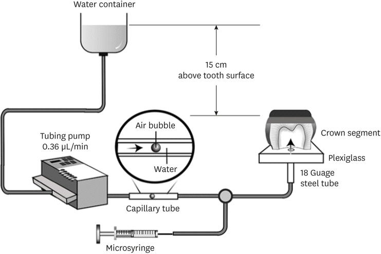
Figure 1 illustrates the dynamic intra-pulpal pressure simulation apparatus used in the current study. The pulpal simulation model used in this study was modified from the protocol of Pashley and Depew [26] with an additional infusion pump. Each crown segment was attached to a plexiglass plate on the pulp side using a cyanoacrylate adhesive (Model Repair II Blue; Dentsply-Sankin, Tochigi, Japan) and the pulp chamber was penetrated by an 18-gauge stainless steel needle. A transparent capillary tube was connected to the stainless steel needle, and the other end of this tube was inserted into a pressure reservoir to maintain an airtight seal and then filled with distilled water. To generate an intra-pulpal pressure of 15 cmH2O, the level of distilled water in the container was adjusted to 15 cm above the flat dentin surface of each crown segment. The pump was set at 0.36 µL/min to simulate the outward fluid flow rate [8]. Dynamic pulpal pressure was maintained throughout the experiment.
Permeability measurement
Forty crown segment-plexiglass plate assemblies were used for the permeability measurement of 4 different surface preparations and 2 self-etching adhesives (n = 5 per group). The dentin smear layer was removed by ultrasonic treatment in a 17% ethylenediaminetetraacetic acid (EDTA) solution for 5 minutes [27] and rinsed with water for 1 minute. Subsequently, each assembly was connected to the pulpal simulation device and underwent the maximum fluid filtration measurement, which was expressed as 100% of its permeability. The fluid flow was measured by monitoring the movement of an air bubble trapped within a capillary tube that was positioned between the pressure reservoir and the crown segment. Images of the air bubble at 0 and 10 minutes under simulated pulpal pressure were obtained and used to measure the distance the air bubble moved by ImageJ software. After measuring the initial maximum permeability, the tube was removed from the needle and the smear layer was re-created on the dentin surfaces using 4 different methods. Three types of tapered cylindrical-shaped dental burs; coarse diamond burs (852G.016, 120–150 µm particle size, diamond point FG; Jota, Rüthi, Switzerland), superfine diamond burs (852EF.016, 20–30 µm particle size, diamond point FG; Jota) or carbide burs (C48L; Jota), and 600-grit SiC abrasive paper (25.8 µm mean particle size; Buehler) were used for surface preparation. A single operator was trained how to apply up to 100 g of pressure using an analytical balance prior to performing the surface preparation [28]. For the bur-prepared groups, the dentin surfaces were ground using the dental burs mounted in a high-speed handpiece with copious water spray for 5 light-pressure strokes. The bur was replaced with a new one after 5 samples were prepared. For the SiC group, the smear layer was created by manually grinding the dentin surfaces with 600-grit SiC paper for 60 seconds under running water [15]. The assembly was then reattached to the model and the fluid filtration rate of the smear layer-covered dentin was measured again. Care was taken not to allow the dentin surfaces to dehydrate throughout the experiment. The percentage of dentin permeability (%P) was calculated using the following equation:

The value derived from this equation exhibited the permeability of the smear layer-covered dentin related to the maximum EDTA-treated permeability, with each tooth serving as its own control.
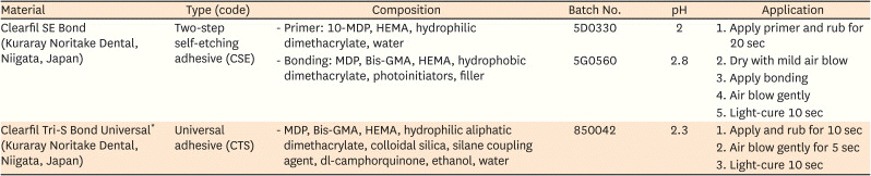
After measuring the smear layer permeability, Clearfil SE Bond (CSE) or Clearfil Tri-S Bond Universal (CTS) (Kuraray Noritake Dental Inc., Niigata, Japan) was applied on the dentin surfaces under simulated pulpal pressure. The compositions and instructions for use of the adhesives tested are listed in Table 1. An LED light-curing unit (Bluephase; Ivoclar Vivadent, Schaan, Liechtenstein) with a power intensity of 1,200 mW/cm2 was used to light-cure each adhesive for 10 seconds. The unit was calibrated before each use. The fluid filtration rate was re-measured and the dentin permeability of each specimen was calculated using the abovementioned equation.
Table 1
The materials used, their compositions, and the application procedures performed in the experiment
µTBS test
The remaining 80 crown segment-plexiglass plate assemblies were employed in this assay. The assemblies were divided into 8 groups according to the 4 different surface preparations and 2 adhesives (n = 10 per group). Smear layer creation and simulation of dynamic intra-pulpal pressure were achieved as described above. A resin composite (Clearfil AP-X ES2; Kuraray Noritake Dental Inc.) was placed on the bonded dentin surface in 2 increments of 2 mm each and cured for 20 seconds. The bonded teeth were removed from the pulp pressure simulation device and kept in distilled water at 37ºC for 24 hours. After storage, each tooth was sectioned occluso-gingivally along the mesio-distal and bucco-lingual axes using a slow-speed diamond saw with constant water coolant. Four resin-dentin beams, approximately 1 × 1 mm in cross section, from the central part of each bonded tooth were used for the non-trimming technique of the µTBS test. The specimens were fixed on a Ciucchi's jig using cyanoacrylate glue. The µTBS test was performed using a Lloyd universal testing machine (Model LR10K; Lloyd Instruments, Fareham, UK) at a crosshead speed of 1 mm/min. The data were recorded and expressed as MPa, and the µTBS values of the specimens from each bonded tooth were averaged [24].
Failure mode analysis
The fractured specimens were mounted on metal stubs. The surfaces were coated with palladium (K500X Sputter coater; SPI Supplies, West Chester, PA, USA) and examined using a scanning electron microscope (JSM 6610LV; JEOL, Peabody, MA, USA) at a 15 kV with 75× magnification to determine the mode of failure. The failure modes were classified as; 1) adhesive at the interface, 2) cohesive in resin either in the adhesive layer or in resin composite, 3) cohesive in dentin, and 4) mixed failure.
Observation of the resin-dentin interface and nanoleakage
The bonded teeth were prepared in the same manner as for the µTBS test (2 teeth per group). After 24 hours of water storage, each bonded tooth was vertically sectioned through the center by a slow-speed diamond saw, obtaining 1-mm thick resin-dentin slabs. Part of each slab was used for the resin-dentin interface observation and the other part for the nanoleakage observation. For the resin-dentin interface observation, the slabs were placed inside an acrylic ring and embedded in epoxy resin. The epoxy-embedded specimens were ground with a series of 800-, 1,000-, and 1,200-grit SiC papers under running water and polished with diamond pastes on polishing cloths down to a 0.25 µm particle size. The specimens were further etched with 10% phosphoric acid for 3 seconds, followed by immersion in 5.25% sodium hypochlorite for 5 minutes [19]. After overnight storage in a desiccator, the specimens were coated with palladium and the resin-dentin interface was observed using a scanning electron microscopy (SEM). For the nanoleakage detection, the resin-dentin slabs were immersed in 50 wt% silver nitrate for 24 hours, followed by exposure in a photo-developing solution for 8 hours under fluorescent light, and then fixed in 10% buffered formalin for 24 hours [29]. The prepared slabs were embedded in epoxy resin, and the specimens were polished as described above. Without the acid-base treatment, SEM observation was performed to detect the nanoleakage zones.
Statistical analysis
Data from the permeability measurement and the µTBS test were analyzed for normal distribution and homogeneity of variance using the Kolmogorov-Smirnov test and Levene's test, respectively. Analysis of Variance (ANOVA) and Dunnett's T3 multiple comparison test were used to analyze the permeability data after different surface preparations and after adhesives application. For the µTBS values, 2-way ANOVA and Duncan multiple comparison test were used. The failure mode distribution was analyzed using Pearson's χ2 test. All analyses were performed using a statistical software system (SPSS 27.0; IBM Corp., Armonk, NY, USA) at the 95% confidence interval.
RESULTS
Permeability measurement
The permeability measurement results are shown in Figure 2. After creating a smear layer on the dentin surfaces using the 4 different armamentaria, the carbide bur and superfine diamond bur groups yielded the lowest permeability (p ≤ 0.035). The highest permeability was found in the coarse diamond bur group; however, it was not significantly different from that of the SiC abrasive paper group (p = 0.115). After adhesive application, although the permeability was reduced in all tested groups, there was no significant difference between the 2 different adhesives (p ≥ 0.444). For both materials, the permeability between the superfine diamond bur, coarse diamond bur, and SiC paper groups was similar (p ≥ 0.373). The permeability of the carbide bur group was the lowest and was also not significantly different from the others (p ≥ 0.114), except for after CTS application where the permeability of the carbide bur group was significantly different from the coarse diamond bur group (p = 0.002).
Figure 2
Mean and standard deviation of dentin permeability (%P) after surface preparation and after application of each tested adhesive. The %P of each tooth is expressed as a percentage of its own maximum permeability. The same uppercase letters indicate no significant difference in permeability after dentin surface preparation (p > 0.05). The same lowercase letters indicate no significant difference in dentin permeability after applying each adhesive (p > 0.05).
SiC, silicon carbide.

µTBS test
The results of the µTBS test are shown in Table 2. Two-way ANOVA revealed that dentin surface preparation and type of adhesive influenced the bond strength (p < 0.001). The CSE group had a significantly higher bond strength compared with the CTS group for the dentin surfaces prepared with SiC paper or carbide bur (p < 0.05). Among the different surface preparations, the µTBS value of both adhesives to dentin prepared with coarse diamond burs was the lowest. The bond strengths of each adhesive were also the highest when bonded to dentin prepared with superfine diamond burs or carbide burs, especially in the CSE group, which was significantly different compared with the coarse diamond bur group (p < 0.05).
Table 2
Microtensile bond strength (µTBS) of the 2 self-etching adhesives to dentin with different surface preparations (n = 10/group)

Failure mode analysis
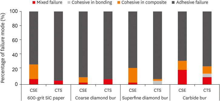
The percentage distribution of the failure modes is shown in Figure 3. None of the specimens demonstrated cohesive failure in dentin. The predominant fracture pattern in every group was adhesive failure (65%–95%). Cohesive failure in the adhesive layer was observed only when CTS was bonded to superfine diamond bur- and carbide bur-prepared dentin. Comparing the 2 adhesives, a significantly higher incidence of cohesive failure in the resin composite was found for CSE.
Observation of the resin-dentin interface and nanoleakage
Representative SEM images of the resin-dentin interface and nanoleakage patterns are displayed in Figures 4 and 5, respectively. For the resin-dentin interface observation, no distinct interaction zone was detected for either tested adhesives (Figure 4). More and longer resin tags were present for CSE compared with CTS. Among the CSE groups, the dentin surfaces prepared by a coarse diamond bur or 600-grit SiC paper demonstrated a few resin tags that were thin and slim (Figure 4A and 4B). In contrast, resin tags on the dentin surfaces prepared by superfine diamond and carbide burs were more abundant and conical (Figure 4C and 4D). For CTS, the resin tags in the coarse diamond bur and 600-grit SiC paper groups were scarce and relatively short (Figure 4E and 4F).
Figure 4
Representative scanning electron microscopy images of the resin-dentin interface of Clearfil SE Bond (upper) and Clearfil Tri-S Bond Universal (lower) at 3,000× magnification. (A, E) Dentin surface prepared with 600-grit SiC abrasive paper. (B, F) Dentin surface prepared with a coarse diamond bur. (C, G) Dentin surface prepared with a superfine diamond bur. (D, H) Dentin surface prepared with a carbide bur.

Figure 5
Representative scanning electron microscopy images of the resin-dentin interface with silver staining of Clearfil SE Bond (upper) and Clearfil Tri-S Bond Universal (lower) at 3,000× magnification. (A, E) Dentin surface prepared with 600-grit silicon carbide (SiC) abrasive paper. (B, F) Dentin surface prepared with a coarse diamond bur. (C, G) Dentin surface prepared with a superfine diamond bur. (D, H) Dentin surface prepared with a carbide bur. Nanoleakage (arrows) were observed in the 600-grit SiC abrasive paper and coarse diamond bur groups for both adhesives.
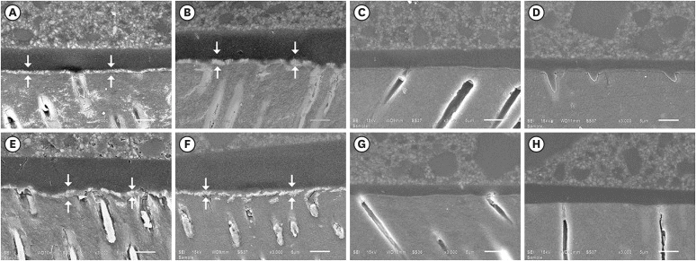
Figure 5 shows the nanoleakage that occurred in each experimental group. Irrespective of the adhesive tested, nanoleakage was obvious within the hybrid layer of the coarse diamond bur and 600-grit SiC paper groups (Figure 5A, 5B, 5E and 5F). In contrast, nanoleakage was not detected in the carbide bur and superfine diamond bur groups using either adhesive (Figure 5C, 5D, 5G and 5H).
DISCUSSION
The present study evaluated the effect of different dentin surface preparations on the bond strength of 2 self-etching adhesives under simulated fluid movement. Our results indicated that the type of surface preparation had a significant effect on dentin permeability. Therefore, the first null hypothesis was rejected.
The presence of a smear layer has been reported to have a significant influence on dentin permeability [30]. A previous study found a 45%–51% reduction in permeability after the smear layer was created using SiC abrasive paper [31]. This is in agreement with the results of our study, where after grinding with 600-grit SiC paper, the permeability was reduced by half. It has been reported that dentin prepared using dental burs demonstrated surfaces covered with a more compact smear layer [1832]. The lower permeability could therefore be expected for the bur-prepared groups. However, the highest permeability was observed in the group prepared using coarse diamond burs. The smear layer characteristics, i.e., thickness and density, should be taken into account for the dentin permeability evaluation.
The smear layer is a mixture of organic and inorganic components created on the tooth surface during mechanical preparation. The inorganic portion consists of hydroxyapatite crystals and minerals from the ground dentin surface [33]. The organic portion consists of fragments of the odontoblastic processes, microorganisms, and denatured collagen fibrils [34]. We hypothesized that the organic portion of the smear layer might play an important role in water diffusion through dentin. A previous study reported that water is essential for stabilizing the collagen triple helix structure [35]. The hydrogen bonds between the collagen and water molecules allow liquid to flow through the dentin and reach the bond surface [36]. Therefore, due to their hydrophilic nature, the collagen fibrils in the smear layer might act as a sponge to absorb water from the underlying dentin surface.
The coarse diamond burs created a more compact and thicker smear layer compared with the superfine diamond or carbide burs; thus, a higher permeability could be anticipated. The superfine diamond burs and carbide burs generated a thinner smear layer that was 50–500 nm thick [32]. The thinner smear layer might be fully saturated with water more readily during surface preparation. Free water in the dentinal tubules then could not move outward during the intra-pulpal pressure simulation. This might explain why low permeability was detected in the superfine diamond bur and carbide bur groups.
When each adhesive was applied and light-cured, hybridized resin tags were formed in the dentinal tubules. In addition, the hybrid layer was also covered by a thin layer of cured adhesive resin. The results of the current study demonstrated that the permeability was reduced to 5%–31% after adhesive application. As noted in the Introduction, one-step self-etching adhesives are hydrophilic and semi-permeable, allowing water to diffuse from the underlying dentin to the adhesive surfaces [3]. It was therefore expected that the CTS permeability should be higher than that of CSE, because the latter was used with a separate hydrophobic bonding resin [37].
However, we found no significant differences in permeability between the 2 adhesives. Thus, the second null hypothesis was not rejected. The so-called universal adhesives were developed to have a more hydrophobic single-component adhesive compared with their predecessors. By incorporating highly cross-linking monomers and minimizing the amount of HEMA, universal adhesives might be sufficiently hydrophobic to be used in a single layer. Furthermore, the 10-methacryloyloxydecyl dihydrogen phosphate (10-MDP), which is a component of CSE and CTS, is the most hydrophobic among the adhesive functional monomers [38]. The inclusion of 10-MDP and modification of the ingredients could optimize the universal adhesive tested to behave hydrophobically as the 2-step self-etching adhesive did after light-curing. This might explain why similar permeabilities were detected between CSE and CTS.
After adhesive application, the results revealed that the carbide bur-bonded dentin demonstrated the lowest permeability. The thin and less compact smear layer produced by the carbide burs might allow each adhesive to easily penetrate deeply into the tubules, eliminating water, and intermingling with the demineralized dentin surface [32]. A resin-dentin interdiffusion zone, along with the overlying cured adhesive could then prevent dentin fluid permeation. Although the permeability in the carbide bur group was the lowest, its µTBS was not the highest. Indeed, it tended to have a lower µTBS compared with that of the superfine diamond bur group, especially when CTS was applied. A previous study reported that the smear layer thickness and tubule openness created by these 2 dental burs were not significantly different [21]. Dissimilarities in dentin smear layer characteristics between diamond particle abrasion and blade cutting action should also be considered, however, this requires further investigation. The SiC abrasive paper and coarse diamond bur groups presented higher permeability values. This could result from the rather thick hydrated smear layers where both adhesives tested might have not been able to fully hybridize. Nanoleakage in the hybrid layer is obvious in the SEM images for both tested groups after resin composite placement.
In the bond strength assay, 2-way ANOVA revealed that the adhesives and the surface preparations influenced the µTBS. Therefore, the third and fourth null hypotheses that the dentin bond strengths would not be affected by the adhesives and the types of smear layer produced were rejected. CSE demonstrated a higher bond strength compared with CTS, especially when bonded to dentin surfaces prepared with 600-grit SiC paper or carbide burs. SiC abrasive paper created a thick smear layer, while that produced by the carbide burs was, although thinner, composed of well-arranged and undisrupted collagen fibrils [18]. The self-etching primer used in CSE, which has a lower pH and viscosity, might dissolve the smear layer and better interact with the underlying dentin compared with CTS. Furthermore, in this study, the CSE primer was intentionally manipulated with active application to enhance the smear layer interaction [3940]. These might be the reasons why CSE outperformed CTS in dentin adhesion. These findings are supported by the failure mode analysis, with CSE demonstrating a significantly higher percentage of cohesive failure in the resin composite. In addition, the resin-dentin interface SEM observation presented more abundant and longer resin tags for the groups bonded with CSE compared with those bonded with CTS. Comparing the diamond burs used, no significant difference in µTBS was observed between 2 adhesives. Deposition of a more compact smear layer might be responsible for the similarity in dentin bond strengths because both mild self-etching adhesives might not perform well over a dense zone of debris.
CSE and CTS demonstrated a similar trend in dentin bond strength with respect to different surface preparations. High µTBS values were detected in the superfine diamond bur and carbide bur groups, followed by the SiC paper, and coarse diamond bur groups. This outcome is supported by the SEM images and the permeability assay results. Nanoleakage was detected within the resin-dentin interdiffusion zone only in the coarse diamond bur and SiC paper groups. In addition, the permeability values of the dentin surfaces prepared with coarse diamond bur and SiC paper were the highest. These results could imply that high water-uptake through the bonded dentin in the coarse diamond bur and SiC paper groups could weaken the resin adhesion. The impact of the smear layer created by SiC paper and coarse diamond burs was more pronounced when bonded with CTS because the resin tags were scarce and tenuous. This could be the reason for the lower bond strength when dentin surfaces prepared with 600-grit SiC paper or coarse diamond burs were bonded, especially using CTS. Long-term dentin adhesion should be further investigated to verify the effect of intra-pulpal pressure on the performances of self-etching adhesives.
CONCLUSIONS
Within the limitations of the current study, we conclude that the permeability of smear layer depends on the instruments used to abrade the dentin surface. For clinicians, the combination of 2-steps self-etching adhesive and surface preparation with either superfine diamond bur or carbide bur is recommended.
 XML Download