INTRODUCTION
Vital tooth bleaching can be divided into in-office (associated or not with a light source) and at-home techniques, and also with the association of these 2 methods. All of these techniques use gels based on hydrogen peroxide or carbamide peroxide. In general, they aim at breaking the chromogen molecules present within the dentin, thus changing the color of the teeth [
1]. In addition to the color change, alterations of micromorphological nature may be associated with the use of bleaching agents, including increased surface roughness, demineralization, changes in enamel hardness and surface morphology [
23]. In-office bleaching involves the direct supervision of a clinician to avoid injuries to the soft tissues and the ingestion of gel, reducing total treatment time and producing quicker result in terms of bleaching efficacy [
456].
It is known that in-office bleaching, using gels with high concentration of hydrogen or carbamide peroxide, can cause hypersensitivity during and after the procedure [
7]. Tooth sensitivity is the most undesirable side effect of tooth bleaching and affects 8% to 66% of patients, usually with a moderate degree of pain in the early stages of treatment [
8]. Recently, alternative protocols that use violet light emitting diode (V-LED) have also been proposed to reduce sensitivity during in-office bleaching procedures [
91011].
Dental bleaching associated with light sources has been extensively studied [
12]. The advantage of light-activated bleaching is the ability to heat the hydrogen peroxide, increasing its decomposition rate into free radicals for the oxidation of complex organic molecules [
13]. However, many randomized clinical studies have shown controversial results, questioning the use of any kind of light sources in bleaching procedures regarding the bleaching efficacy [
46]. In addition, some studies have shown that light-activated bleaching can increase tooth sensitivity due to the release of more free radicals that reach the pulp and tooth heating [
1415]. A recent systematic review has not shown any superior bleaching efficacy with the use of light activation in different in-office bleaching protocols when compared to those that did not use light sources [
12].
Violet light has a wavelength that varies between 405 and 410 nm. The wavelength range of the violet light corresponds to the absorption peak of the pigment molecules, which are greatly reactive to light, promoting the breakdown of the bonds present in the molecular chains that form the pigments present in the tooth structure [
10111617]. Violet light does not seem to promote demineralization effect on the enamel or cause alterations in enamel morphology [
1618]. V-LED can be used with and without bleaching gel during in-office bleaching [
9111617]. The application of V-LED alone, without gel application, would reduce the post-bleaching sensitivity, making the patient’s postoperative period more comfortable [
16]. However, the results regarding its efficacy have not yet been fully demonstrated. Studies have shown that the use of V-LED alone can have a bleaching effect, but to a lesser extent than when used in association with carbamide or hydrogen peroxide-based gels [
111617]. In a clinical study, the group that was subjected to tooth bleaching with V-LED showed significantly more color change than the group that did not use the violet light [
9]. In another clinical study, V-LED improved the bleaching results when associated with hydrogen peroxide [
16]. Nonetheless, although the results are promising, additional studies need to be carried out to confirm the efficacy of bleaching techniques associated with V-LED.
There is still a lack of evidence related to efficacy and safety of this new V-LED device [
16]. No studies have yet been found that showed the occurrence and magnitude of pulpal temperature increase with LED violet light in the bleaching protocols. Therefore, the objectives of the present study are: i) to evaluate the efficacy of different in-office bleaching protocols associated or not with V-LED; and ii) to evaluate the change in pulp temperature during in-office bleaching associated with V-LED with and without gel application. The hypotheses tested are: i) the V-LED would influence the efficacy of in-office bleaching protocols; and ii) the V-LED would increase the pulp temperature, regardless the application of bleaching gel.
MATERIALS AND METHODS
Bleaching efficacy of different protocols associated with LED violet light
Forty freshly extracted bovine incisors free of enamel defects or evident discoloration were disinfected in chloramine solution (1%) for 24 hours. Then were kept refrigerated (< 4°C) in distilled water, which was changed weekly for a month, until the beginning of the experiments. The shades of the teeth were assessed prior to the experiments with a spectrophotometer (EasyShade Advance, Vita Zahnfabrik, Bad Säckingen, Germany), and were classified as A1, B2 or A2. Before the bleaching procedures, the teeth were cleaned with pumice and then were distributed into 4 groups (
n = 10), according to the in-office bleaching protocol to be used, as described in
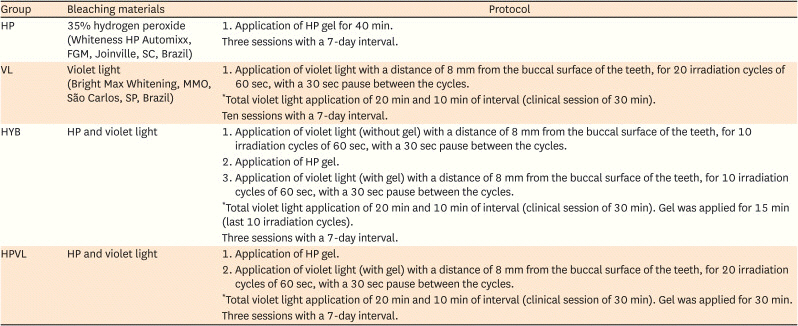
Table 1 (VL – V-LED; HP – 35% hydrogen peroxide [control group]; HYB – hybrid protocol, V-LED applied without gel for 10 irradiation cycles followed by V-LED applied with gel for another 10 irradiation cycles; and HPVL – peroxide and V-LED applied in 20 irradiation cycles).
Table 1
Summarized bleaching products and protocols used

|
Group |
Bleaching materials |
Protocol |
|
HP |
35% hydrogen peroxide (Whiteness HP Automixx, FGM, Joinville, SC, Brazil) |
1. Application of HP gel for 40 min. |
|
Three sessions with a 7-day interval. |
|
VL |
Violet light (Bright Max Whitening, MMO, São Carlos, SP, Brazil) |
1. Application of violet light with a distance of 8 mm from the buccal surface of the teeth, for 20 irradiation cycles of 60 sec, with a 30 sec pause between the cycles. |
|
*Total violet light application of 20 min and 10 min of interval (clinical session of 30 min). |
|
Ten sessions with a 7-day interval. |
|
HYB |
HP and violet light |
1. Application of violet light (without gel) with a distance of 8 mm from the buccal surface of the teeth, for 10 irradiation cycles of 60 sec, with a 30 sec pause between the cycles. |
|
2. Application of HP gel. |
|
3. Application of violet light (with gel) with a distance of 8 mm from the buccal surface of the teeth, for 10 irradiation cycles of 60 sec, with a 30 sec pause between the cycles. |
|
*Total violet light application of 20 min and 10 min of interval (clinical session of 30 min). Gel was applied for 15 min (last 10 irradiation cycles). |
|
Three sessions with a 7-day interval. |
|
HPVL |
HP and violet light |
1. Application of HP gel. |
|
2. Application of violet light (with gel) with a distance of 8 mm from the buccal surface of the teeth, for 20 irradiation cycles of 60 sec, with a 30 sec pause between the cycles. |
|
*Total violet light application of 20 min and 10 min of interval (clinical session of 30 min). Gel was applied for 30 min. |
|
Three sessions with a 7-day interval. |
The V-LED used (Bright Max Whitening, MMO, São Carlos, SP, Brazil;
https://mmo.com.br/bright-max-whitening/!#) has a power of 1.2 W, produced by 4 super V-LEDs (each with 300 mW) with a wavelength from 405 to 410 nm, irradiance 112 mW/cm
2, target area 10.7 cm
2 and total energy per session of 1,440 J [
9].
A 6 × 6 mm squared shallow groove was created with a spherical diamond bur (FG1012, KG Sorensen, Cotia, SP, Brazil) on the center of the buccal surface of each tooth to standardize the area where the color readings were performed. The baseline color measurements were conducted immediately before the first bleaching application.
Before the bleaching procedure, the specimens were gently dried to remove excess moisture. To standardize the amount of gel, an even 2 mm thick layer of bleaching gel was applied to the buccal enamel surface of the teeth. The teeth were kept in 100% humidity at 37°C for the time in contact with the bleaching gel. After each application, the teeth were washed thoroughly under tap water for complete removal of the gel. The teeth were stored in individual containers immersed in distilled water at 37°C in the interval between applications and until the end of the experiment [
1920]. After the last bleaching gel application, the specimens were treated with topical fluoride gel.
The bleaching efficacy was evaluated by using an objective color measurement with a spectrophotometer (EasyShade Advance, Vita Zahnfabrik). All color readings were performed within the previously defined area on the center of the buccal surface of the teeth by an experienced clinician previously trained in a pilot study. A research laboratory was chosen for the procedures, having standard temperature, humidity, and illumination conditions. Color readings were conducted at baseline (prior to bleaching), and then repeated at 7, 14, 21 and 70 days after the first bleaching application.
For the spectrophotometric evaluation, the specimens were gently dried with absorbent paper, and placed on a flat surface with a standard white background. The spectrophotometer was calibrated before the readings, and was always positioned at a 90° angle to the surface. The
CIEL*
a*
b tridimensional color space was used, where
L* indicates luminosity axis (
L* = 0 is black and
L* = 100 is white),
a* represents the greenness (-
a*) and redness (+
a*) axis, and
b* represents the blueness (-
b*) and yellowness +
b* axis [
21].
Color stability was assessed by calculating the color difference between each time interval and baseline. Both the equations
(Eq. 1) and the Δ
E00 (Eq. 2) were used in this study [
21]:
where ΔL*, Δa*, and Δb* are the difference between a pair of color coordinates measure at baseline and each time interval.
where ΔL′, ΔC′, and ΔH′ are the differences in lightness, chroma, and hue for a pair of color measurements (baseline and each time interval) in CIEDE2000. RT is a rotation function that accounts for the interaction between chroma and hue differences in the blue region. The weighting functions SL, SC, and SH adjust the total color difference for variation in the location of the color difference pair at the L*, a*, and b* coordinates, and the parametric factors kL, kC, and kH are correction terms for experimental conditions. In the present study, kL, kC, and kH were set to 1.
The 50:50% acceptability thresholds (ATs) for
CIEL*
a*
b* (
) and
CIEDE2000 (Δ
E00) were 2.7 and 1.8, respectively [
22].
The
CIEL*
a*
b color space-based whitening index (
WID) was calculated for each assessment time according to the formula [
23]:
The Δ
WID was calculated by the difference between the indices at baseline and each time interval (Δ
WID = |Δ
WID2 - Δ
WID1|). The whiteness difference threshold for acceptable bleaching effect was 2.60
WID units [
24].
The efficacy of tooth bleaching was determined based on the interpretation of visual thresholds, adopting a classification system from 1 to 5, derived from research results on perceptibility threshold (PT) and AT [
24]. In this system, index 1 corresponds to ineffective bleaching (< PT) and index 5 to excellent bleaching efficiency (> AT × 3).
Normality and homogeneity of variances of the color difference data were evaluated by Shapiro-Wilk test and Levene’s test. As both p values were > 0.05, data were statistically analyzed by 2-way repeated measures analysis of variance and Tukey’s test. The correlations among , ΔE00 and ΔWID were determined by Pearson’s correlation coefficient. All analyses were performed with a 5% significance level. Power analysis was performed and values of 0.8 were obtained for each of the double interactions (, ΔE00, ΔWID).
Determination of pulpal temperature
To assess the change in pulpal temperature, 3 healthy human maxillary central incisors were used. The teeth were obtained by direct donation, after approval by the Institutional Review Board of Universidade Positivo (CAAE: 16867519.1.0000.0093, approval protocol #3.456.559). Endodontic access was made with spherical diamond burs on the palatal surface of the tooth, following the pulp chamber. The teeth had their dental pulps removed, the pulp chambers cleaned and filled with a heat absorption zinc oxide-based thermal paste (Implastec, Votorantim, São Paulo, SP, Brazil) to allow the conduction of heat, similar to what would happen with pulp tissue. Type K thermocouples (Tm-902c) were placed inside the pulp chamber, immersed in the paste, touching the dentin in the region at the center of the buccal surface of the teeth. The thermocouples were kept in position and the access to the pulp chamber was sealed with light-curing resin (Top Dam, FGM, Joinville, SC, Brazil). These thermocouples were connected to a digital thermometer to determine the temperature in °C. The environmental temperature was maintained at 23°C and humidity between 30% and 50%. Temperature evaluation was carried out in a thermal heater (water bath), with the roots of the teeth immersed in water at 37°C. The buccal faces of the incisors were positioned 8 mm from the tip of the VL. The teeth were irradiated with the VL for 30 minutes (20 irradiation cycles of 60 seconds, with a pause of 30 seconds between cycles). The temperature was recorded every 30 seconds. The values of temperature and temperature variation in relation to the initial temperature for each specimen were calculated.
The same 3 maxillary central incisors were used for pulpal temperature determination analyzes, performed without and with the application of the bleaching gel on the buccal surface of the teeth, in an even 2 mm-layer. An interval of one hour was used between the temperature measurements with and without gel.
Normality (Shapiro-Wilk test) and homogeneity of variances (Levene’s test) of the pulpal temperature and temperature variation (ΔT) data, without and with the application of the bleaching gel, were evaluated. As both p values were > 0.05, data were statistically analyzed by Student’s t-test, with a significance level of 5%. Power analysis was performed and values of 0.9 were obtained for pulpal temperature and temperature variation.
RESULTS
The
, Δ
E00 and Δ
WID results showed statistically significant differences for groups (
p < 0.001), time (
p < 0.001) and the double interaction group × time (
p < 0.001). The data for
and Δ
E00 are described in
Tables 2 and
3. For the HP bleaching protocol, 7 days showed color change (
and Δ
E00) values not different from and 14 days, and lower than 21 and 70 days, also not different from each other. For the HPLV group, 7 days showed
and Δ
E00 values lower than 21 and 70 days, which were also not different from each other. At 7, 21 and 70 days, HYB showed color change values (
and ΔE
00) statistically lower than HPLV. At 14 and 21 days
, HP presented color change values statistically lower than HPLV. At all evaluated times, HPLV showed the highest
and ΔE
00. All
and ΔE
00 values were greater than the 50:50% acceptability limit for
CIEL*
a*
b* (
) and
CIEDE2000 (ΔE
00), respectively of 2.66 and 1.77 units. The VL bleaching protocol did not show significant differences comparing the 4 follow-up times. VL group also obtained significantly lower
and ΔE
00 values than the other 3 groups at all times. For this protocol, all
and ΔE
00 values were lower than the 50:50% acceptability limit for
CIEL*
a*
b* (
) and
CIEDE2000 (ΔE
00).
Table 2
Means and standard deviations of for the different bleaching protocols

|
Bleaching protocol |
|
|
7 days |
14 days |
21 days |
70 days |
|
VL |
1.9 ± 1.0Ac
|
1.9 ± 0.8Ac
|
2.3 ± 1.0Ac
|
2.0 ± 0.8Ac
|
|
HP |
7.0 ± 1.6Bab
|
6.6 ± 2.0Bb
|
8.6 ± 2.1Ab
|
9.1 ± 2.1Aab
|
|
HYB |
5.0 ± 2.3Db
|
6.6 ± 2.5Cb
|
9.4 ± 1.9Aab
|
8.1 ± 1.9Bb
|
|
HPVL |
8.7 ± 3.4Ca
|
9.7 ± 2.7BCa
|
11.4 ± 1.2Aa
|
10.6 ± 1.7ABa
|
Table 3
Means and standard deviations of ΔE00 for the different bleaching protocols

|
Bleaching protocol |
ΔE00
|
|
7 days |
14 days |
21 days |
70 days |
|
VL |
1.2 ± 0.6Ac
|
1.2 ± 0.5Ac
|
1.6 ± 0.5Ac
|
1.3 ± 0.4Ac
|
|
HP |
4.2 ± 0.8Ba
|
4.0 ± 0.9Bb
|
5.2 ± 1.0Ab
|
5.5 ± 0.9Aab
|
|
HYB |
2.7 ± 1.1Db
|
3.6 ± 1.3Cb
|
5.2 ± 1.1Ab
|
4.5 ± 1.1Bb
|
|
HPVL |
5.0 ± 2.3Ca
|
5.6 ± 1.9BCa
|
6.7 ± 1.0Aa
|
6.2 ± 1.3ABa
|
For the HP bleaching protocol, 7 days showed whiteness changes (Δ
WID) values not different from 14 days; and lower than 21 and 70 days, which were not different from each other. For the HPLV group, 7 days showed Δ
WID lower than 14, 21 and 70 days. At all times, HYB and HPLV showed whiteness changes values; and HP showed the highest Δ
WID. The VL bleaching protocol showed significantly lower Δ
WID at 70 days when compared to 21 days. VL group also presented significantly lower Δ
WID values than the other 3 groups. All Δ
WID means were greater than the whiteness difference threshold of 2.60 units (
Table 4), except for VL group at 14 and 70 days.
Table 4
Means and standard deviations of ΔWID for the different bleaching protocols

|
Bleaching protocol |
ΔWID
|
|
7 days |
14 days |
21 days |
70 days |
|
VL |
2.8 ± 2.1ABc
|
2.4 ± 2.3ABb
|
4.1 ± 2.0Ab
|
1.5 ± 1.4Bc
|
|
HP |
10.3 ± 2.4Ba
|
10.2 ± 2.7Ba
|
13.1 ± 2.9Aa
|
13.7 ± 2.7Aa
|
|
HYB |
6.0 ± 3.1Cbc
|
8.3 ± 3.4Ba
|
12.1 ± 2.6Aa
|
10.2 ± 2.5ABb
|
|
HPVL |
8.8 ± 3.4Bab
|
10.2 ± 3.6Aa
|
11.9 ± 5.4Aa
|
11.2 ± 5.6Aab
|
Correlations between all parameters (, ΔE00 and ΔWID) were performed. Strong positive significant correlations were found between and ΔE00 (R = 0.982 and p < 0.001), and ΔWID (R = 0.917 and p < 0.001) and ΔE00 and ΔWID (R = 0.898 and p < 0.001).
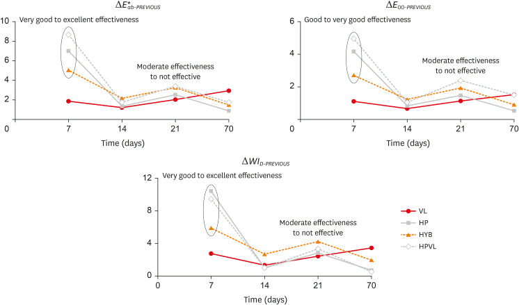
Figure 1 shows the bleaching efficiency classification, based on the interpretation of visual thresholds, for the follow-up times in relation to the immediately previous time for the evaluated groups for all the parameters used (
, Δ
E00-PREVIOUS and Δ
WID-PREVIOUS). It can be seen that, except for VL group, all protocols showed very good to excellent effectiveness in the first week. After this period, the effectiveness decreased significantly, to moderate or not effective (in relation to the immediately previous time).
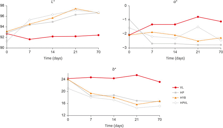
Figure 2 shows the
L*,
a* and
b* coordinates for the evaluated groups and follow-up times. For HP, HYB and HPVL groups, the specimens tended to become lighter (increased
L*), greener (-
a*) and bluer (-
b*). VL specimens maintained the
L* and
b* values, and increase
a* values overtime.
Figure 1
, ΔE00-PREVIOUS and ΔWID-PREVIOUS showing the bleaching efficiency classification for the follow-up times in relation to the immediately previous time for the evaluated groups.
VL, violet light emitting diode (V-LED); HP, 35% hydrogen peroxide; HYB, hybrid protocol, V-LED applied without gel for 10 irradiation cycles followed by V-LED applied with gel for another 10 irradiation cycles; HPVL, hydrogen peroxide and V-LED applied in 20 irradiation cycles.

Figure 2
L*, a* and b* coordinates for the evaluated groups and follow-up times. One should note that the a* values are negative and the x-axis is set at −3.
VL, violet light emitting diode (V-LED); HP, 35% hydrogen peroxide; HYB, hybrid protocol, V-LED applied without gel for 10 irradiation cycles followed by V-LED applied with gel for another 10 irradiation cycles; HPVL, hydrogen peroxide and V-LED applied in 20 irradiation cycles.

The results for pulpal temperature and temperature variation (Δ
T) with and without the application of the bleaching gel are shown in
Figure 3. It can be observed that there were statistically significant differences when comparing the data with and without gel, for both the absolute temperature (
p = 0.001) and the temperature variation (
p < 0.001), with higher temperatures and Δ
T for the group with the gel application.
Figure 3
Data for pulpal temperature and temperature variation with without and the application of the bleaching gel. For both parameters, values followed by the same letters are statistically similar (p > 0.05).
SD, standard deviation.

DISCUSSION
The first hypothesis of the present study, that the violet light would influence the efficacy of in-office bleaching protocols, was rejected, since the 2 protocols with violet light (HYB and HPVL) presented similar results to the control group (only hydrogen peroxide). In addition, the group in which only violet light was used showed low efficacy compared to the other protocols.
The present study evaluated alternatives to the in-office dental bleaching protocol to identify an approach that could maintain the bleaching efficacy and reduce side effects, using violet light for the partial or total breakdown of chromogens [
10111617]. Among the gels for in-office bleaching, those based on hydrogen and carbamide peroxide at high concentrations (above 30%) are the most used. In general, gels with a high concentration of peroxides are preferred for the in-office technique because they accelerate the process and promote good clinical results in the first applications [
25].
Therefore, this study evaluated the efficacy of several in-office bleaching protocols, with a high hydrogen peroxide concentration gel, associated or not with the use of V-LED. Our results corroborate previous reports, showing that violet light may not influence the efficacy of hydrogen peroxide gels, possibly due to the high availability of peroxides in dental tissue, which can reduce or mask the action of violet light [
11].
It is also important to mention the results observed herein with the isolated use of violet light, without association with the bleaching gel. The
value is less than the 50:50% acceptability limit for
CIEL*
a*
b* (2.66 units), but it is greater than the 50:50% perceptibility limit for
CIEL*
a*
b* (1.22 units) [
22]. This shows that the result obtained in this group was a color change noticeable to the human eye, but very discreet, when compared to the other bleaching protocols tested herein. It is worth mentioning that the present study used healthy teeth without any artificial pigmentation prior to bleaching. Differently, the study by Gallinari
et al. [
11], performed previous pigmentation with a mixture of black tea and Coke for 6 days before bleaching. Therefore, the use of teeth with a more saturated color and with a greater amount of chromogens may have influenced the bleaching results, especially in the group that did not use the gel.
In the present study, in the group that used only violet light, the protocol with 10 sessions was used, with a 7-day interval, evaluating the efficacy of the treatment throughout the whole period. Generally, when using protocols with bleaching gel, 3 applications are made at intervals of one week. According to
Table 2, it can be seen that the color change occurred in the first 3 weeks (
of 1.9 in the first and second weeks and 2.3 in the third), with little to no change after this period.
It should be noted that, for all bleaching protocols used, the efficacy was greater in the first week of application, in which the results remained stable or slightly increased in the second and third weeks. From a clinical point of view, the benefit of the desired result must be balanced with the possible deleterious effects of repeated gel applications and possible increase in pulpal temperature. In cases of teeth with little pigmentation or that require small color changes, just one in-office application can be enough to achieve the desired esthetic result.
The present study used several parameters to determine the color change and the bleaching efficacy.
is well known in Dentistry to determine color difference. However, the use of the
CIEDE2000 formula and the respective ΔE
00 has been encouraged as a more suitable tool for assessing the color of resin-based materials. In addition to the color change, it is important to evaluate a specific parameter to determine the whiteness level. Many whitening indexes have been described, but a new whitening index for dentistry (
WID) has been proposed based on the
CIEL*
a*
b*color space, which has a better correlation with visual perception compared to other whitening indexes [
2326]. In the present study, Δ
WID showed significant whitening in the first week and the results have been maintained since then. Thus, Δ
WID is in agreement with Δ
E data, as it can be seen in the strong significant positive correlations with
(
R = 0.917) and with ΔE
00 (
R = 0.898).
Further, extracted bovine incisors were used in this study as substitutes for human teeth, since it is difficult to collect a sufficient number of freshly extracted human incisors in good condition. The area chosen for evaluation had an enamel thickness of approximately 1 mm, which is comparable to the enamel average thickness of sound human maxillary incisors. It is well established in the literature that the physical and chemical properties of bovine teeth resemble those of human teeth [
2728]. Also, bovine teeth have already been used in many
in vitro dental bleaching studies [
27282930].
The second hypothesis, that the violet light would influence pulpal temperature change with and without the application of the bleaching gel, was accepted. It is known that light activation during the bleaching procedures can result in thermal damage to the pulp tissue. In a classic study with rhesus monkeys, it was reported that a temperature increase of 2.8°C inside the pulp chamber can lead to a reversible inflammatory response, whereas increases of 5.6°C and 11°C compromise the pulp vitality in 15% and 60% of cases, respectively. In the same study, temperature rises above 16.6°C caused pulp necrosis in 100% of the evaluated teeth [
31]. Another study also reported similar values, indicating that a temperature of 42°C can be critical for the pulp tissue when maintained for 1 minute [
32]. However, conflicting reports about increased pulpal temperature and tissue damage are also found in the literature. Baldissara
et al. [
33] suggested that an increase in pulpal temperature from 8.9°C to 14.7°C may not cause pulp pathology. Therefore, due to conflicting reports, further studies are needed to establish precise correlations between the increase in temperature promoted by the new light sources available on the market and their implications the vitality of pulp tissue. Although no histological evaluation was performed in the present study, the increase in temperature with the use of the V-LED could have caused pulp damage, since, on average, an increase in pulpal temperature between 7°C and 9°C was observed and maintained throughout the 30 minutes of intermittent V-LED exposure. Although the increase in pulpal temperature may not lead to histological changes in pulp tissue, it is believed that light-activated bleaching can lead to greater risk and/or intensity of tooth sensitivity. A systematic review confirmed this hypothesis for low concentration bleaching gels [
12].
Another significant factor that must be taken into account in relation to the increase in pulpal temperature is the use of protocols with and without the application of bleaching gel. According to the manufacturer of the V-LED, protocols without the application of gel should be used for patients with a previous history of postoperative sensitivity. However, even without gel application, significant increases in temperature can lead to postoperative sensitivity. It is interesting to note that, when using the bleaching gel, the increase in pulpal temperature can be even greater. The literature reports that the bleaching gel applied to the buccal surface of the teeth can act as an insulator, reducing the temperature rise in the pulp chamber, in comparison with no gel application [
34]. In the present study, the application of the gel had the opposite effect, working as a deleterious factor, promoting a greater increase in the pulpal temperature. One of the explanations for such a significant rise in temperature may be the activation time. In this study, the exposure time was quite long, of 30 minutes, which comprised 20 irradiation cycles of 60 seconds, with a pause of 30 seconds between cycles. Perhaps the pause of 30 seconds between irradiation cycles is too short and not enough to promote an adequate heat loss for the dental structure, causing a cumulative effect of temperature increase. According to
Figure 3, it can be seen that, in the group without the application of the gel, there was an increase in temperature during the first 9 minutes and that after this time the temperature stabilized and eventually started to decrease. In the groups that used gel, the pulpal temperature tends to have a continuous increase, surpassing the temperature of the group without gel after 12 minutes and maintaining a slight increase until the 24 minutes, when it stabilizes at a higher level until the end of the experiment.
It has been demonstrated in the literature that the use of a simulated pulpal circulation system during
in vitro pulpal temperature evaluation can present lower values of temperature increase than under conditions in which a closed system is used [
35]. The higher temperature values observed in the present study can be partially explained by the use of a closed system design; though the roots of the teeth were immersed in temperature-controlled water throughout the experiment. Also, in a study that compared
in vivo and
in vitro pulp temperature increase using premolars exposed to a LED curing light, it was observed that significantly higher pulp temperature increase values were found for the
in vitro model than for the
in vivo model, regardless of exposure mode.
In vitro values were 60% to 80% higher than those observed
in vivo [
36]. Applying this 60% difference (observed in the condition of 60 seconds at high power in the study by Runnacles
et al. [
36] in our mean temperature values, the Δ
T of 7.2°C would be equivalent to 4.3°C (without gel) and the Δ
T of 8.9°C would be equivalent to 5.3°C (with gel). However, it is important to highlight some important differences in the methodology of this study and that of Runnacles
et al. [
36]. The later used upper premolars, a Polywave LED curing light and pulp circulation system in the
in vitro model [
36]. The present study used upper central incisors, a V-LED and a system with the roots immersed in temperature-controlled water. It is also important note that the present results cannot be directly extrapolated to clinical conditions, in which pulp pressure and circulation are present and play an important role in temperature regulation.
Two previous studies evaluated postoperative sensitivity in in-office bleaching with or without the use of the V-LED. The first is a case series in which the dental sensitivity of 6 patients was assessed after the following bleaching protocols: at-home bleaching with 10% carbamide peroxide; in-office bleaching with 17.5% hydrogen peroxide and treatment with a placebo gel. The V-LED had no effect on the pain, but it increased the threshold for detecting thermal changes in the teeth that were irradiated [
10]. The second is a clinical study with 50 participants divided into 2 groups: 2 bleaching sessions of 30 minutes each with 35% carbamide peroxide, with an interval of 7 days, without violet light; and the same protocol associated with the V-LED (20 irradiation cycles of 60 seconds with an interval of 30 seconds), 30 minutes each session. Only 2 participants in the group in which the V-LED was used (8%) reported moderate sensitivity on the seventh day (5 and 6 scores on the visual analog scale). However, statistically, no significant difference was observed between groups for this outcome [
9].
The present study had some limitations. This was an in vitro study that used bovine incisors to evaluate bleaching efficacy. Only one hydrogen peroxide was used, and the results may not be generalized for other bleaching agents and concentrations. Color and whiteness changes were assessed for a short period of time and it is not clear how chromatic stability would remain over longer periods than those evaluated. Also, the in vitro pulpal temperature evaluation was performed with a closed system design, without the use of a simulated pulpal circulation. There are limited data in the literature indicating the effect of light and heat activation during bleaching procedures on increasing pulpal temperature, especially with regard to the violet light device. Therefore, more studies are needed showing histologically the possible pulpal damage, in addition to clinical studies evaluating postoperative sensitivity with the use of these LEDs.
Although raising the temperature of the pulp chamber during the light-activated bleaching process can impair pulp tissue vitality and health, tissue regeneration processes can help pulp survival and maintain vitality. In addition, the local blood flow, the flow of dentin fluid and the surrounding periodontal tissues can buffer the heat transferred to the pulp [
37]. Nevertheless, the effects of light-activated bleaching on pulp health should be further studied clinically.


 PDF
PDF Citation
 Print
Print


 XML Download