

 PDF
PDF Citation
 Print
Print


INTRODUCTION
Recent advances in radiological technology and increased demand for brain examinations in health screening have led to an increase in the accidental detection of unruptured intracranial aneurysms (UIAs). Both surgical and endovascular treatments have been actively performed for UIAs. Surgical clipping is generally associated with higher rates of perioperative morbidity and aneurysm obliteration and lower rates of aneurysm recurrence compared to endovascular coiling [35].
The incidence of moderate to severe postcraniotomy headache (PCH) has been reported to be as high as 80% [12]. However, acute PCH is frequently overlooked and undertreated in clinical practice because systemic or neuraxial administration of analgesics, particularly opioids, can cause sedation and hinder neurological examinations [15]. In addition, the use of local anesthetics rather than opioid for pain control is emphasized in enhanced recovery after surgery [3,10,24]. Therefore, scalp nerve block (SNB) has been increasingly used as an effective alternative to systemic administration of opioids in PCH. SNB is known to effectively reduce PCH, opioid consumption, inflammation, and postoperative nausea and vomiting (PONV) in conventional craniotomy patients [1,2,17,41,42,45].
In recent years, enhanced recovery after surgery, including craniotomy, has been in the spotlight in perioperative management, as it has shown to improve postoperative outcomes in terms of complications and hospital stays [11,22,23,34,39]. The quality of recovery (QoR)-40 is a patient-reported questionnaire based on five dimensions of health (emotional state, physical comfort, psychological support, physical independence, pain), and is widely used as a tool for assessing postoperative QoR quantitatively over a period of 24 hours [14,25]. The QoR-40K, the Korean version of the QoR-40, offers a high level of validity, reliability, and clinical utility for evaluating postoperative QoR in Korean populations [20]. However, there have been no clinical studies investigating the effects of SNB on the QoR in minicraniotomy patients, especially using such questionnaire.
Low postoperative QoR-40 scores were associated with PCH and postoperative complications, including PONV, in cranial surgery patients [21]. Based on this, this study hypothesized that SNB would improve the QoR by reducing PCH, opioid consumption, and PONV in the early postoperative period in minicraniotomy patients. This study compared the QoR-40 scores, severity of PCH, use of analgesics, incidence and severity of PONV, and length of hospital stay between patients with and without SNB after minicraniotomy for clipping of UIAs.
MATERIALS AND METHODS
Ethics
This double-blind, parallel-group, randomized, and placebo-controlled trial was approved by Seoul National University College of Medicine/Seoul National University Hospital Institutional Review Board (number : 2005-232-1130, date : August 11, 2020) and registered at the Clinical Research Information Service (number : KCT0005354, date : August 26, 2020). This study was conducted according to Good Clinical Practice guidelines and this paper adhered to the applicable Consolidated Standards of Reporting Trials guidelines.
Subjects
Patients aged 20–79 years who were scheduled for an elective single keyhole minicraniotomy for clipping of UIAs at single academic tertiary hospital were included. Patients with symptomatic aneurysms, allergies to local anesthetics or analgesics, chronic use of opioids, a history of craniotomy or scalp incision, other diseases (scalp disease, cancer, dementia, mood disorder, psychiatric disorder), or American Society of Anesthesiologists physical status ≥3 were excluded. Patients unable to communicate with their healthcare provider or complete a written version of the QoR-40K were also excluded.
Randomization and blinding
Randomization software (Random Allocation Software version 1.0.0; Isfahan University of Medical Sciences, Isfahan, Iran) was used for block randomization with a block size of four. By an anesthesiologist not involved in this study, a random allocation sequence was created, concealed in an opaque envelope, and checked just after anesthetic induction. Patients were assigned to the SNB (SNB using ropivacaine with epinephrine) and control (SNB using normal saline) groups at a 1 : 1 ratio based on the random allocation sequence. The drugs for SNB were prepared by the same anesthesiologist so as to be indistinguishable. The anesthesiologists who performed SNB, neurosurgeons, patients, and researchers who investigated perioperative variables, including the QoR-40 scores, were blinded to group assignment.
SNB
At the end of surgery, landmark-guided SNB was performed using aseptic technique by one of the four attending anesthesiologists who had performed more than 50 SNBs. SNB was induced using 0.75% ropivacaine with 1 : 200000 epinephrine and normal saline in the SNB and control groups, respectively. These solutions were prepared in three 10-mL syringes labeled as “study drug” so that their composition was unknown to the anesthesiologist who performed SNB. To cover the area of surgical incision and skull clamp, seven scalp nerves, including the supratrochlear, supraorbital, zygomaticotemporal, auriculotemporal, great auricular, lesser occipital, and greater occipital nerves, were blocked bilaterally by injecting 2–3 mL of the solution using a 25-gauge needle. The supratrochlear and supraorbital nerves were blocked at the eyebrow, near the superomedial margin of the orbit and above the palpable supraorbital notch, respectively [26]. The zygomaticotemporal nerve was blocked between the superolateral margin of the orbit and the posterior margin of the zygomatic arch [26]. The auriculotemporal nerve and the posterior branch of the great auricular nerve were blocked at the level of the tragus, near the pulsating superficial temporal artery and on the mastoid process, respectively [26]. The greater and lesser occipital nerves were blocked at the medial and lateral thirds of the superior nuchal line, respectively [26]. SNB-related adverse events such as infection, hematoma formation, facial nerve palsy, local anesthetic systemic toxicity, and severe hemodynamic instability (a change more than 20% in mean arterial pressure or heart rate) were noted if present.
Anesthesia
Patients were monitored with non-invasive blood pressure measurement, electrocardiography, and peripheral oxygen saturation in the operating room. Total intravenous anesthesia was induced and maintained using target-controlled infusion of propofol and remifentanil. The upper airway was topically anesthetized using a 10% lidocaine spray during mask ventilation to minimize hemodynamic response to tracheal intubation. The patient’s radial artery was catheterized to continuously monitor invasive blood pressure. After anesthetic induction, intravenous dexamethasone (5 mg) were administered for PONV prophylaxis. The effect site concentrations of propofol and remifentanil were adjusted to keep bispectral index and mean blood pressure within 40–60 and 80–120% of its preoperative baseline, respectively. Intravenous acetaminophen (1000 mg), fentanyl (50 µg), and ramosetron (300 µg) were administered 30 minutes before the end of surgery. At the end of surgery, intravenous patient-controlled analgesia (IV-PCA; total volume, 100 mL; fentanyl concentration, 15 µg/mL; ramosetron concentration, 3 µg/mL; continuous infusion rate, 0 mL/h; bolus injection volume, 1 mL; lockout time, 10 minutes) was established. After the end of surgery, all patients were extubated in the operating room and transferred to the postanesthesia care unit.
Surgery
All craniotomies and clipping of UIAs were performed by a skilled neurosurgeon. A skull clamp with three pins (MAYFIELD® Skull Clamps; Integra LifeSciences, Princeton, NJ, USA) was used to immobilize the patient’s head. The surgical approach for clipping of UIAs was based on the location and direction of the aneurysms, spatial relationship among the aneurysms, brain parenchyma, and bony structures, and neurosurgeon’s preference. Four types of keyhole minicraniotomy were considered : supraciliary supraorbital, frontolateral supraorbital, lateral supraorbital, and minipterional approaches [8,16]. Surgical complications, including intraoperative premature aneurysmal rupture and postoperative stroke with neurological deficit, were recorded if present.
Postoperative management
IV-PCA was maintained until the patient requested its removal. Before resuming sips of water, intravenous acetaminophen (1000 mg) was administered every 6 hours, while intravenous fentanyl (50 µg) was used for rescue analgesia. After resuming sips of water, oral acetaminophen (650 mg) was administered every 6 hours, while oral aceclofenac (100 mg) and tramadol (50 mg), transdermal fentanyl patches (25–100 µg/h), and intravenous fentanyl (25 µg) were used in order for rescue analgesia. For PONV control, intravenous ramosetron (300 µg) and metoclopramide (10 mg) were administered up to twice a day as first-line and second-line antiemetics, respectively.
Outcomes
The primary outcome measure was the QoR-40 scores measured 1 day postoperatively. As secondary outcome measures, the QoR-40 scores were measured 2–3 days postoperatively, at hospital discharge, and 1 month postoperatively and the severity of PCH, total amount of IV-PCA consumed, total number of rescue analgesics administered, and incidence and severity of PONV were evaluated 3, 6, 9, and 12 hours and 1–3 days postoperatively. The time from the end of surgery to the first administration of rescue analgesics and length of hospital stay were also investigated. The severity of PCH was assessed using a numeric rating scale (NRS; 0, no pain; 10, worst pain imaginable). The severity of PONV was divided into three grades (mild, no requirement for antiemetic administration; moderate, relief after antiemetic administration; severe, no relief even after antiemetic administration).
Sample size calculation
In a prospective observational study, the mean and standard deviation of the QoR-40 scores measured 1 day postoperatively in cranial surgery patients were 160 and 19, respectively [21]. Under the assumption that a 10% increase in the mean QoR40 scores measured 1 day postoperatively in minicraniotomy patients with SNB is clinically significant, a minimum sample size of 46 patients (23 patients per group) was calculated with α of 0.05 and β of 0.2. Considering a drop rate of 20%, at least 56 patients (28 patients per group) were required.
Statistical analysis
All statistical analyses were performed using statistical software (SPSS version 25; IBM Corp., Armonk, NY, USA). The normality of data distribution was determined using ShapiroWilk test. Categorical variables were presented as numbers (proportions) and compared using Pearson’s chi-squared test with continuity correction or Fisher’s exact test depending on the expected frequency of cells. Continuous variables were presented as means (standard deviations) or medians (interquartile ranges) and compared using Student’s t-test or MannWhitney U test according to the normality of their data distribution. The time effect of the QoR-40 scores within the groups and the time × group effect of the QoR-40 scores was evaluated using repeated measures analysis of variance. To determine whether there would be differences in the effects of SNB according to the type of minicraniotomy, a subgroup analysis was performed by dividing patients into those who underwent minicraniotomy with muscle incision (lateral supraorbital and minipterional approaches) and those who underwent minicraniotomy without muscle incision (supraciliary and frontolateral supraorbital approaches). A p-value less than 0.05 was considered statistically significant.
RESULTS
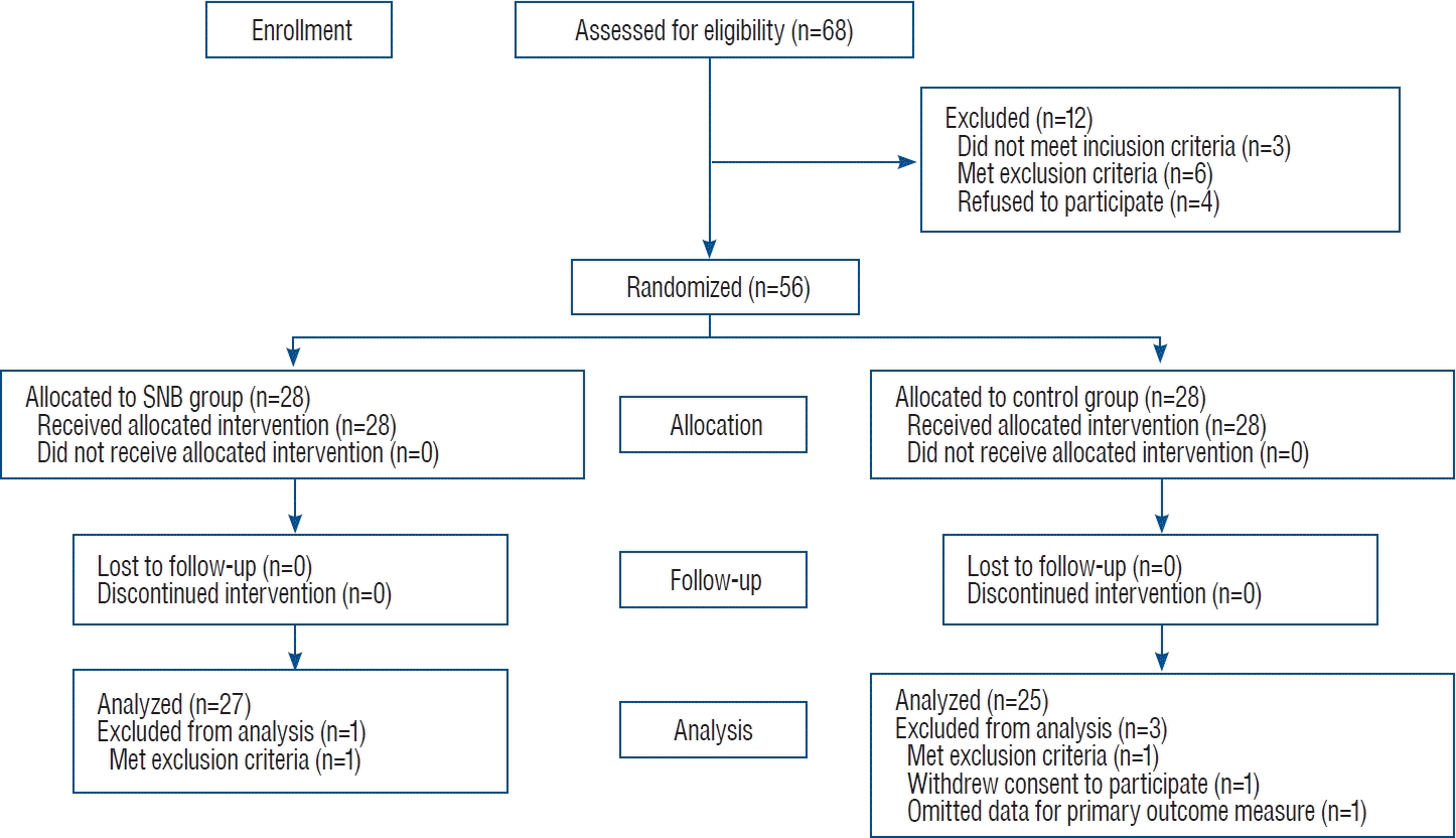
A total of 56 patients were enrolled between October 2020 and November 2021 (Fig. 1). Four patients (two exclusion criteria, one withdrawn consent to participate, and one missing data for the primary outcome measure) were excluded from the analysis. There were no significant differences in demographic and intraoperative variables between the SNB and control groups except for a longer surgical duration in the SNB group (2.6±0.6 vs. 2.1±0.5 hours, p=0.002; Table 1).
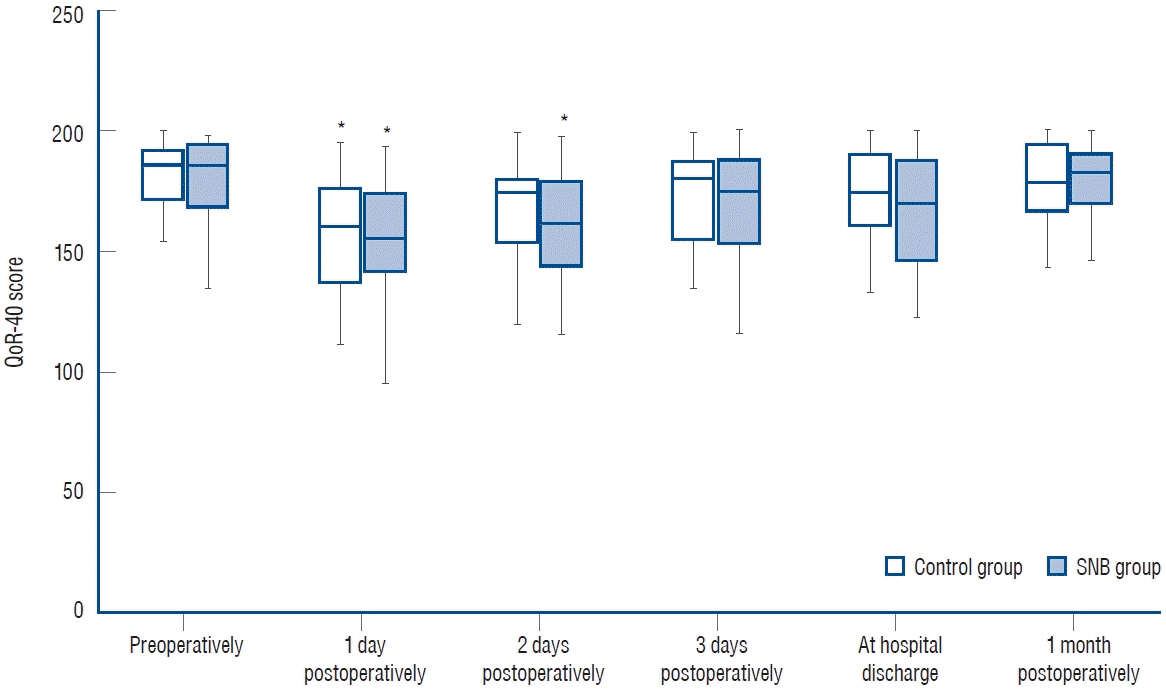
All QoR-40 scores, including those measured 1 day postoperatively (155.0 [141.0–176.0] vs. 161.0 [140.5–179.5]; median difference, -5.0; 95% confidence interval, -18.0 to 9.0; p=0.464; Table 2), were not significantly different between the SNB and control groups. The QoR-40 scores showed no significant time × group (p=0.469) effect, whereas a significant time (p<0.001) effect within the groups. The QoR-40 scores measured 1 (155.0 [141.0–176.0] vs. 185.0 [167.0–195.0], p<0.001) and 2 (161.0 [142.0–179.0] vs. 185.0 [167.0–195.0], p<0.001) days postoperatively and those measured 1 (161.0 [140.5–179.5] vs. 187.0 [174.0–191.5], p<0.001) day postoperatively were significantly lower than those measured preoperatively in the SNB and control groups, respectively (Fig. 2).
PCH was significantly less severe 3 (NRS; 3.0 [2.0–4.0] vs. 5.0 [3.5–5.5], p=0.029), 9 (NRS; 3.0 [2.0–4.0] vs. 3.0 [2.5–5.5], p=0.048), and 12 (NRS; 3.0 [2.0–4.0] vs. 4.0 [3.0–5.0], p=0.035) hours postoperatively in the SNB group (Table 3). The total amount of IV-PCA consumed was also significantly less 3 hours postoperatively in the SNB group (2.0 [1.0–4.0] vs. 4.0 [2.0–5.0] mL, p=0.044). There were no significant differences in the total number of rescue analgesics administered, time to the first administration of rescue analgesics, incidence and severity of PONV, and length of hospital stay between the SNB and control groups. In both groups, neither SNB-related adverse events nor surgical complications were observed.
In the subgroup analysis, all demographic and intraoperative variables, perioperative QoR-40 scores, and postoperative outcomes were not significantly different between the SNB and control groups in patients who underwent craniotomy with muscle incision (Supplementary Tables 1-3). In patients who underwent minicraniotomy without muscle incision, the SNB group showed significantly less severe PCH until 6 hours postoperatively and significantly less total amount of IV-PCA consumed until 12 hours postoperatively, whereas the SNB group had a significantly longer surgical duration and a significantly higher incidence of PONV 6 hour postoperatively. Other demographic and intraoperative variables, perioperative QoR-40 scores, and postoperative outcomes did not significantly differ between the two groups in these patients.
DISCUSSION
The importance of postoperative QoR is increasingly emphasized, but methods to improve the QoR after neurosurgery, especially craniotomy, have not yet been fully investigated [34]. To the best of our knowledge, this is the first study evaluating the effects of SNB on the QoR after craniotomy using a self-rated questionnaire. In this randomized controlled trial, SNB did not improve the QoR-40 scores but reduced PCH and IV-PCA consumption during the first few hours postoperatively in patients who underwent a minicraniotomy for clipping of UIAs.
No significant improvement in the QoR-40 scores in the SNB group may be because PCH and PONV account for relatively small proportions in the QoR-40. In the QoR-40, the number of direct questions for PCH and PONV are only 3 out of 7 and 3 out of 12 in the dimensions of pain and physical comfort, respectively [25]. Therefore, it is difficult to show a statistically significant difference in postoperative QoR-40 scores unless there is a dramatic amelioration of PCH and PONV. However, in this study, the maximum median difference in the severity of PCH was only 1 (NRS) on the day of surgery, while the incidence and severity of PONV did not significantly differ between the SNB and control groups. Furthermore, significant differences in surgical duration between the two groups could affect the QoR in the early postoperative period. Compared to conventional craniotomy, minicraniotomy generally has the advantage of reducing intraoperative loss of cerebrospinal fluid and blood as surgical duration is about half as short [8,16,27,28]. However, long surgical duration in the SNB group may have offset these advantages of minicraniotomy, causing greater discomfort and hindering early ambulation after surgery, and may have ultimately counterbalanced the benefits of SNB on postoperative QoR [31,38]. In addition, contrary to our expectation that SNB might prevent persistent PCH, SNB did not significantly affect the QoR-40 scores 1 month postoperatively [5,6,13].
As mentioned earlier, the analgesic effect of SNB, although statistically significant, was not clinically significant during the first 12 hours postoperatively in this study. This may be because the surgery was performed through a minimally invasive approach. PCH mainly originates in superficial structures, including the scalp and pericranial muscles [9]. The keyhole minicraniotomies performed in this study may have caused less trauma to the skin and temporalis muscles, resulting in reduced PCH compared to the conventional pterional craniotomy [29,40]. Thus, the analgesic effect of SNB may not have been noticeable as PCH had already been surgically minimized. Furthermore, multimodal analgesia, including IV-PCA, preemptive administration of intravenous fentanyl and acetaminophen at the end of surgery, periodic administration of acetaminophen after surgery, and active rescue analgesia, was provided equally in both groups except for SNB. Thus, it is possible that PCH was largely controlled even without SNB in the control group. Nevertheless, the control group had significantly severe PCH despite significantly more IV-PCA consumption up to 3 hours postoperatively, suggesting that SNB had significant analgesic effect in the immediate postoperative period.
Contrary to previous findings that SNB could help reduce PONV by reducing opioid consumption, the incidence and severity of PONV in this study were comparable between the SNB and control groups despite significantly more IV-PCA consumption up to 3 hours postoperatively in the control group [17,41]. This could be explained by significantly longer surgical duration in the SNB group with a mean difference of 30 minutes compared to the control group. Longer surgical duration is associated with an increased risk of PONV and a 30-minute increase in surgical duration may increase the risk of PONV by 60% [4,7,38]. Moreover, longer surgical duration in craniotomy patients also results in greater loss of cerebrospinal fluid, which is another risk factor for PONV [31,38]. Since the incidence of PONV was slightly higher in the SNB group, although not statistically significant, the negative effects of longer surgical duration on PONV may overweigh the positive effects of SNB.
In the subgroup analysis, the analgesic effect of SNB was significant during the first 12 hours postoperatively in patients who underwent minicraniotomy without muscle incision, but not in patients who underwent minicraniotomy with muscle incision. The reason for these findings is thought to be that blocking the zygomaticotemporal and auriculotemporal nerves are more essential to reduce the pain from temporalis muscle incision in the latter patients. However, the zygomaticotemporal nerve is known to have a significantly higher failure rate of landmark-guided SNB than other scalp nerves including the supratrochlear, supraorbital, auriculotemporal, lesser occipital, and greater occipital nerves, because it has many anatomical variations, such as wide distribution of emergence point form the temporalis fascia, diverse course, and various number and orientation of accessary branches, and is distributed deep below the skin [18,32,36]. In addition, the auriculotemporal nerve, which communicates with the zygomaticotemporal nerve and also innervates part of the temporalis muscles, was reported to have the second highest failure rate of landmark-guided SNB among the aforementioned six scalp nerves [32,36]. Therefore, there was a possibility that the pain from temporalis muscle incision was not effectively reduced by SNB in some patients who underwent minicraniotomy with muscle incision in this study.
This study has several limitations. First, the sample size was insufficient to compare other postoperative outcomes, particularly in the subgroup analysis, between the SNB and control groups with statistically sufficient power. Additional studies are necessary to verify the effects of SNB on postoperative outcome, especially QoR-40 scores, with statistically sufficient power in each minicraniotomy. Second, the effectiveness of landmark-guided SNB may have been greatly influenced by anatomical variations in the target nerves and surrounding structures. Although there have been no studies comparing landmark-guided and ultrasound-guided SNBs, ultrasound guidance may improve the effectiveness of SNB [37,44]. Third, the pattern of postoperative recovery may be different in patients undergoing a craniotomy for other procedures which can directly damage the brain parenchyma, such as the resection of brain tumors and arteriovenous malformations. Thus, the results of this study may not be reproducible in such patients. Fourth, in this study, SNB was performed at the end of surgery using 0.75% ropivacaine with 1 : 200000 epinephrine to maximize its analgesic effect in the postoperative period. Results may differ if the timing of SNB and the type and dose of local anesthetics and adjunctives used for SNB are different from those of this study. In particular, the use of local anesthetics with a much longer duration of action, such as liposomal bupivacaine, for SNB may further improve the QoR after craniotomy by reducing PCH for a longer time. Fifth, in this study, the sites of skull clamp were not noted and the pain from skull clamp was not investigated separately from the pain from surgical incision. In addition, because SNB was performed uniformly targeting bilateral seven scalp nerves in this study, it was difficult to know how much and long SNB reduced the pain from skull clamp. Further studies are needed to determine which scalp nerves need to be mainly blocked to effectively reduce the pain from skull clamp. Lastly, the QoR-40 was used in this study because it was the only validated tool for evaluating postoperative QoR in Korean populations at the time of this study [20]. However, there are various other tools for assessing postoperative QoR, including the QoR-15, a shortened version of the QoR-40, and Postoperative QoR Scale, and results may vary if these tools are used instead of the QoR-40 [19,30,33,43].
CONCLUSION
In conclusion, this study shows that SNB did not significantly improve postoperative QoR-40 scores despite reductions in PCH and opioid consumption in the early postoperative period in patients who underwent a minicraniotomy for clipping of UIAs. Further clinical study is needed to find methods to significantly improve the QoR after minicraniotomy.
 XML Download