This article has been
cited by other articles in ScienceCentral.
Abstract
Foreign body ingestion is commonly seen in children. However, occasionally it may also be seen among adults and is often associated with intellectual disability, psychiatric disorders, and alcoholism. Ingestion of a magnetic foreign body may cause complications such as gastrointestinal tract perforation, wherein emergency endoscopic removal of the foreign body is generally required. Here, we report a rare case of a 59-year-old male with an intellectual disability and psychiatric disorder in whom metallic objects in the stomach cavity were accidentally discovered during abdominal CT. Esophagogastroduodenoscopy revealed several metallic objects attached to two magnets, which had been ingested several years before and had remained in the stomach cavity. The magnets and metallic objects were safely removed endoscopically using rat-tooth forceps without complications.
Go to :

Keywords: Foreign body, Magnet, Stomach, Endoscopy
INTRODUCTION
Foreign body ingestion is common in children, but it is also seen in adults. In adults, most cases of foreign body ingestion are accidental and related to food such as fish bones, causing food bolus and bone impaction. However, intentional ingestion of foreign bodies usually occurs in individuals with intellectual disabilities, psychiatric disorders, and alcoholics.
1 Most swallowed foreign bodies pass spontaneously (80–90%) through the gastrointestinal tract (GI). However, 10–20% of such cases require endoscopic removal. Rarely, approximately 1% of patients may require surgery to remove foreign bodies and treat complications such as GI tract perforation or obstruction.
2 These complications usually occur when patients swallow large or sharp-pointed objects.
3,4 However, the same complications can also occur after ingesting magnetic foreign bodies.
The guidelines of the European Society of Gastrointestinal Endoscopy (ESGE) for treatment of the ingestion of foreign bodies such as sharp-pointed objects, magnets, and batteries recommend that an urgent therapeutic esophagogastroduodenoscopy (EGD) be performed within 24 hours. When medium-sized, blunt foreign bodies are ingested, it is recommended that a non-urgent therapeutic EGD be performed within 72 hours.
2
Although several cases in the literature describe the urgent treatment of foreign body ingestion, we found a case of foreign body ingestion without complications such as GI perforation. This was accidentally discovered 25 years after the patient had ingested a ballpoint pen.
5 Here, we report a similar case of foreign body ingestion that was accidentally discovered several years after a patient swallowed magnets and metallic objects which posed no complications in the intervening years between ingestion to removal.
Go to :

CASE REPORT
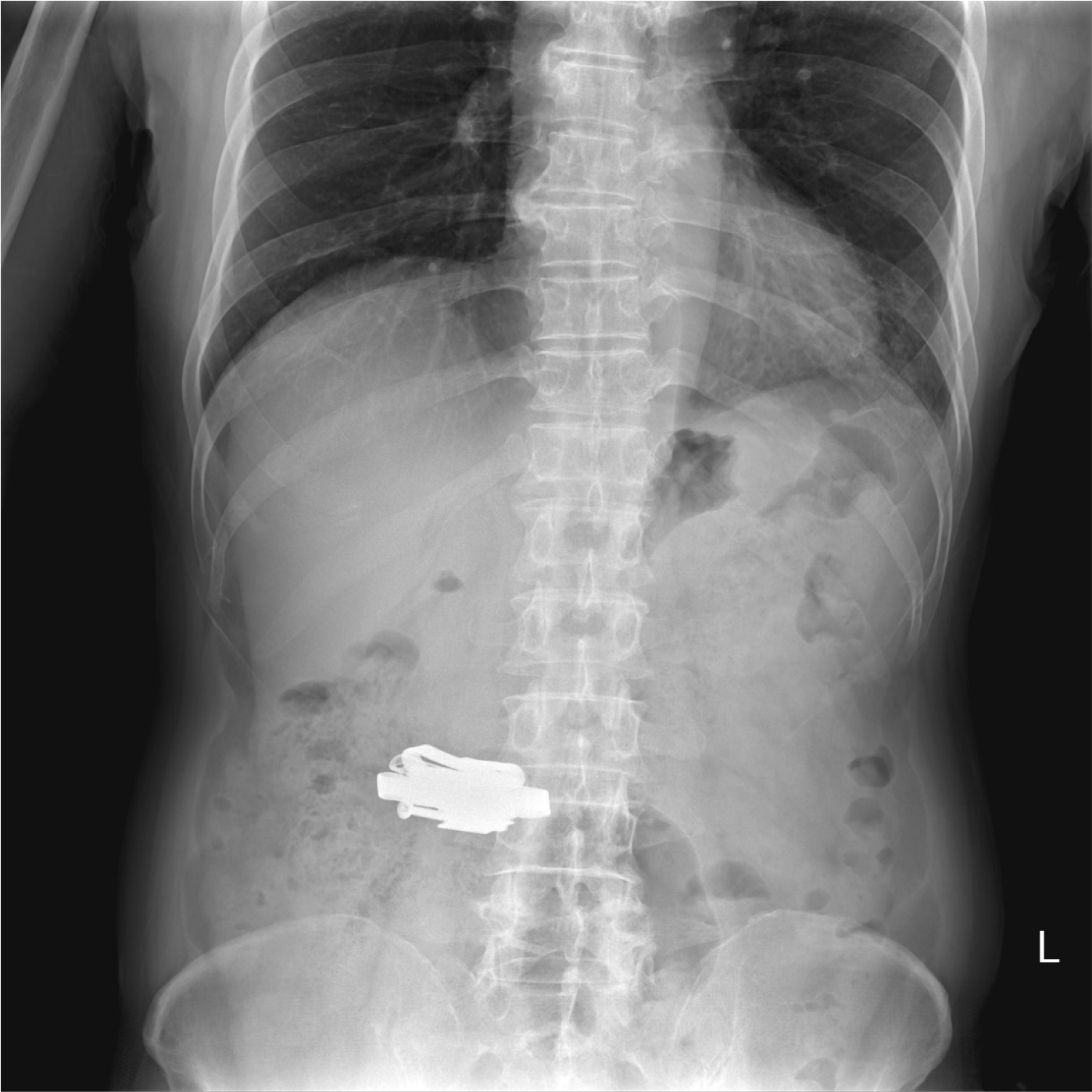
A 59-year-old male with a history of intellectual disability and schizophrenia was referred to our department for the treatment of a metallic foreign object present in the stomach that was incidentally discovered during abdominal CT performed to evaluate decreased renal function. Besides the presence of the foreign object, no other abnormalities were noted on the abdominal CT. The patient had complained of mild intermittent abdominal discomfort for several years but had no other specific symptoms. The metallic object was about 6 cm in size as confirmed by an abdominal X-ray (
Fig. 1). The patient was hospitalized for the surgical removal of the object.
 | Fig. 1Plain abdominal X-rays show a metallic mass in the stomach. 
|
On admission, his vital signs were stable; blood pressure was 130/80 mmHg, pulse rate was 73 beats/minute, respiratory rate was 20 breaths/minute, body temperature was 36.1°C, and oxygen saturation was 96% in room air. He was alert and did not appear to be ill. No abnormal findings were observed on physical examination. Laboratory examinations also showed normal findings; the white blood cell count was 7,240/mm3, hemoglobin level was 14.0 g/dL, platelet count was 264,000/mm3, and C-reactive protein level was 0.4 mg/dL.
EGD revealed a metallic object in the stomach, which was approximately 6 cm in size. When we poked the metallic object with forceps, some pieces of metal separated from it. Based on this observation, we concluded that several metal pieces had agglomerated together to form a larger metallic mass, contrary to our initial judgment of the object being a single piece. We removed the larger metal pieces one by one using rat-tooth forceps, and the remaining small metal pieces were removed together using a net (
Fig. 2).
 | Fig. 2Metallic mass found in the gastric cavity during esophagogastroduodenoscopy (EGD). (A) The metallic mass shows a clearly visible screw, along with food material. (B) Removal of the larger metal pieces was performed with rat-tooth forceps. (C) The remaining small metal pieces were removed together using a net. 
|
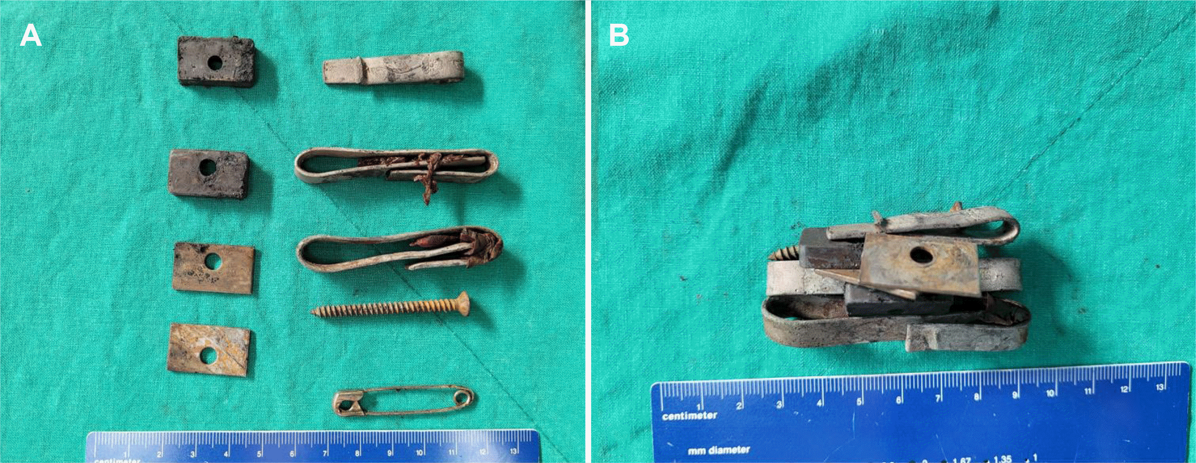
Upon observation of the foreign bodies after removal, we found seven metal pieces and two magnets and reconstructed a conformation in which the metal pieces were agglomerated around the magnets (
Fig. 3). The metallic mass was likely to have been in this or a similar conformation on discovery. Upon subsequent questioning, the patient recalled that he had swallowed the metallic objects all at once several years ago. After the removal of the foreign bodies, the patient exhibited no symptoms or abnormalities on radiography. He was discharged from the hospital one day after the endoscopic foreign body removal and is undergoing follow-up evaluations at the outpatient clinic.
 | Fig. 3Individual pieces of the metallic mass. After endoscopic foreign body removal, we found (A) several metal pieces and 2 magnets. (B) These metallic objects were agglomerated around the two magnets in the stomach cavity in a conformation similar to that shown. 
|
Go to :

DISCUSSION
Here, we report a case of foreign body ingestion by an adult in which metallic objects bound to two magnets remained in the stomach for several years without causing any complications. These foreign bodies were accidentally discovered on abdominal CT during an evaluation of decreased renal function and were removed endoscopically.
Ingestion of magnetic foreign bodies is relatively common in children but is rare in adults. Known risk factors for the ingestion of magnetic foreign bodies in adults include a history of intellectual disability, psychiatric disorder, and alcoholism. Our patient had a history of intellectual disability and schizophrenia.
A single magnet on ingestion can pass through the GI tract without causing complications, whereas the ingestion of multiple magnets can bind between the intestinal loops. Therefore, these foreign bodies cannot pass through with normal motility and can cause pressure necrosis, resulting in abscess formation or GI perforation.
6 In addition, mucosal injury can occur within 8 hours of magnetic foreign body ingestion.
7 In the literature, we found a case report detailing the ingestion of two magnetic objects by a 21-year-old male, which resulted in intestinal obstruction.
8 In another case, the ingestion of multiple magnets by a 28-year-old patient with learning difficulties resulted in ileocolic fistulation and cecal perforation.
9 Both cases required urgent treatment. As magnet ingestion can lead to serious gastrointestinal complications, as exemplified by the aforementioned case studies, the ESGE guidelines recommend urgent therapeutic EGD within 24 hours of magnet ingestion.
According to a review article on the GI damage caused by multiple magnet ingestion, the treatment depends on their location. When the magnets are located above the gastric pylorus, they can be removed using endoscopy. When the magnets are located below the gastric pylorus, the treatment of choice is exploratory laparotomy.
6 In this case, because the mass was located above the gastric pylorus, it was successfully removed using endoscopy.
Single or several magnets ingested at short time intervals are likely to pass through the GI tract without complications. However, multiple magnets ingested over long time intervals are likely to cause complications such as gastric obstruction, fistula, and perforation.
6 In this case, two magnets caused other metallic objects to clump together and remain in the patient’s gastric cavity without passing into the duodenum. It could be inferred that the objects were swallowed at short intervals based on the fact that the swallowed magnets and metallic objects were agglomerated. In addition, since the metallic objects were relatively blunt, they did not cause perforation.
In cases of magnetic foreign body ingestion, there is a very high risk of complications, such as intestinal obstruction or perforation. While there is a plethora of previous studies detailing the urgent treatment of multiple magnet ingestion, to the best of our knowledge, there have been no reported cases of multiple magnet ingestion without complications for several years in adults. This is a rare case of a successful endoscopic magnetic foreign body removal performed several years after ingestion, thus providing an exception to the ESGE treatment guidelines for magnet ingestion. This case can revise our current understanding of the durability of the human GI tract and provide clinicians with a possibility for consideration in the diagnoses of unlikely cases in the future.
Go to :


 PDF
PDF Citation
 Print
Print


 XML Download