Abstract
Odontogenic keratocysts (OKCs) located in the maxillae have rarely been reported in the literature. Standard treatment modalities for OKC range from marsupialization to marginal resection. However, most of the studies on OKC treatment have been related to mandibular OKCs. The anatomical structure and loose bone density of the maxillae and the empty space of the maxillary sinus could allow rapid growth of a lesion and the ability to tolerate tumor occupancy in the entire maxilla within a short period of time. Therefore, OKCs of the maxillae require more aggressive surgery, such as resection. As an alternative, this report introduces a modified Carnoy’s solution, a strong acid, as an adjuvant chemotherapy after cyst enucleation. This report describes the clinical outcomes of enucleation using a modified Carnoy’s solution in patients with large OKCs on the posterior maxillae. In three cases, application of a modified Carnoy’s solution had few side effects or morbidity. Each patient was followed for four to six years, and none showed any signs of recurrence. In conclusion, adjuvant treatment with a modified Carnoy’s solution can be considered a treatment option capable of reducing the recurrence rate of OKC in the maxillae.
An odontogenic keratocyst (OKC) is a cystic lesion derived from the dental lamina1. OKCs appear on radiographs as unilocular or multilocular lesions with a scalloped margin. In 25%-40% of cases, an unerupted tooth lies within the lesion2. OKCs account for 11% of all jaw cysts and are found most often in the mandibular ramus or angle region3, although there have been a few reports of OKCs in the maxillae.
OKCs are known for their high recurrence rate (from 20%-62%)3 and aggressive nature, as they can grow quite large before any symptoms manifest. A distinguishing feature of an OKC is the daughter cyst, which is formed by budding of the basal layer into the surrounding connective tissue4. Since this feature is related to the high rate of recurrence, several potential treatment modalities have been explored to eradicate these cysts, including modalities as aggressive as resection, especially due to the maxilla’s thin cortical lining. Others have advocated for the use of radical surgical tactics in conjunction with bone resection5. Radical excision may prevent recurrence, but the procedure is associated with a high morbidity rate and should be considered only as a last resort.
Blanas et al.6 suggested that application of Carnoy’s solution to the cystic cavity for three minutes after enucleation reduces the rate of recurrence to 1.6% (comparable to resection); this approach was more effective than a simple enucleation, which carries a recurrence rate of 17%-56%. Carnoy’s solution is a chemical cauterizing agent that has been used to treat various cysts and tumors of an aggressive nature in the oral and maxillofacial surgery area7. Carnoy’s solution functions as a tissue fixative and dehydrating agent due to its ability to penetrate the tissues and fix the proteins, effectively preserving the tissue architecture. This fixation process helps to eliminate any residual cystic epithelial lining and prevent its proliferation. Second, the solution has a cauterizing effect on the remaining cystic tissue. It acts as a keratolytic agent by breaking down and dissolving the cystic lining and has a destructive effect on epithelial cells, which helps to eliminate any remaining cells that could potentially lead to recurrence. The Carnoy’s solution is composed of 1 g of ferric chloride dissolved in 6 mL absolute alcohol, 3 mL chloroform, and 1 mL glacial acetic acid. Due to the carcinogenic effect of the chloroform, it has been suggested that a modified Carnoy’s solution consisting of 1 g of ferric chloride dissolved in 6 mL absolute alcohol and 1 mL glacial acetic acid be used. The study of Donnelly et al.8 found no significant difference in recurrence rate or distribution of time to recurrence between OKCs treated with Carnoy’s solution or a modified Carnoy’s solution. This report aims to demonstrate the clinical outcomes of enucleation using a modified Carnoy’s solution in patients with large OKCs located in the posterior maxillae.
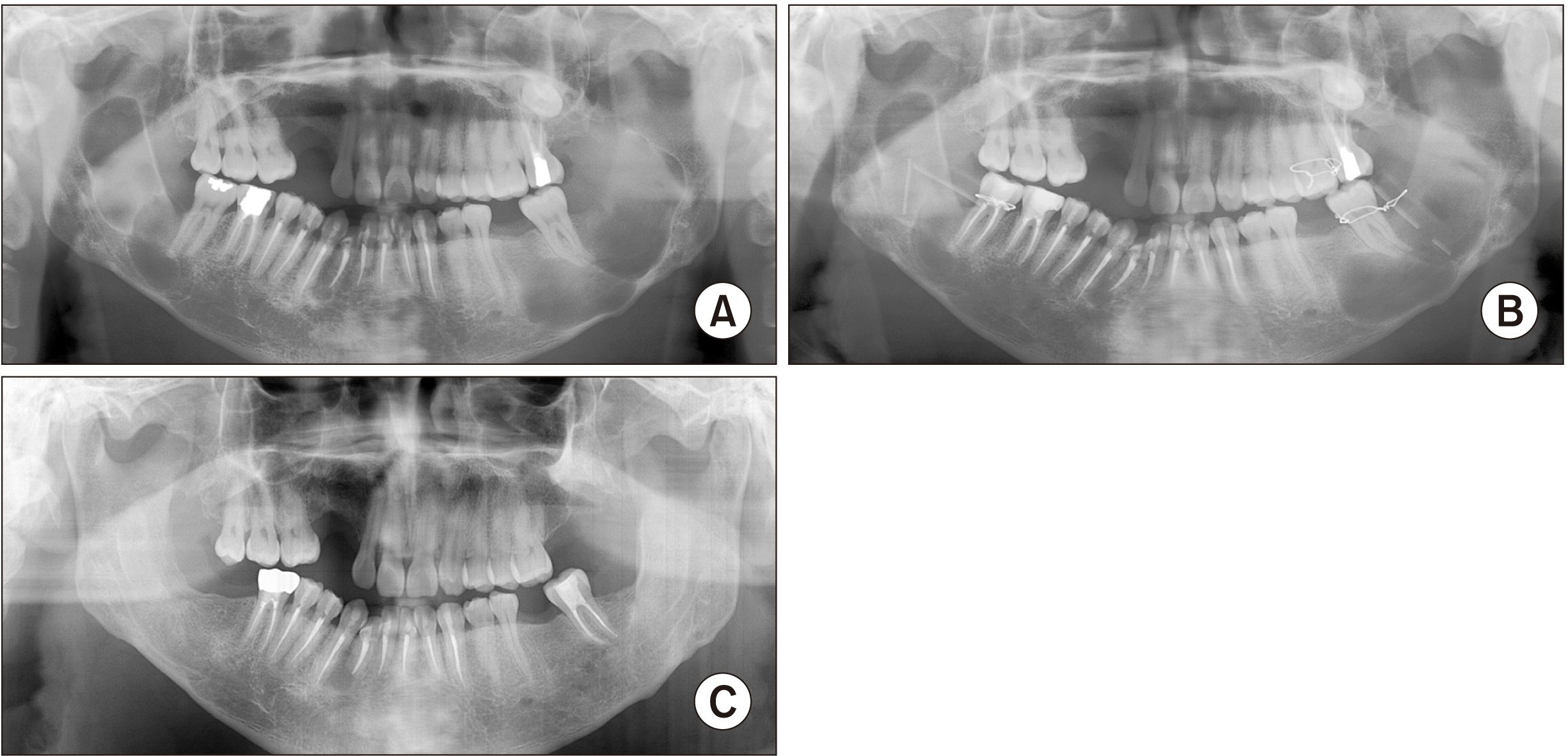
A 37-year-old male was referred by a local clinic to the Department of Oral and Maxillofacial Surgery for multiple cystic lesions in his jaws. His medical history was collected at the initial visit but was not contributory. The patient was later diagnosed with nevoid basal cell carcinoma syndrome. A panoramic radiograph revealed radiolucent lesions on the left posterior maxilla and both posterior mandibles.(Fig. 1. A) Computed tomography (CT) scans showed well-defined expansive lesions in the maxillae and mandibles. The patient was scheduled for an incisional biopsy and marsupialization to reduce their volume.(Fig. 1. B) The biopsy confirmed OKC. The patient was prescribed irrigation twice a week with normal saline solution for six months. Six months after marsupialization, the patient was scheduled for cyst enucleation of the left maxilla and both sides of the mandible under general anesthesia. The modified Carnoy’s solution was soaked into 2×2-inch gauze and applied for three minutes after removal of the lesions. After copious saline irrigation, primary closure was achieved. The permanent biopsy confirmed OKC.
There were no remarkable complications after the surgery. The patient was followed for four years with no signs of any recurrence.(Fig. 1. C)
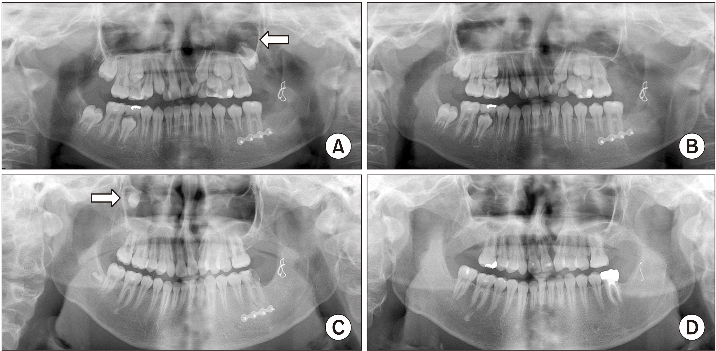
A 10-year-old male presented to the clinic complaining of swelling of the left mandible angle. The patient had undergone a cleft palate surgery at 2 years old and was diagnosed with nevoid basal cell carcinoma syndrome. He was under observation after the OKC surgery on both posterior mandibles. After eight months, the patient developed OKC on the left posterior maxilla. The patient underwent cyst enucleation and extraction of #27.(Fig. 2. A) A modified Carnoy’s solution was applied to 2×2-inch gauze and applied for five minutes after enucleation. After copious saline irrigation, primary closure was achieved.(Fig. 2. B)
The patient was followed for three years, at which point another OKC was found on the right posterior maxilla.(Fig. 2. C) The patient underwent cyst enucleation and extraction of #17 and #18. A modified Carnoy’s solution was applied to 2×2-inch gauze for five minutes after enucleation. The patient was followed for five years and showed no signs of recurrence.(Fig. 2. D)
A 10-year-old female patient was referred from the pediatric dentistry department, and her chief concern was a cystic lesion on the mandibular anterior area. The patient had no relevant prior medical history. The patient had undergone cyst enucleation under general anesthesia earlier the same year. At the follow-up visit, the patient presented with a new cystic change in the #17, #37, and #47 areas. The patient underwent cyst enucleation without the use of a modified Carnoy’s solution. In 2016, the patient developed cystic lesions on both posterior maxillae.(Fig. 3. A) The patient underwent cyst enucleation in those areas; this time, a modified Carnoy’s solution was soaked in 2×2-inch gauze and applied to each area for three minutes. After copious saline irrigation, primary closure was achieved. Postoperatively, the patient was followed for six years with no signs of recurrence.(Fig. 3. B)
Standard treatment modalities for OKC range from marsupialization to marginal resection. Among OKC’s various surgical methods, we selected cyst enucleation simultaneously with the use of a Carnoy’s solution. Since cyst enucleation alone does not guarantee complete removal of the lesion, Carnoy’s solution was also applied with the hope that it would prevent recurrence caused by the remaining cyst epithelium after enucleation. Within the limitations of this case series, the minimally invasive surgery with Carnoy’s solution could be effective to treat OKC in the maxillae.
The study of Blanas et al.6 found recurrence rates of 1.85% for marginal resection, 4.8% for enucleation with Carnoy’s solution, and 30.8% for enucleation and adjunctive measures other than Carnoy’s solution. Alchalabi et al.9 suggested that complications and morbidities originating from the application of Carnoy’s solution occurred less frequently and were less serious than those associated with resection, while there was no recurrence, which is equal to the recurrence rate of resection. OKC mainly occurs in the mandible and rarely in the maxilla. Accordingly, there is a lack of literature describing treatment protocols. Lal et al.10 have suggested that the extent of surgical resection should be dictated by the age of the patient, the site of the lesion, and the involvement of surrounding vital structures. Certainly, the most common treatment for aggressive benign lesions associated with OKCs is maxilla resection with a margin of safety. The anatomical structure, loose bone density of the maxilla, and empty space of the maxillary sinus could allow rapid growth of the lesion and lead to tumor occupancy in the entire maxilla within a short period of time. Therefore, OKCs of the maxilla require more aggressive surgery such as resection11-13. However, this procedure is known to significantly reduce quality of life due to its serious side effects. The trend is now to attempt minimally invasive surgery to save as much of the vital structures of the maxilla as possible and to treat the condition more conservatively.
In this report, OKCs in the maxilla were successfully treated with no recurrence using a minimally invasive enucleation surgery and the application of a modified Carnoy’s solution. As part of their surgical procedures, patients were initially treated with marsupialization to decompress the size of the cyst. Enucleation was carried out only after it was determined that there was no further increase in radiopacity from the panoramic radiograph and cone-beam CT imaging. A modified Carnoy’s solution was applied to the bony bed of the cyst by wetting the gauze for three to five minutes after enucleation. Copious saline irrigation was performed after Carnoy solution application to ensure that the solution was completely washed out. Each patient was followed for four to six years with no signs of recurrence.
In our cases, the modified Carnoy’s solution was applied to the bony bed of the enucleated site with soaked gauze to avoid chemical injury on the surrounding tissue. The solution was applied for three to five minutes depending on the size of the cyst. Within the small sample size limitation of this study, we observed that the modified Carnoy’s solution was effective at preventing the recurrence of OKC not only in the mandible, but also in the maxilla. We accordingly suggest that surgeons can safely use a modified Carnoy’s solution as an adjunct therapy to treat OKC in the maxilla. In subsequent studies, more samples will need to be included, and the follow-up period should be extended. In conclusion, conservative treatment with a modified Carnoy’s solution could be considered a viable option for reducing the recurrence rate of an OKC in the maxilla. Since it is a minimally invasive approach, the patient’s quality of life is likely to improve significantly following the procedure.
Notes
References
1. Maurette PE, Jorge J, de Moraes M. 2006; Conservative treatment protocol of odontogenic keratocyst: a preliminary study. J Oral Maxillofac Surg. 64:379–83. https://doi.org/10.1016/j.joms.2005.11.007. DOI: 10.1016/j.joms.2005.11.007. PMID: 16487797.

2. Myoung H, Hong SP, Hong SD, Lee JI, Lim CY, Choung PH, et al. 2001; Odontogenic keratocyst: review of 256 cases for recurrence and clinicopathologic parameters. Oral Surg Oral Med Oral Pathol Oral Radiol Endod. 91:328–33. https://doi.org/10.1067/moe.2001.113109. DOI: 10.1067/moe.2001.113109. PMID: 11250631.

3. Chirapathomsakul D, Sastravaha P, Jansisyanont P. 2006; A review of odontogenic keratocysts and the behavior of recurrences. Oral Surg Oral Med Oral Pathol Oral Radiol Endod. 101:5–9. discussion 10. https://doi.org/10.1016/j.tripleo.2005.03.023. DOI: 10.1016/j.tripleo.2005.03.023. PMID: 16360602.

4. Khan AA, Qahtani SA, Dawasaz AA, Saquib SA, Asif SM, Ishfaq M, et al. 2019; Management of an extensive odontogenic keratocyst: a rare case report with 10-year follow-up. Medicine (Baltimore). 98:e17987. https://doi.org/10.1097/md.0000000000017987. DOI: 10.1097/MD.0000000000017987. PMID: 31860950. PMCID: PMC6940056.

5. Kaczmarzyk T, Mojsa I, Stypulkowska J. 2012; A systematic review of the recurrence rate for keratocystic odontogenic tumour in relation to treatment modalities. Int J Oral Maxillofac Surg. 41:756–67. https://doi.org/10.1016/j.ijom.2012.02.008. DOI: 10.1016/j.ijom.2012.02.008. PMID: 22445416.

6. Blanas N, Freund B, Schwartz M, Furst IM. 2000; Systematic review of the treatment and prognosis of the odontogenic keratocyst. Oral Surg Oral Med Oral Pathol Oral Radiol Endod. 90:553–8. https://doi.org/10.1067/moe.2000.110814. DOI: 10.1067/moe.2000.110814. PMID: 11077375.

7. Jo HJ, Kim HY, Kang DC, Leem DH, Baek JA, Ko SO. 2020; A clinical study of inferior alveolar nerve damage caused by Carnoy's solution used as a complementary therapeutic agent in a cystic lesion. Maxillofac Plast Reconstr Surg. 42:16. https://doi.org/10.1186/s40902-020-00257-4. DOI: 10.1186/s40902-020-00257-4. PMID: 32509707. PMCID: PMC7248162.

8. Donnelly LA, Simmons TH, Blitstein BJ, Pham MH, Saha PT, Phillips C, et al. 2021; Modified Carnoy's compared to Carnoy's solution is equally effective in preventing recurrence of odontogenic keratocysts. J Oral Maxillofac Surg. 79:1874–81. https://doi.org/10.1016/j.joms.2021.03.010. DOI: 10.1016/j.joms.2021.03.010. PMID: 33901451.

9. Alchalabi NJ, Merza AM, Issa SA. 2017; Using Carnoy's solution in treatment of keratocystic odontogenic tumor. Ann Maxillofac Surg. 7:51–6. https://doi.org/10.4103/ams.ams_127_16. DOI: 10.4103/ams.ams_127_16. PMID: 28713736. PMCID: PMC5502516.

10. Lal B, Kumar RD, Alagarsamy R, Shanmuga Sundaram D, Bhutia O, Roychoudhury A. 2021; Role of Carnoy's solution as treatment adjunct in jaw lesions other than odontogenic keratocyst: a systematic review. Br J Oral Maxillofac Surg. 59:729–41. https://doi.org/10.1016/j.bjoms.2020.12.019. DOI: 10.1016/j.bjoms.2020.12.019. PMID: 34272109.

11. Altas E, Karasen RM, Yilmaz AB, Aktan B, Kocer I, Erman Z. 1997; A case of a large dentigerous cyst containing a canine tooth in the maxillary antrum leading to epiphora. J Laryngol Otol. 111:641–3. https://doi.org/10.1017/s0022215100138198. DOI: 10.1017/S0022215100138198. PMID: 9282204.

12. Albright CR, Hennig GH. 1971; Large dentigerous cyst of the maxilla near the maxillary sinus: report of case. J Am Dent Assoc. 83:1112–5. https://doi.org/10.14219/jada.archive.1971.0414. DOI: 10.14219/jada.archive.1971.0414. PMID: 5286148.

13. Cho JY, Nam KY. 2012; Expansile dentigerous cyst invading the entire maxillary sinus: a case report. J Korean Assoc Oral Maxillofac Surg. 38:245–8. https://doi.org/10.5125/jkaoms.2012.38.4.245. DOI: 10.5125/jkaoms.2012.38.4.245.

Fig. 1
Radiographs of Patient No. 1 (37-year-old male). A. A radiograph showing large radiolucent lesions on the left posterior maxilla and both posterior mandibles. B. A radiograph showing marsupialization to reduce size. C. A radiograph taken four years after surgery and showing no recurrence.

Fig. 2
Radiographs of Patient No. 3 (10-year-old female). A. A radiograph showing radiolucent lesions on both posterior maxillae (arrows). B. A radiograph taken six years after surgery, showing no recurrence.

Fig. 3
Radiographs of Patient No. 2 (10-year-old male). A. A radiograph showing a patient with odontogenic keratocyst (OKC) on the left posterior maxilla (arrow). B. A panoramic view after cyst enucleation and extraction of #27. C. A radiograph taken three years after surgery, showing an additional OKC on the right posterior maxilla (arrow). D. A radiograph showing no recurrence throughout the follow-up period of three years (mandible) and five years (maxilla).


 PDF
PDF Citation
 Print
Print


 XML Download