Abstract
Generally, if the size of a lip cancer defect exceeds 30% of the lower lip, a local flap or free flap is recommended. However, defects up to 50% of the lower lip in size have been reconstructed successfully by primary closure without a local flap or free flap. In one case, an 80-year-old male farmer who had smoked for more than 50 years presented with squamous cell carcinoma of the lower lip and underwent mass resection and supraomohyoid neck dissection. The defect accounted for almost 2/3 of the lower lip and was repaired by primary closure with V-shaped resection. Biopsy results confirmed pT2N0cM0 stage II disease with clear margins. In another case, a 68-year-old male also presented with squamous cell carcinoma of the lower lip and underwent mass resection. The defect accounted for about half the size of the lower lip but was repaired by primary closure with V-shaped resection. Both patients experienced no discomfort while eating or speaking and were satisfied with the cosmetic and functional outcomes with no evidence of recurrence. Thus, direct closure can be considered even in large lower lip cancers.
Lip cancer is the most common malignant oral lesion, accounting for 23.6% to 30% of oral cancers. Lip squamous cell carcinoma (SCC) is the most common type of lip cancer, and the most common area is the lower lip, comprising approximately 90%1. The lip plays important roles in facial esthetics, communications, and oral functions. As the lip is an anatomically cutaneous and oral mucosa overlap site, lip SCC has a high probability of nodal metastasis and poor prognosis compared with cutaneous SCC, but it has a good prognosis compared with oral mucosa SCC2. Cancer resection causes a defect that can lead to major alterations in normal lip appearance and function3. Optimal reconstruction of lip defects is thus very important.
Lip defects can be reconstructed in many ways. Primary closure is the simplest, most intuitive technique that provides direct restoration of the orbicularis oris muscle4. Skin graft can be considered for a partial thickness defect; however, graft contraction and color mismatch may occur4. For defects in the range of one-third to two-thirds the size of the lip, rotation methods such as Abbe flaps or advancement methods such as Karapandzic flaps are mainly used5. A distant free flap provides a wider range of reconstruction options in defects more than two-thirds the size of the lip5. A flap, however, may lead to color mismatch, decreased mucosal sensation, thinning of the lip, or excessive lip fullness5-10.
Primary closure has been generally recommended in cases in which the defect size is less than 30% of the lower lip11. However, a recent study showed good results of primary closure in lower lip defects accounting for up to 50%-60% of lower lip size4,12.
This report describes two cases in which satisfactory results were obtained by primary closure of a large defect of the lower lip related to lip SCC resection. Among the techniques described above, we chose primary closure over time-consuming local flap procedures because of the old age of our patients. Elderly patients are prone to complications of long operations such as delirium. Also, a lower lip defect is easier to repair with a primary closure, particularly in elderly patients with greater orbicularis oris muscle function and tissue laxity13,14.
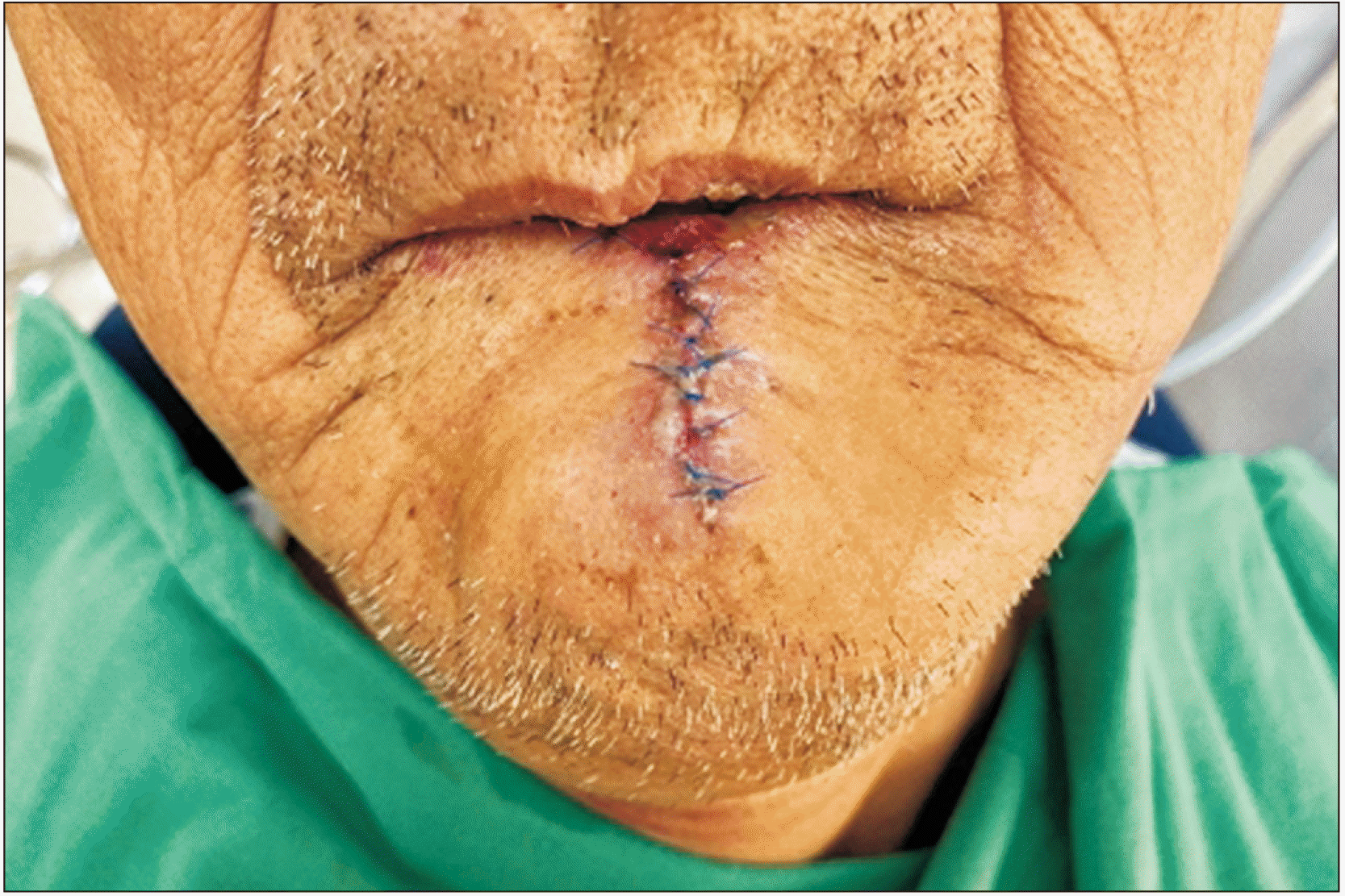
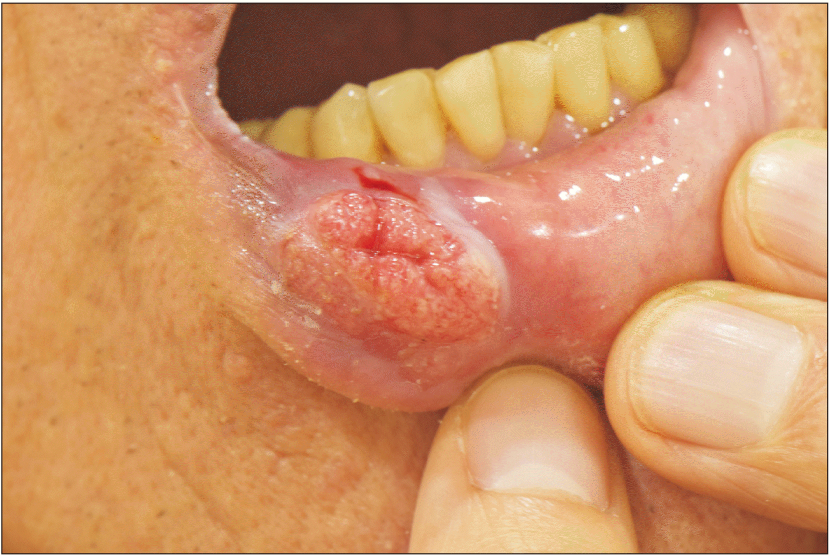
An 80-year-old man who had been diagnosed with SCC of the lower lip through an incisional biopsy at another hospital was referred to the Department of Oral and Maxillofacial Surgery at Seoul National University Dental Hospital. The patient was a farmer and had smoked since the age of 30. Although the exact timing was not known, he reported often feeling a tingling sensation in his lower lip area. At the first visit, on physical and radiological examination, the tumor of the lower lip was about 2.5 cm×2 cm in size and crossed the wet-dry border in the oral cavity.(Fig. 1) On enhanced computed tomography (CT), suspected metastatic lymph node lesions were observed in the left neck at the level Ia and Ib areas. The mass was excised with a safety margin using a V-shaped incision under general anesthesia, and supraomohyoid neck dissection was performed.(Fig. 2) Specimens for frozen biopsy were submitted to the Department of Oral Pathology, and all specimens had negative margin. Primary closure of the lower lip defect was performed.(Fig. 3) The biopsy results confirmed pT2N0cM0 stage II disease.(Fig. 4) The minimum margin was 0.5 cm, and no metastasis was observed in the 12 sampled regional lymph nodes. At 14 months postoperative, no recurrence was observed on physical or radiographic examination. The patient experienced no discomfort while eating or speaking and was satisfied with cosmetic and functional outcomes.
A 68-year-old man was referred to the Department of Oral and Maxillofacial Surgery at Seoul National University Dental Hospital due to redness and swelling of the lower lip. He reported that these symptoms had been going on for two years without spontaneous pain. The first incisional biopsy confirmed the lesion as epithelial dysplasia.(Fig. 5) The lesion, however, did not heal properly and increased in size.(Fig. 6) After 3 months of close follow-up, the last incisional biopsy result showed SCC. Mass resection with a safety margin using a V-shaped incision was performed under general anesthesia. Primary closure of the lower lip defect was performed, and the surgical resection margin was clear. Four years postoperative, no recurrence was observed, and the patient was satisfied with the cosmetic and functional outcomes.
As the lower lip also plays an important functional role in mastication, swallowing, pronunciation, and emotional expression, resection and reconstruction of lip defects after surgical excision should consider normal shape and sphincter function15.
Lip defects can be reconstructed with a variety of techniques such as primary closure, skin graft, and local flaps. Primary closure provides direct restoration of the orbicularis oris muscle, and the tissue remains sensate4. However, it may cause microstomia or unpleasant redundant vermilion16. Skin graft is for a partial thickness defect. It has the risk of graft contraction and color mismatch4. A local flap or a free flap is generally recommended in cases where the defect size comprises more than 30% of the lower lip. It provides a wider range of reconstruction options than primary closure. However, the technique may result in color mismatch, change in hair growth direction, and decreased mucosal sensation5,6,8. Thinning of the lip or excessive lip fullness from flap overadvancement may also occur8-10.
A recent study has suggested primary closure of lower lip defects accounting for up to 50%-60% of lower lip size, especially in elderly patients4,12. Even microstomia in patients treated with primary closure can be managed with postoperative physical therapy including lip stretching12. Here we showed that linear closure is a functionally and esthetically satisfying technique for lower lip defects that require resection of not only half but even up to two-thirds of the lip. These cases demonstrate that even large lower lip defects can be successfully reconstructed through primary closure without a local flap or free flap, both of which require a longer duration of surgery and hospitalization period, higher financial burden, and the possibility of donor site morbidities.
It may have been helpful to measure lip strength or lip endurance using the IOPI (Iowa Oral Performance Instrument) in order to objectively evaluate lip function17. However, the patients did not experience any discomfort and were satisfied in terms of esthetics and function and thus declined further evaluation.
As an old saying goes, “Prevention is better than a cure.” It is important to notice and remove lip lesions in the early stages before they grow to a size that requires invasive reconstruction procedures. The highest risk factor of lip cancer is ultraviolet (UV) radiation, but other risk factors include smoking, alcohol use, and human papilloma virus2. Because the patient in Case 1 was a farmer, outdoor activities and UV exposure were likely common. He had also smoked for about 50 years, and thus he had many risk factors for lip cancer. When the patient first presented to our hospital, the exophytic mass was already larger than 2 cm; on the axial view of enhanced CT, it appeared to be 2.5 cm long, which was more than 50% the size of the lower lip. The presentation was likely delayed because the patient did not experience much discomfort. Therefore, dentists should carefully monitor patients for lip lesions, especially high-risk patients, for timely detection. Actinic cheilitis (AC), plasma cell cheilitis, and viral warts should be considered in the differential diagnosis of lip SCC, as up to 95% of lip SCCs originate from AC18,19. AC is a chronic inflammatory process leading to the loss of the sharp border of the lip and appears atrophic or erosive on examination20,21. Leukoplakia, which is defined as a white patch with undefined risk of transformation, is the most common form of AC and the most common change that precedes SCC21. Initially lip SCC is asymptomatic and difficult to distinguish from AC and demonstrates leukoplakia or erythroplakia, with atrophic plaques. In advanced stages, it can cause pain with exudate ulcers or may present as a bleeding, friable, verrucous, or exophytic mass21.
In conclusion, the size criteria for primary closure of lower lip defects can be expanded from less than 30% to up to two-thirds of the lip in elderly patients. By applying this method to patients with a larger lesion, we can shorten the duration of surgery and the hospitalization period as well as reducing financial burden and the possibility of donor site morbidities. Careful monitoring of patients at high risk for lip cancer is also crucial, since it is important to remove lesions at an early stage.
Notes
Authors’ Contributions
S.B.Y. performed data collection and wrote the manuscript. I.-J.K. helped to draft the manuscript. H.M. approved the final manuscript.
References
1. Ozturk K, Gode S, Erdogan U, Akyildiz S, Apaydin F. 2015; Squamous cell carcinoma of the lip: survival analysis with long-term follow-up. Eur Arch Otorhinolaryngol. 272:3545–50. https://doi.org/10.1007/s00405-014-3415-6. DOI: 10.1007/s00405-014-3415-6. PMID: 25467011.

2. Bota JP, Lyons AB, Carroll BT. 2017; Squamous cell carcinoma of the lip-a review of squamous cell carcinogenesis of the mucosal and cutaneous junction. Dermatol Surg. 43:494–506. https://doi.org/10.1097/dss.0000000000001020. DOI: 10.1097/DSS.0000000000001020. PMID: 28157733.

3. Matin MB, Dillon J. 2014; Lip reconstruction. Oral Maxillofac Surg Clin North Am. 26:335–57. https://doi.org/10.1016/j.coms.2014.05.013. DOI: 10.1016/j.coms.2014.05.013. PMID: 25086695.

4. Boukovalas S, Boson AL, Hays JP, Malone CH, Cole EL, Wagner RF. 2017; A systematic review of lower lip anatomy, mechanics of local flaps, and special considerations for lower lip reconstruction. J Drugs Dermatol. 16:1254–61. PMID: 29240861.
5. Malard O, Corre P, Jégoux F, Durand N, Dréno B, Beauvillain C, et al. 2010; Surgical repair of labial defect. Eur Ann Otorhinolaryngol Head Neck Dis. 127:49–62. https://doi.org/10.1016/j.anorl.2010.04.001. DOI: 10.1016/j.anorl.2010.04.001. PMID: 20822758.

6. Krunic AL, Weitzul S, Taylor RS. 2005; Advanced reconstructive techniques for the lip and perioral area. Dermatol Clin. 23:43–53. v–vi. https://doi.org/10.1016/j.det.2004.08.009. DOI: 10.1016/j.det.2004.08.009. PMID: 15620618.

7. Baker SR. 2007. Local flaps in facial reconstruction. 2nd ed:Mosby;DOI: 10.1016/B978-0-323-03684-9.50023-2.
8. Neligan PC. 2013. Plastic surgery. 3rd ed:Elsevier;DOI: 10.1016/j.det.2004.08.009.
9. McCarn KE, Park SS. 2005; Lip reconstruction. Facial Plast Surg Clin North Am. 13:301–14. viihttps://doi.org/10.1016/j.fsc.2004.11.005. DOI: 10.1016/j.fsc.2004.11.005. PMID: 15817408.

10. Anvar BA, Evans BCD, Evans GRD. 2007; Lip reconstruction. Plast Reconstr Surg. 120:57e–64e. https://doi.org/10.1097/01.prs.0000278056.41753.ce. DOI: 10.1097/01.prs.0000278056.41753.ce. PMID: 17805106. PMCID: PMC10484365.

11. Demirdover C, Vayvada H, Ozturk FA, Yazgan HS, Karaca C. 2019; A new modification of fan flap for large lower lip defects. Scand J Surg. 108:172–7. https://doi.org/10.1177/1457496918798211. DOI: 10.1177/1457496918798211. PMID: 30178718.

12. Sanniec KJ, Carboy JA, Thornton JF. 2018; Simplifying lip reconstruction: an algorithmic approach. Semin Plast Surg. 32:69–74. https://doi.org/10.1055/s-0038-1645882. DOI: 10.1055/s-0038-1645882. PMID: 29765270. PMCID: PMC5951711.

13. Soliman S, Hatef DA, Hollier LH Jr, Thornton JF. 2011; The rationale for direct linear closure of facial Mohs' defects. Plast Reconstr Surg. 127:142–9. https://doi.org/10.1097/prs.0b013e3181f95978. DOI: 10.1097/PRS.0b013e3181f95978. PMID: 21200208.

14. Harris L, Higgins K, Enepekides D. 2012; Local flap reconstruction of acquired lip defects. Curr Opin Otolaryngol Head Neck Surg. 20:254–61. https://doi.org/10.1097/moo.0b013e3283557dcf. DOI: 10.1097/MOO.0b013e3283557dcf. PMID: 22894993.

15. Park CH, Kwon TK, Hong SJ, Chu HR. 2008; Reconstruction after resection of lower lip squamous cell carcinoma. Korean J Otorhinolaryngol Head Neck Surg. 51:471–6.
16. Ishii LE, Byrne PJ. 2009; Lip reconstruction. Facial Plast Surg Clin North Am. 17:445–53. https://doi.org/10.1016/j.fsc.2009.05.007. DOI: 10.1016/j.fsc.2009.05.007. PMID: 19698922.

17. Jeong DM, Shin YJ, Lee NR, Lim HK, Choung HW, Pang KM, et al. 2017; Maximal strength and endurance scores of the tongue, lip, and cheek in healthy, normal Koreans. J Korean Assoc Oral Maxillofac Surg. 43:221–8. https://doi.org/10.5125/jkaoms.2017.43.4.221. DOI: 10.5125/jkaoms.2017.43.4.221. PMID: 28875136. PMCID: PMC5583196.

18. Lupu M, Caruntu A, Caruntu C, Boda D, Moraru L, Voiculescu V, et al. 2018; Non-invasive imaging of actinic cheilitis and squamous cell carcinoma of the lip. Mol Clin Oncol. 8:640–6. https://doi.org/10.3892/mco.2018.1599. DOI: 10.3892/mco.2018.1599. PMID: 29725529. PMCID: PMC5920479.

19. Elmas ÖF, Metin MS, Kilitçi A. 2019; Dermoscopic features of lower lip squamous cell carcinoma: a descriptive study. Indian Dermatol Online J. 10:536–41. https://doi.org/10.4103/idoj.idoj_435_18. DOI: 10.4103/idoj.IDOJ_435_18. PMID: 31544072. PMCID: PMC6743386.

20. Wood NH, Khammissa R, Meyerov R, Lemmer J, Feller L. 2011; Actinic cheilitis: a case report and a review of the literature. Eur J Dent. 5:101–6. DOI: 10.1055/s-0039-1698864. PMID: 21228959. PMCID: PMC3019754.

21. Vieira RA, Minicucci EM, Marques ME, Marques SA. 2012; Actinic cheilitis and squamous cell carcinoma of the lip: clinical, histopathological and immunogenetic aspects. An Bras Dermatol. 87:105–14. https://doi.org/10.1590/s0365-05962012000100013. DOI: 10.1590/S0365-05962012000100013. PMID: 22481658.

Fig. 1
Preoperative clinical photo and enhanced computed tomography (CT) view. A tumor mass approximately 2.5 cm in diameter crosses the wet-dry border of the oral cavity. A, B. Clinical photos. C. CT axial view. D. Magnetic resonance imaging axial view.
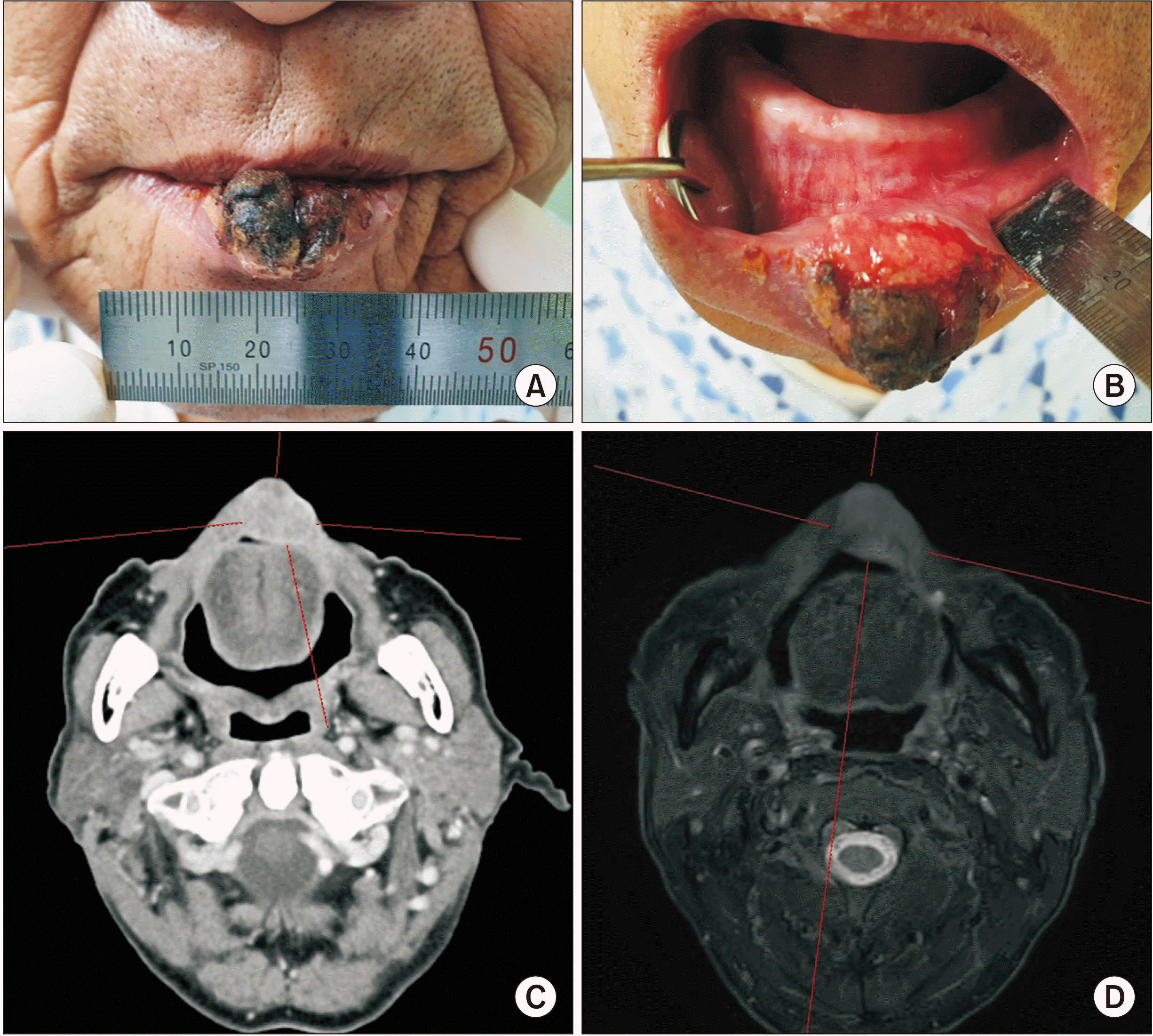
Fig. 2
Intraoperative clinical photo. The mass was excised with a safety margin using a V-shaped incision. A, B. Clinical images before operation. C, D. Clinical images after operation.
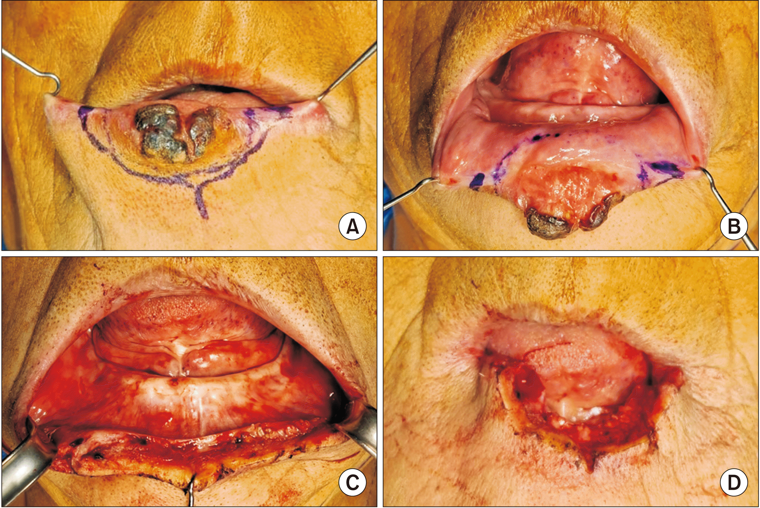
Fig. 4
Histopathologic view of the main mass. Keratin pearls can be identified in the histopathologic image (H&E staining, A: ×4, B: ×100).
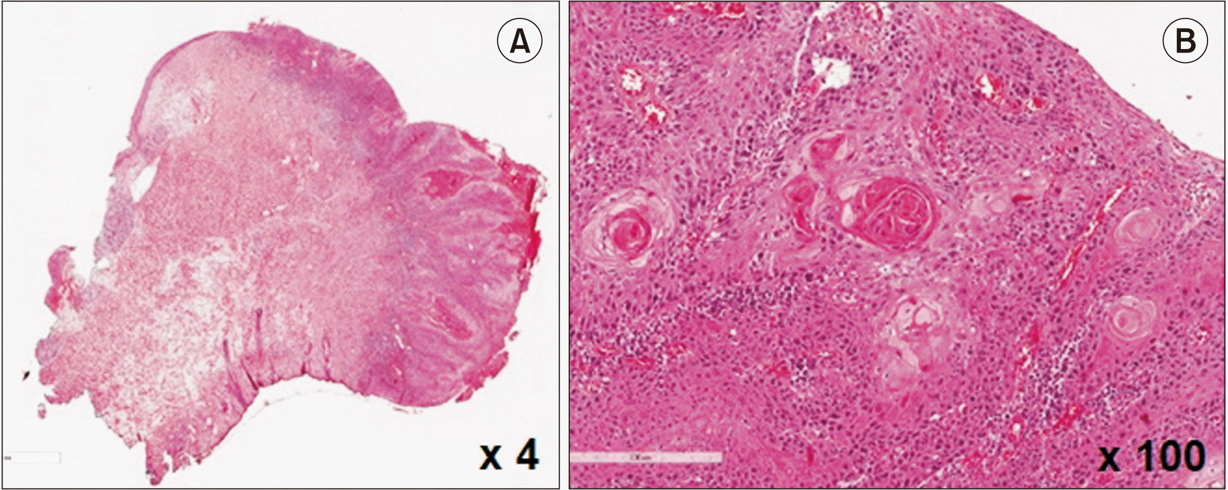
Fig. 5
The H&E stained histopathologic specimen from the incisional biopsy performed on the first visit (A) and the main mass (B) in Case 2. A. At first the lesion was diagnosed as dysplasia due to lack of tumor invasion of the basement membrane. B. The main mass from the surgery was diagnosed as squamous cell carcinoma (H&E staining, A: ×12.5, B: ×12.5).


 PDF
PDF Citation
 Print
Print


 XML Download