

 PDF
PDF Citation
 Print
Print


I. Introduction
Fractures of the anterior wall of the maxillary sinus are common in maxillofacial surgery. The anterior superior alveolar nerve (ASAN) and the middle superior alveolar nerve (MSAN) are branches of the maxillary division of the trigeminal nerve that course along the lateral walls of the maxillary sinus after branching off the infraorbital nerve in the floor of the orbit1.
We hypothesize that injury to the anterior wall of the sinus results in injury to the ASAN and MSAN. The available literature documents ASAN and MSAN injuries in the context of patients presenting with neurosensory deficits (NSD) of the teeth and gingiva on the ipsilateral side of the injury2-4. However, objective assessments of deficits resulting from maxillary sinus fractures are lacking.
Zuniga and Essick5 provided clinical guidelines for testing NSD. Evaluation of patients with NSD starts with clinical examination and patient interviews to understand the nature of the altered sensation. Next, a series of clinical neurosensory tests (NST) are performed to evaluate severity of nerve injury. Each test is specific to a type of nerve fiber6. The tests are as follows:
• Level A: Evaluation of two-point discrimination (TPD) and directional discrimination. This test evaluates receptor density and large myelinated fiber function. Disruption of the connective tissue component of the nerve results in abnormalities at this level.
• Level B: Contact detection that tests for large myelinated fiber function.
• Level C: Pain sensitivity that tests for Aδ and C fiber function.
A large nerve is composed of a combination of large and small myelinated and unmyelinated fibers. Hence, a combination of different neurosensory tests is required for global assessments of nerve injury. TPD and fine test discrimination (FTD) values were the primary outcomes. Secondary outcomes were thermal detection thresholds, vibration detection thresholds, electric pulp test results, and radiographic signs. The specific study aims were to:
(1) Estimate the prevalence of post-traumatic NSD related to maxillary anterolateral wall fractures using NST.
(2) Observe patterns of associations between primary and secondary outcomes.
II. Materials and Methods
1. Study participants and design
This was a prospective study conducted among patients aged 18-70 years with maxillofacial fractures that were reported to our institute between February 2021 and October 2022. Patients with unilateral fractures of the anterolateral wall of the maxillary sinus with or without other facial bone fractures were included. Patients who were included also presented with bilaterally intact maxillary anterior and premolar teeth, with no evidence of dentoalveolar fracture or dental pathology. Patients with traumatic brain injury or who could not have their teeth subjected to NSTs due to comorbidities were excluded from the study. NSTs were carried out before any surgical interventions within one week of trauma and at 2-month follow-up. This study was approved by the Institutional Ethical Committee of JSS Dental College and Hospital (protocol No. 37/2020) and informed consent was obtained from all patients. Before neurosensory testing the following data were collected:
(1) Demographic information of the patient and details of the injury sustained.
(2) Signs and symptoms of head injury.
(3) Subjective assessment of the sensory complaint via patient interview.
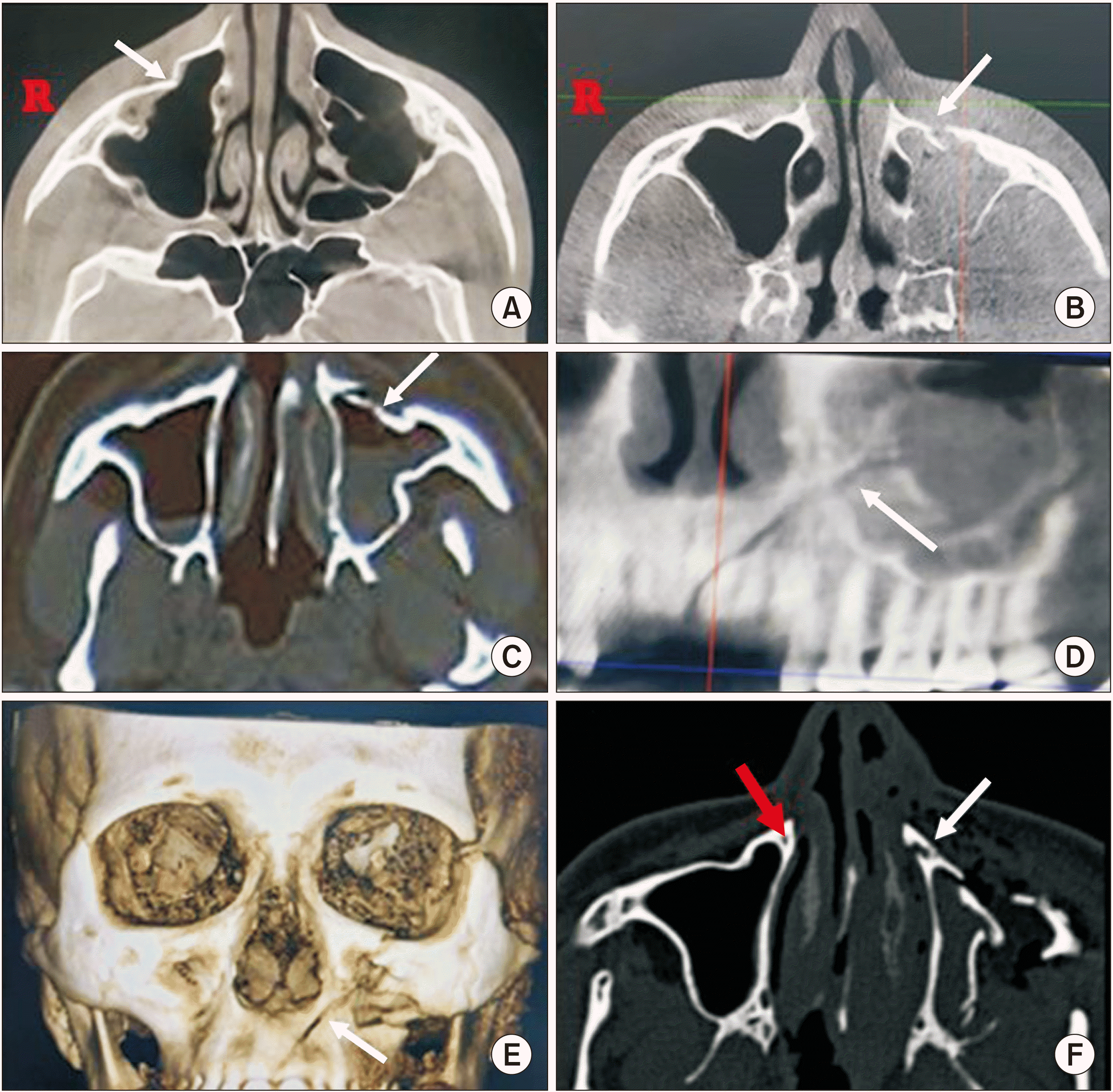
(4) Radiographic examination including computed tomography (CT) or cone-beam CT (CBCT) to assess for hemosinus, depression, displacement, or discontinuity of the maxillary anterior wall fracture.(Fig. 1)
Radiographic fracture patterns were assessed by the following criteria:
(1) Discontinuous: No large change in the volume of the maxillary sinus. Presence of fracture line without any change in the position of the fracture segments.(Fig. 1. B)
(2) Depressed: Inward displacement of the fracture segments at the point of fracture associated with a mild decrease in the volume of the maxillary sinus.(Fig. 1. A)
(3) Displaced: The fractured segment of bone is completely disconnected from the antral wall with a large change in the volume of the sinus.(Fig. 1. C)
(4) Involvement of canalis sinuosus (CS).(Fig. 1. F)
2. Neurosensory testing
Testing was carried out in the following sequential order:
(1) TPD was performed using Baseline 2-point Discrim-A-Gon and FTD was performed using Baseline Tactile Semmes-Weinstein type monofilaments 5-piece hand kit. The smallest diameter filaments in this monofilament kit can be used to test fine touch sensation, and the largest diameter filament, with a 300-g force, can be used to test for pain and deep pressure sensation. TPD and FTD were carried out on the labial and buccal gingival regions supplied by the ASAN and the MSAN, respectively. Readings were compared between the traumatized side (test) and the intact side (control). TPD and FTD tests were performed using the method of limits procedure. For the TPD test, the difference between the patient’s response on the test and control sides was recorded in millimeters. Similarly, for FTD test responses were recorded in terms of the difference in grams force. If patients could perceive the 3.61-size monofilament during FTD, it was assumed that their perception had recovered to the threshold7. For TPD testing a difference of ≥2 mm from the contralateral side was considered significant. The least possible value that can be measured was 2 mm for TPD and 0.07 g for FTD. Both TPD and FTD values were used for classifying the patients into 5 groups: normal, mild, moderate, severe, and anesthetic, according to the clinical guidelines proposed by Zuniga and Essick5. The characteristics of each group were as follows:
• Normal: No loss of TPD as compared to normal side.
• Mild: Only TPD threshold increased on affected side.
• Moderate: Both TPD and FTD thresholds increased on the affected side, while retaining the ability to detect fine touch sensation.
• Severe: Loss of FTD thresholds. Only deep pressure and pin prick sensation intact on the affected side. Patients respond to sense of touch only with 300 g force on the mucosa.
• Anesthetic: Complete loss of both pin prick and deep pressure sensation on affected side. No response even with 300 g force on the oral mucosa.
(2) Vibrotactile testing (VT) was performed using tuning forks at 512 Hz, 256 Hz, and 128 Hz on both the gingiva and teeth starting with the lowest frequency. If patient perception was dull or absent the response was recorded as NO and for normal perception, the response was recorded as YES.
(3) Thermal detection threshold (TDT) of the mucosa were carried out using a cotton pellet saturated with NEOENDO NEOSNOW endodontic spray (Orikam Healthcare India). Normal perception was recorded as YES and decreased or no response was recorded as NO.
(4) The Waldent electric pulp tester was used for electric pulp testing (EPT), and cotton pellets saturated with NEOENDO NEOSNOW endodontic spray were used for cold vitality testing (CVT) on teeth.
All experiments were conducted as per the manufacturer’s instructions. Statistical analysis was performed using IBM SPSS Statistics software (ver. 25.0; IBM). P<0.05 was considered significant.
III. Results
1. Demographic assessment
A total of 39 participants fit the inclusion criteria, of which 3 were lost to follow-up. The mean age of the study participants was 35±14 years, of which 97.4% (38 participants) were male and 2.6% (1 participant) was female. The most common mode of injury was road traffic accidents at 74.4% (29 participants), followed by self-fall at 17.9% (7 participants) and other modes of injury at 7.7% (3 participants). The type of trauma associated with anterior wall fractures was isolated unilateral zygomatico maxillary complex (ZMC) fractures (56.4%), ZMC fractures in conjunction with other trauma (38%), and isolated anterior wall fractures (5.6%). The treatment administered included open reduction and internal fixation under general anesthesia (35 patients, 89.8%), conservative management (2 patients, 5.1%), and no treatment (2 patients, 5.1%). All patients who had complaints of NSD were put on oral neuroprotective medications as deemed appropriate by the operating surgeon.
2. Subjective assessment
A total of 44% of patients examined had altered sensations. The most common complaint was hypoesthesia (33.6%) followed by anesthesia (7.8%) and dysesthesia (2.6%). Reduced gingival responses to TPD and FTD were seen in all patients who reported anesthesia and dysesthesia. In comparison, 10%-15% of individuals who reported hypoesthesia were found to have normal responses to FTD and TPD.(Table 1)
3. Assessment of TPD and FTD – primary outcomes
Immediately following trauma, the TPD threshold was increased on the traumatized side by 5±5 mm in comparison to the control. At two months there was only a 2.3±1 mm difference, and this improvement was significant (P=0.007, McNemar’s test). Similarly, the mean FTD difference was 40.3 g in the immediate period, which significantly dropped to 9.0 g at the two-month follow-up (P=0.04, McNemar’s test). The mean value for change was 34.4±109.1 g (Q1=0 g, Q3=1.60 g). Measurements of FTD and TPD were identical in the areas innervated by the ASAN and MSAN.
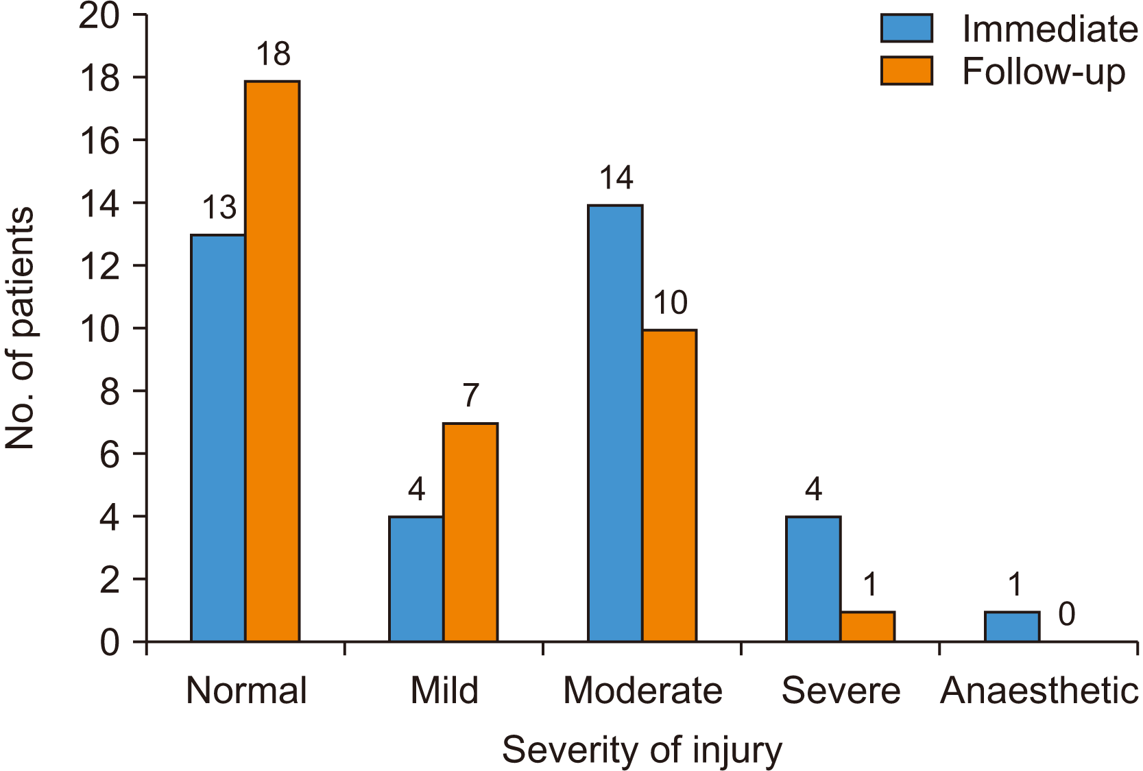
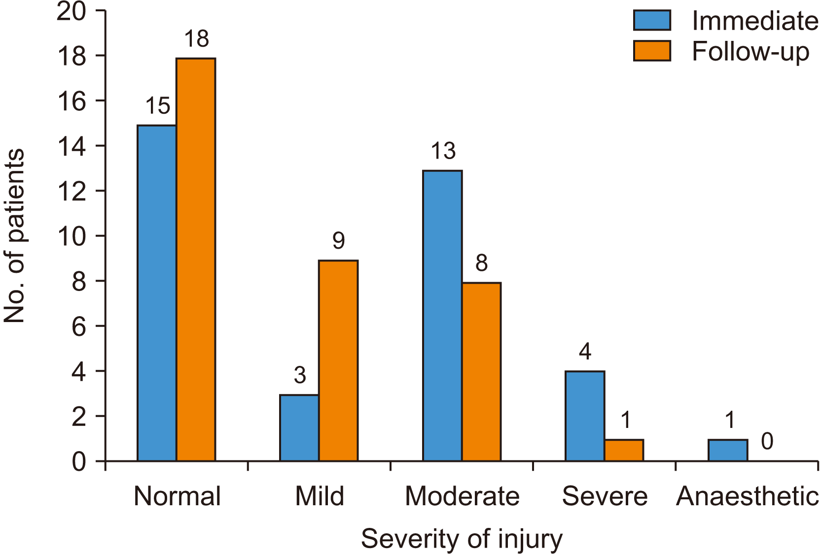
The distributions of severity of nerve injury according to Zuniga’s and Essick’s guidelines are given in Fig. 2 and 3 for ASAN and MSAN, respectively. At two months, it was noted that there had been little to no overall restoration of baseline sensation in any group. The incidence rates of NSD between the ASAN and MSAN groups did not differ substantially either immediately following trauma or at two-month follow-up. Nerve injury in patients was most often moderate, followed by mild, severe, and anesthetic types.
4. Assessment of Aβ fibers (dynamic stimuli) of the gingiva and periodontal ligament – VT
Dullness with vibration stimuli was present only for anesthetic type injuries. Only two patients had deficits related to VT at two-month follow-up. Of these two patients, one had deficit immediately following injury that continued at two months. The other individual did not present with VT deficit immediately following injury but developed it after 2 months of follow-up. The change was not significant for either ASAN and MSAN groups at 2 months.(Table 2)
5. Assessment of Aδ and C fibers of the gingiva – TDT
The loss of temperature discrimination was significantly associated with severe types of nerve injuries (P=0.007, Pearson’s chi-square test). The overall improvement for the group was not significant (paired t-test). No significant difference was observed between ASAN and MSAN groups.(Table 3)
1) EPT (Aδ fibers)
Some individuals in the MSAN (8 patients) and ASAN (5 patients) groups had invalid EPT findings, i.e., they had no sensibility to the electrical stimuli in the non-traumatized side resulting in the inability to calculate threshold values. These participants were excluded when evaluating the significance values for the test. For the anterior teeth, 33.4% of individuals showed no response to the test, 12.8% did not have a valid test and 53.8% had a valid response to the test (n=39). Of the patients with valid responses, 20.5% showed a higher threshold as compared to the normal contralateral side. For posterior teeth, 25.6% had no response and 53.8% had valid responses. The EPT values were 5.9±3.9 units for the anterior teeth and 8.5±5.0 units for the posterior teeth immediately after injury. This number changed to 5.2±5.3 units for the anterior teeth and 6.2±7.6 units for the posterior teeth on follow-up. As a group, significant improvement was present in ASAN (P=0.008, McNemar’s test) but not in MSAN. At two months follow-up, 83.3% of ASAN and 77.8% of MSAN individuals were responsive to the EPT and only 8.3% of the tests were not valid. Of the responsive patients, 27.8% of ASAN and 36.1% of MSAN individuals showed persistent altered sensation.(Table 4)
2) CVT (C fibers)
A total of 13 patients (36.1%) reported altered sensations to CVT in the anterior and posterior teeth regions immediately after trauma. At the two-month follow-up 6 patients had regained normal sensation, and 7 patients retained altered sensation of the tooth. Overall, 80.6% of patients tested regained normal sensation at two months (29 patients). There was no difference between the anterior and posterior teeth. The improvement over 2 months was significant (P=0.031 paired t-test).(Table 5)
7. Radiographic interpretation
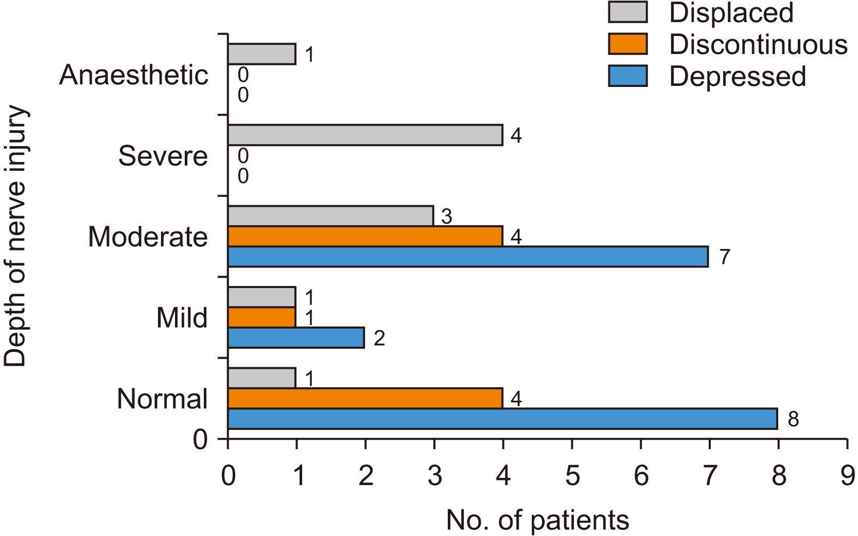
The distribution of patients with various radiographic patterns of maxillary sinus wall fractures is depicted in Fig. 4. The most severe nerve injury when comparing ASAN and MSAN was taken as the final diagnosis. The most common type of fracture pattern was depressed fractures.(Fig. 1. A)The severity of NSD was greater in fractures involving the CS.(Fig. 1. F)
IV. Discussion
In this study, we quantified nerve injuries of the ASAN and the MSAN sustained by patients after maxillary anterior wall fractures. Injury to the anterior wall primarily arises from trauma, although it can also occur as a result of surgical procedures such as Caldwell-Luc surgery, implant placement, or antral tumors1. By quantifying nerve injury patterns, we can effectively predict anatomically significant areas on the anterior wall that should be carefully avoided during surgical interventions as well as prognosis. Such comprehensive understanding facilitates better surgical planning and patient outcomes.
The clinical guidelines for assessment of abnormal sensibilities for the oral and perioral sites established by Zuniga and Essick5 are based on previous observations that Aβ (myelinated) fibers, responsible for mechanoreception, are more susceptible to compression and ischemia than the Aδ (myelinated) and C (unmyelinated) fibers (pain, temperature)8. Clinically, testing for loss of TPD using the disc discriminator is followed by testing for loss of touch and pain sensation using Semmes-Weinstein monofilament kit. In this study, 50% of patients with gingival loss of mechanical sensibilities presented with increased thresholds to both fine touch and TPD, i.e., moderate degree of nerve injury. Such patients had complaints of graphesthesia (i.e., loss of shape perception) and loss of texture discrimination due to losses in peripheral innervation density and injury to both the epi- and perineurium9. Patients with only one type of deficit, TPD or FTD (i.e., mild nerve injury) were not associated with subjective loss of sensation. In the follow-up period, the improvement of the threshold for TPD and FTD was significant but a complete return to normal sensation was not observed in our sample. Other authors reported similar recovery at three months for the infraorbital nerve8,10. When these tests are performed individually, Vriens and Moos8 considered TPD to be the more sensitive test in diagnosing NSD. However, in our study we found FTD to be more sensitive.
There is a link between the recovery of mechanical thresholds and temperature perception. Patients with severe nerve injuries were found to have a significant chance of having concomitant loss of temperature discrimination because nerve fibers carrying temperature stimuli are more resistant to a traumatic injury. Pedemonte and Basili11 suggests that evaluating thermal perception thresholds may help predict the prognosis of a nerve injury.
Mechanical perception thresholds of the PDL ligament help when evaluating the innervation of Aβ fibers. Recovery of these fibers is expected at 5 months after anterior wall fractures12. Classically, dynamic stimuli such as vibrations have been used to evaluate these fibers. This may not be the ideal test for teeth, as vibrations can be transmitted through the bone to the maxilla and mandible via the TMJ13. This could be why very few patients tested positive for NSD with tuning forks for VT. We also observed very low sensitivity for this test. Based on our results, we suggest alternative testing modalities for mechanical threshold measurement for periodontal ligaments such as modified SWM14.
Pulpal sensibility tests have been used to assess nerve injuries of the teeth after trauma to the maxillary antrum by several authors15,16. The dental pulp is a sensory organ that conducts impulses transmitted to it via the conduction of enamel and the dentinal fluid. It is innervated by both Aδ and C fibers, which are responsible for sharp shooting pain and dull pain, respectively17. In some cases of pulpal injury, Aδ fibers lose sensibility while C fibers, being more resistant to hypoxia, may still function18. Hence, a more accurate description of the sensation felt by the patient needs to be recorded when performing these tests. EPT is sensitive for identifying vital teeth 75% of the time and CVT has a sensitivity of 91.6%. One hundred percent of vital teeth tested with both CVT and EPT respond to at least one test19. Hence, a combination of these tests is suggested when evaluating NSD of the teeth. EPT is more technique sensitive, which may be the reason for the invalid tests found in this study. The difference in EPT values between the MSAN and ASAN can be attributed to the volume of pulpal tissues present. Thicker enamel and dentine will necessitate a higher electrical stimulus for perception. We found that there was a significant improvement in pulpal sensibilities over two months but did not observe complete return to baseline. Despite the loss of tooth sensitivity being associated with severe gingival injury, a significant association between the two could not be drawn. Other authors have proposed that re-innervation of teeth can start as early as 6 weeks after injury and continue until no later than 1 year20. Tajima12 reported a significant difference between the recovery time for pulpal, periodontal ligament, and gingival sensations after malar bone fractures.
There were no significant differences in sensibilities between ASAN and MSAN for any NSTs we performed. There are two possible reasons for this. The first is that MSAN occurs as a distinct branch in only 60%-70% of all individuals21. In individuals without MSAN, the branches of ASAN are more likely to innervate the premolars than the posterior superior alveolar nerve (PSAN). Hence, injury to ASAN branches after anterior wall fractures also leads to NSD of the premolars. The second possible reason is the presence of the superior dental plexus, which is formed by the confluence of the ASAN, MSAN, and PSAN. Hence, NSD of ASAN and MSAN could be poorly differentiated by the patient.
Fracture lines that pass lower on the maxilla may result in a greater incidence of NSD, as the dental plexus is located here12. In this study, individuals with anesthesia or with response to only deep pressure stimuli had fracture lines passing through the canine fossa region and involving the CS (Fig. 1. D-F), as a result of direct injury to the ASAN. Displaced fractures of the maxillary antrum also resulted in severe forms of nerve injuries.
The main limitation of this study is the lack of long-term follow-up, which could not be achieved in milder and moderate groups due to poor patient compliance. Despite this drawback, we standardized testing modalities of NSD and suggested improvements in techniques for future research.
V. Conclusion
NSDs after maxillary anterior wall fractures are common. Severe NSD of intraoral structures is associated with displaced fractures of the canine fossa and direct injury to the CS. Loss of temperature discrimination predicts the severity of nerve injuries. Sensibilities of teeth appear to recover independently of the severity of gingival NSD.
 XML Download