

 PDF
PDF Citation
 Print
Print


INTRODUCTION
Bone-turnover markers (BTMs) have been defined as “biochemical products measured in blood or urine, and which reflect the metabolic activity of bone but which themselves have no function in controlling skeletal metabolism” [1]. BTMs are markers of bone formation and bone resorption. Bone-formation markers are produced during various stages of bone formation. They are synthesized and secreted by osteoblasts, and their concentrations in the blood are thought to reflect the bone-formation rate [2]. BTMs, which are designated as resorption markers, are products of bone-collagen degradation or enzymes secreted by osteoclasts during bone resorption. Their concentrations in the blood and urine reflect the bone-resorption rate [2]. The BTMs in current clinical use or that show promise are listed in Table 1.
Table 1
Bone-turnover markers currently in common use
![]()
A joint working group between the International Osteoporosis Foundation (IOF) and the International Federation of Clinical Chemistry and Laboratory Medicine (IFCC) designated serum β-isomerized carboxy-terminal telopeptide of collagen type-I (s-β-CTX; referred to hereafter in the text as “CTX”) as the standard bone-resorption marker and serum procollagen type-I N-terminal propeptide (s-PINP) as the standard bone-formation marker, recommending that they be used in all future clinical studies [1]. Due to the lack of standardization between currently available commercial assays for each marker, the joint IOF-IFCC working group called for standardization (or harmonization, if appropriate) of the available assays for CTX and PINP [2]. The Kidney Disease: Improving Global Outcomes (KDIGO) organization recommends bone-specific alkaline phosphatase (B-ALP) as a marker for monitoring chronic kidney disease-mineral and bone disorder (CKD-MBD) [3]. B-ALP may also be useful for diagnosing and managing osteomalacia [4]. B-ALP together with PINP are useful biomarkers for diagnosing Paget’s disease of the bone, and PINP and CTX can be used in the management of hyperparathyroidism [5, 6]. Bone remodeling is usually strikingly increased in Paget’s disease of the bone (a condition that is uncommon in Asia-Pacific populations) or osteomalacia and rickets (which are not uncommon in the region), such that total ALP testing alone is usually adequate for assisting with the diagnosis (without the need for a more specific BTM). Table 2 provides an overview of changes usually observed in BTMs in common metabolic bone diseases in the untreated state.
Table 2
Usual directions of bone-turnover marker changes seen in common conditions
![]()
Go to : 
BTMS DURING THE LIFE COURSE IN HEALTH AND DISEASE
Bone-remodeling rates and BTMs, which reflect them, change with age and are highly variable in childhood. BTMs are much more abundant in children than in adults, reflecting the fact that increased bone modeling occurs during bone growth [7]. The increased BTM concentrations are accentuated during infancy and the pubertal growth spurt (which occurs earlier in girls than in boys) [8]. BTM concentrations reach a stable nadir once skeletal maturity is attained during the third decade of life [9]. BTMs may be useful for clinically investigating bone turnover in children with bone diseases (such as rickets and growth-hormone deficiency) or genetic conditions (such as osteogenesis imperfecta) [10, 11].
Pediatric reference intervals for PINP and CTX
Establishing reference intervals for pediatric populations is particularly challenging. Pediatric reference intervals for serum PINP (Roche Diagnostics GmbH, Mannheim, Germany or Immunodiagnostic Systems [IDS] PLC, Tyne and Wear, UK) and plasma CTX (Roche systems only) assays were recently proposed based on a systematic review of published data [12]. These values are listed in Table 3 and are not population-specific but may be useful until local reference intervals can be established.
Table 3
Suggested pediatric reference intervals for plasma PINP (automated Roche Diagnostics and Immunodiagnostic Systems assays) and β-CTX (Roche Diagnostics assay only)*
*The results shown in this table were reprinted from [12] with permission.
![]()
The systematic review indicated that plasma PINP concentrations were generally high in newborns and infants of both sexes and decreased to a nadir from the ages of 3 to 9 yrs. Between the ages of 9 and 14 yrs, the plasma PINP concentration generally increased (being slightly higher in males) and then decreased gradually during the late teens and early twenties, reaching adult concentrations in the mid-twenties. The reference intervals for males aged 19–26 yrs have not been determined. Plasma CTX concentrations are relatively low in newborns of both sexes, remain low for up to 5 yrs, and then increases slightly from 5 to 9 yrs. A major increase in plasma CTX concentrations was found at 9 yrs, with peak concentrations observed between 9 and 14 yrs in females; however, even higher concentrations were reached in males between 13 and 15 yrs. After peak pubertal concentrations occurred, the CTX concentrations decreased gradually in the late teens and early twenties, reaching adult concentrations in the mid-twenties. The reference intervals for males between 19 and 26 yrs have also not been determined for plasma CTX.
Premenopausal reference intervals for PINP and CTX
Bone turnover in women starts to increase during the perimenopausal period, generally manifesting as an increase in BTMs after 45 yrs of age [13]. Bone-remodeling rates in postmenopausal women were significantly higher than those in premenopausal women [14]. BTMs in premenopausal women were higher than those in men <50 yrs of age, and menopausal women had higher BTMs than men >50 yrs of age, necessitating sex-specific age-related reference intervals [15].
Reference intervals for PINP and CTX have been published in multiple studies in several countries, mostly for premenopausal women in Caucasian populations in countries such as the United Kingdom, France, Belgium, the United States of America, Italy, Denmark, Australia, and Saudi Arabia [16]. Few reference-interval studies on BTMs have been conducted with Asian populations. A Korean study of 287 premenopausal women reported a reference interval for serum CTX (Roche assay) of 101–632 ng/L with a median of 251 ng/L [17]. Serum PINP concentrations were not measured. Another Korean study of healthy males and females included 85 females (aged 30–39 yrs), and a reference interval of 19–83 μg/L was reported with a median PINP concentration of 40 μg/L in the premenopausal cohort [18]. CTX concentrations were not measured in that study. Data from a study of 406 premenopausal Chinese women in Shanghai revealed a reference interval for serum PINP (Roche assay) of 14–59 μg/L with a median of 33 μg/L and a reference interval for serum CTX (Roche assay) of 112–497 ng/L with a median of 210 ng/L [19]. The results of another study of Chinese women showed higher upper reference limits for both PINP and CTX, which may be explained by the inclusion of women of different ages (including menopausal women) [20]. Another reference-interval study conducted with Chinese subjects was designed to examine subjects aged ≥50 yrs, and hence, the results were not relevant for establishing a premenopausal female reference interval [21]. The Japan Osteoporosis Society Guidelines state that PINP and CTX values are similar in Japanese and western populations [22]. Similarly, the consensus statement on the use of BTMs for short-term monitoring of osteoporosis treatment in the Asia-Pacific region indicated that BTM reference intervals, despite minor differences, may be similar across races and geographical regions [23].
The above consensus statement was based on published studies and suggested universal reference intervals for plasma CTX and PINP concentrations in premenopausal women (Table 4) [17-20]. In contrast, the Korean Society for Bone and Mineral Research consensus statement on the use of BTMs for monitoring osteoporosis treatment provided premenopausal reference intervals for PINP (19–83 µg/L, median: 40 g/L) and CTX (36–899 ng/L, median: 279 ng/L) [24]. While awaiting more reference-interval studies on BTMs in Asia-Pacific populations, these reference intervals may be used in the interim. These values could be applied to automated Roche assays for CTX and PINP as well as the automated IDS assay for PINP, but not for CTX [16]. Inter-assay differences for other assays indicate that assay-specific reference intervals would have to be used for CTX with the automated IDS assay and for both PINP and CTX (with other methods) until their agreement with the above assays is verified. The importance of premenopausal reference intervals revolves around the recommendation that treatment targets for antiresorptive therapy in osteoporosis should be based on median premenopausal values. In a previous publication, the recommended targets were 35 g/L for PINP and approximately 300 ng/L for plasma CTX [25]. The inter-assay biases that necessitate assay-specific targets and the need for standardization or harmonization were discussed in that publication in detail [25].
Table 4
Consensus premenopausal reference intervals for plasma PINP and β-CTX in the Asia-Pacific region [21]
| Analyte | Reference interval |
|---|---|
| PINP | 15–70 µg/L |
| β-CTX | 100–700 ng/L |
![]()
Establishing accurate age- and sex-specific BTM reference intervals for specific populations may be useful for initially assessing patients with metabolic bone disease, the presence of which may be indicated with values well outside the reference intervals [26]. However, most patients with postmenopausal or age-related osteoporosis have BTM values within the reference intervals, albeit in the upper half of the interval [27].
Go to :
PREANALYTICAL CONSIDERATIONS
Biological variations in BTMs were the subject of much attention during the early days of evaluating BTMs for clinical applications [28]. Initially, markers were measured in the urine, although further research indicated that urine measurements show significantly higher biological variations when measuring the same markers in blood. However, modern BTMs measured in blood and the long-established serum marker ALP (both total and bone-specific) exhibit relatively low and clinically acceptable biological variations for routine applications [25]. Some BTMs are more affected by food and circadian rhythms than others, and it is standard practice to take blood samples for BTM measurements in the fasting state in the morning. BTM concentrations may increase 6–12 months after a fracture due to local remodeling and healing of the fracture and, hence, may be misleading regarding overall skeletal remodeling [25]. Detailed preanalytical specifications for individual analytes have been published by the American Society for Bone and Mineral Research and may be used as an authoritative resource for appropriate preanalytical practices for BTM measurements [29], as reflected in the Korean Society for Bone and Mineral Research consensus statement [24].
Go to :
APPLICATION OF BTM MEASUREMENTS FOR MANAGING OSTEOPOROSIS
Three consensus statements have been published in recent years to address the clinical application of BTM measurements in managing osteoporosis in the Asia-Pacific region [22-24], from which the following recommendations were drawn.
Diagnosing osteoporosis
Osteoporosis is diagnosed based on the bone-mineral density (BMD) and is defined by a BMD t-score of −2.5 or lower at the hip or spine [30]. However, dual-energy X-ray absorptiometry is required for BMD measurements and is not widely available in many developing countries in the Asia-Pacific region, particularly in rural areas [31]. A clinical diagnosis of osteoporosis can be made without BMD measurements if there is a history of minimal trauma fracture. The IOF has estimated that osteoporosis is greatly underdiagnosed and undertreated in the Asia-Pacific region, even in patients who have already experienced bone fractures and are at a high risk of further fractures [32]. Although BTMs are, on average, higher in patients with osteoporosis than in controls, they are not useful for diagnosing osteoporosis in individual patients [1, 2].
BTM measurements may still be useful for the initial assessment of patients with osteoporosis, even though most patients with uncomplicated postmenopausal osteoporosis have BTMs within the reference intervals, albeit with a slight upward shift, resulting in BTM concentrations within the upper half of the reference intervals in most patients [1, 2, 27]. The utility of reference intervals in identifying patients with secondary osteoporosis has not been adequately studied; however, pathological processes should be considered in patients with osteoporosis and BTMs outside the corresponding reference intervals [15, 26]. The degree of abnormality outside the BTM reference interval indicates the extent to which it is necessary to look for a secondary cause. For example, a Z-score >3 leads to a high degree of suspicion of a secondary cause for the development of osteoporosis, such as primary hyperparathyroidism, thyrotoxicosis, multiple myeloma, or Cushing’s syndrome [15, 26].
Predicting bone loss or fractures in untreated patients
Increased BTM concentrations observed around the time of menopause are thought to reflect increased remodeling and bone loss at this life stage and are associated with an increased risk for fractures in later life [33, 34]. The relationships seen in population studies are not strong enough in individual patients for BTMs to be used in clinical practice to predict bone loss or fractures [1, 2]. Moreover, the interrelationship between BTMs and risk factors currently included in fracture-risk algorithms must be determined before a decision to include BTMs in fracture risk calculators such as the Fracture Risk Assessment Tool (FRAX) can be made [35]. In addition, baseline BTM concentrations have not proven useful for selecting osteoporosis treatments, despite the theoretical attractiveness of selecting antiresorptive therapies for patients with increased BTMs or anabolic therapies for patients with low bone-turnover osteoporosis [36].
Monitoring osteoporosis treatment
Osteoporosis treatment is generally classified as antiresorptive or anabolic. Antiresorptive therapies primarily act by inhibiting osteoclast activity, which leads to the inhibition of bone resorption, as reflected by a decrease in concentrations of bone-resorption markers [1, 2]. Due to “coupling” between resorption and formation, reduced resorption is later followed by reduced bone formation, which is reflected by a smaller decrease in bone-formation markers. The net effect of these actions is an increase in bone mass and strength, leading to a reduced risk for fractures [1, 2]. Anabolic therapies stimulate osteoblast activity, leading to increases in formation, mass, and strength of bones [1, 2], as reflected by major increases in bone-formation markers followed by smaller increases in bone-resorption markers.
Antiresorptive treatment
The changes observed in BTMs following oral and parenteral antiresorptive therapies, such as oral alendronate and risedronate, annual intravenous zoledronic acid infusions, and subcutaneous denosumab injections every 6 months, are well described. The utility of measuring BTM changes in predicting fracture-risk reduction following treatment led to the development of guidelines for using BTMs to monitor such therapies [22-24]. BTMs are mainly monitored with oral therapies to confirm and enhance adherence, as suboptimal adherence to long-term oral therapies is well recognized and the efficacy of therapy is dependent on adherence [37, 38]. Plasma CTX may be measured 3 months after oral antiresorptive therapy is initiated, and PINP may be measured 6 months after such therapy is initiated [38]. A significant change should be observed if a patient adheres to the therapy and if the medication is adequately absorbed. An adequate response to plasma CTX is defined as an absolute decrease of >100 ng/L (based on the reference change value [RCV] at the median pre-treatment CTX concentration) with a target of approximately <300 ng/L (assay- and population-dependent) [16, 23, 37]. Regarding PINP, the RCV was 10 g/L and the target was <35 g/L (population-dependent) [16, 23, 37]. A lack of an adequate response may indicate non-adherence to therapy (including inadequate calcium and vitamin D intake) or a prompt search for a secondary cause of osteoporosis, which may require specific treatment [38]. Osteoporosis may be associated with lower bone turnover and BTM concentrations in patients with diabetes than in patients without diabetes. In patients whose baseline BTMs are already below the treatment target, the treatment response is ascertained by the observation of a significant decrease in BTMs [39].
Since plasma CTX and total-PINP assays are affected by renal failure (assessed based on a glomerular filtration rate [eGFR] of <30 mL/min/1.73 m2), these markers are unsuitable for patients with advanced chronic kidney disease (CKD), and this issue is addressed below. Total-PINP assays detect both intact PINP molecules and monomeric PINP fragments, which accumulate in advanced CKD and provide artificially increased values when the eGFR is <30 mL/min/1.73 m2) [25]. In contrast, intact-PINP assays measure intact PINP molecules only and are not affected by renal failure (Fig. 1). The measurement of tartrate-resistant acid phosphatase 5b (TRACP-5b), an enzyme secreted by osteoclasts that reflects osteoclast numbers, has been recommended in Japan for clinical practice in patients with osteoporosis (Table 5) [22].
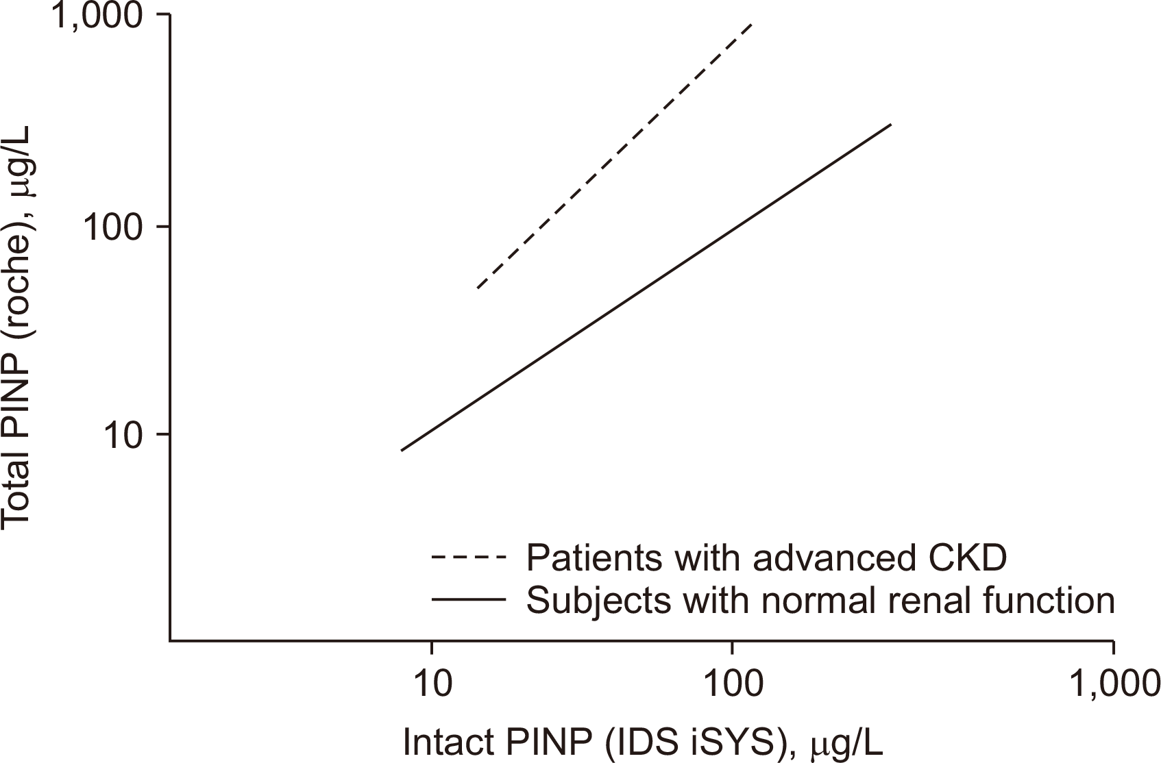 | Fig. 1Diagrammatic depiction of relationship between results obtained using an automated intact-PINP assay (IDS iSYS) and an automated total-PINP assay (Roche) in subjects with normal renal function and patients with advanced CKD. X- and Y-axis values are logarithmically spaced. This is a diagrammatic depiction, not real values from a study.
Abbreviations: PINP, procollagen type-I N-terminal propeptide; IDS, Immunodiagnostic Systems; CKD, chronic kidney disease.
|
Table 5
Japanese Osteoporosis Society reference intervals for B-ALP and TRACP-5b [20]
![]()
Anabolic therapy
Anabolic therapy (such as teriparatide, which is self-administered daily by patients) may be monitored by measuring the bone-formation marker PINP to confirm adherence and the appropriate injection technique. A significant increase in PINP >10 μg/L at 1 month or more after the initiation of treatment confirms the effectiveness of treatment [23, 40].
Monitoring the offset of treatment effects
Bisphosphonate action persists for a period after therapy cessation (months with risedronate and years with alendronate and zoledronic acid), and the offset of activity is slow, with a gradual increase in BTM concentrations to the baseline [41, 42]. Oral alendronate therapy may be discontinued after 5 yrs in the absence of incident fractures if the BMDs have improved [43]. Whilst BTM measurements at the end of treatment do not predict future fractures [44, 45], in practice, BTMs are often used to monitor the offset of the treatment effect and provide guidance for reinstituting treatment once the BTMs increase above the treatment target. Similarly, BTMs have been used to monitor the offset of treatment effects following zoledronic acid infusion to determine the timing of the next infusion [43]. These practices, although not evidence based, are well recognized.
The cessation of denosumab therapy, which is administered every 6 months via subcutaneous injection, is followed by a rebound increase in BTMs to well above baseline by approximately 9–12 months after the last injection [46]. These changes are of interest because the cessation of denosumab therapy has been associated with multiple vertebral fractures in some patients [47]. These outcomes have led to the recommendation that if denosumab therapy is discontinued, then treatment with a potent bisphosphonate (such as alendronate or zoledronic acid) should be instituted at least in the short term to attenuate the increase in bone turnover, and BTMs can be used to monitor the effectiveness of such therapy [48].
Go to :
BTMS IN CKD AND OSTEOPOROSIS OF RENAL OSTEODYSTROPHY (ROD)
BTMs that are cleared by the kidneys may accumulate in the circulation in patients with advanced CKD (eGFR <30 mL/min/1.73 m2). Measuring BTMs in the blood (and urine if the patient is still producing urine) may give misleading results by not accurately reflecting bone turnover. Plasma CTX and total PINP concentrations are both affected by renal failure, and their measurements are not recommended in patients with an eGFR of <30 mL/min/1.73 m2 [49, 50]. Intact-PINP assays are not affected by renal failure, making them potentially suitable for use in patients with advanced CKD [50]. The KDIGO organization recommends B-ALP and parathyroid hormone (PTH) as markers for assessing bone turnover in patients with CKD. Another promising marker is the bone-resorption marker, serum TRACP-5b [51-53].
Patients with CKD (even early CKD) have a higher risk for fractures than subjects with normal renal function, and patients on dialysis have four times the risk [3, 53]. ROD in CKD is characterized by abnormal bone turnover, mineralization defects, and reduced bone volumes [3]. ROD may be associated with high-turnover disease due to secondary hyperparathyroidism or low-turnover (adynamic bone) disease. Both conditions can lead to low bone strength and an increased risk for fractures [3]. However, most major osteoporosis-treatment trials have excluded patients with advanced CKD, and sparse evidence exists supporting the treatment of patients with osteoporosis and advanced CKD. Adynamic bone disease is often observed in ROD, precluding the use of antiresorptive therapies [53]. Ideally, ROD can be diagnosed through a bone biopsy, which is an invasive procedure with a significant risk of adverse events. The risk of fracture in CKD is highest with extreme PTH concentrations, and the KDIGO recommends maintaining serum PTH concentrations between two and nine times the upper reference limit of a particular PTH assay [3]. Most patients with CKD have PTH concentrations between these extremes, which does not exclude the presence of adynamic bone disease or a high-turnover state [53]. Low-turnover bone disease may occur even when serum PTH concentrations are up to five times the upper reference limit [52].
In the absence of bone biopsy guidance, BTM and PTH concentrations may be useful for guiding osteoporosis therapy. The results of a few studies have pointed to the use of B-ALP to minimize the possibility of adynamic bone disease before starting antiresorptive therapy. B-ALP mass assay results of >20 g/L (Ostase) or activity measurements of >30 U/L (Quidel) may be useful for ruling out low bone-turnover disease in CKD before initiating antiresorptive therapy in ROD [51, 52, 54] in the absence of vitamin D deficiency (which can increase B-ALP due to osteomalacia) or a very high total ALP activity (which can interfere significantly with B-ALP assays). Alternatively, a B-ALP of <30 μg/L (40 U/L) may be useful for ruling out high bone-turnover disease before commencing anabolic therapy for ROD [53, 54]. TRACP5b shows great promise for future use in assessing ROD [52]. In theory, as a resorption marker that is unaffected by renal clearance, TRACP5b should be most useful for confirming/ruling out high-turnover disease before initiating antiresorptive therapy in ROD. Further studies comparing TRACP5b with B-ALP and, importantly, bone-biopsy histomorphometric data and prospective studies on fracture-risk predictions are required for confident applications. Intact-PINP assays may also be useful for assessing ROD in patients, possibly for diagnosing low-turnover diseases; however, further studies are required.
This narrative review was limited to literature published in the English language, and a systematic literature search using specific keywords was not conducted. The articles selected included original research publications, review articles, and guidelines, which were not limited by the date of publication. However, the small number of published reference interval studies for BTMs from the Asia-Pacific region and the lack of well-designed studies covering most countries in the region limit our ability to recommend targeted population-specific reference intervals for most patient groups in the region.
Go to :
FUTURE DIRECTIONS AND CONCLUSIONS
BTMs play several roles in the clinical management of bone diseases. Although the diagnosis of osteoporosis is not based on laboratory testing, including BTM analysis, such testing is useful for diagnosing several metabolic bone diseases that may contribute to the development of osteoporosis. BTMs are most widely used to monitor osteoporosis therapy, including assessing the offset of effects after therapy cessation. Newer BTMs unaffected by renal failure show promise for assessing ROD to assist in treatment decisions for patients with high fracture risks. Standardizing or harmonizing assays for each marker would aid in applying universal guidelines and decision limits. In this regard, the IFCC and IOF Committee on Bone Metabolism (C-BM) have indicated that PINP measurements are harmonized between the automated assays (Roche Cobas and IDS iSYS) when the eGFR is >30 mL/min/1.73 m2 [55] and that the harmonization of other PINP assays is required. No commercial plasma CTX assays are currently harmonized, but this is being pursued by the IFCC-IOF C-BM [55]. Population-specific reference intervals are needed in most jurisdictions in the Asia-Pacific region. Ideally, future international multicenter clinical trials should include populations from this region also. Such studies have the potential to improve the targeted use of selected BTMs tailored to individual patients, using suitable decision limits for specific situations.
Go to :
 XML Download