Journal List > Korean Circ J > v.53(10) > 1516084296

Kim and Kim: Sex Differences in Coronary Artery Disease: Insights From the KoRean wOmen’S chest pain rEgistry (KoROSE)

Author's summary

Interest in sex differences in coronary artery disease (CAD) is growing, with data mostly from Western studies. This review summarizes findings from the KoRean wOmen’S chest pain rEgistry (KoROSE), illustrating differences in CAD between sexes. Postmenopausal women have higher cardiovascular risk due to reduced estrogen. Women with CAD tend to be older with more risk factors and face more diagnostic delays due to atypical symptoms. Despite similar in-hospital outcomes, women have more bleeding complications post-intervention and show differences in long-term prognosis. Moreover, women use fewer protective drugs, undergo fewer procedures, and participate less in clinical research. By acknowledging these differences, personalized treatments can improve prognosis.

Abstract

Interest in sex differences in coronary artery disease (CAD) has been steadily increasing. Concurrently, most of the data on these differences have primarily been Western-oriented. The KoRean wOmen’S chest pain rEgistry (KoROSE), started in 2011, has since published numerous research findings. This review aims to summarize the reported differences between men and women in CAD, integrating data from KoROSE. Cardiovascular risk in postmenopausal women escalates dramatically due to the decrease in estrogen levels, which normally offer cardiovascular protective effects. Lower estrogen levels can lead to abdominal obesity, insulin resistance, increased blood pressure, and endothelial dysfunction in older women. Upon analyzing patients with CAD, women are typically older and exhibit more cardiovascular risk factors than men. Diagnosing CAD in women tends to be delayed due to their symptoms being more atypical than men’s. While in-hospital outcome was similar between sexes, bleeding complications after percutaneous coronary intervention occur more frequently in women. The differences in long-term prognosis for CAD patients between men and women are still a subject of ongoing debate. Pregnancy and reproductive factors also play a significant role as risk factors for cardiovascular disease in women. A notable sex disparity exists, with women found to use fewer cardiovascular protective drugs and undergo fewer interventional or surgical procedures than men. Additionally, women participate less frequently than men in clinical research. Through concerted efforts to increase awareness of sex differences and mitigate sex disparity, personalized treatment can be provided. This approach can ultimately improve patient prognosis.

INTRODUCTION

Coronary artery disease (CAD) is the leading cause of death worldwide.1) Effective prevention relies on early detection and management of risk factors such as high blood pressure, dyslipidemia, hyperglycemia, smoking, and obesity.2)3)4)5)6)7) Controlling these risk factors has been the basis for CAD prevention.8)9)10) Although the prognosis of CAD has improved with the development of many therapeutic drugs and diagnostic methods, CAD-related morbidity and mortality rates are still high.11) Furthermore, it is suggested that a significant number of patients without traditional risk factors suffer from CAD,12)13) stimulating interest in non-traditional risk factors as potential areas for enhancing CAD prognosis.
Interest in sex differences in CAD has grown, acknowledging that these differences play a critical role in cardiovascular health. Historically, studies largely focused on men, leaving a deficit of data on women’s clinical characteristics, prognosis, and treatment responses.14) Recently, developed Western nations have increased focus on women’s heart disease, implementing sex-specific treatment and reducing disparities.15) This has led to a significant decrease in CAD-related mortality among women in the United States (US).16) In 2011, the Women’s Heart Disease Research Working Group in Korea started the KoRean wOmen’S chest pain rEgistry (KoROSE) to study sex differences in CAD. This review explores the differences among Korean CAD patients by utilizing both domestic and international data, along with the findings from the KoROSE database.

KoRean wOmen'S chest pain rEgistry

The KoROSE was established in 2011 by the Women’s Heart Disease Research Working Group to study heart disease in women.17) With 32 Korean cardiovascular centers participating, the registry aimed to determine clinical characteristics and prognosis of women with CAD in Korea. Men were also included to identify sex differences. Among the patients who visited the outpatient department due to chest pain, those who required a coronary angiogram (CAG) for additional evaluation of angina pectoris were identified by the cardiologist and subsequently enrolled in the registry. Most patients underwent invasive CAG (ICA), but some were evaluated with computed tomography angiography. Patients with unstable vital signs, end-stage renal disease, chronic obstructive lung disease, and certain other conditions were excluded. Prior to ICA, patients usually underwent transthoracic echocardiography and noninvasive stress tests. Data collected included anthropometric information, reproductive factors, occupation, cardiovascular risk factors, current medications, physical activity, chest pain characteristics, and results of examinations. Information on mortality and various cardiovascular events during follow-ups was also gathered. As of March 2023, approximately 3,700 subjects have been registered in the KoROSE database, with ongoing enrollment.

EPIDEMIOLOGY OF CARDIOVASCULAR DISEASE

Cardiovascular disease (CVD) is a major health concern globally, particularly for women. In 2019, there were an estimated 275 million cases of CVD in women globally, and CVD is responsible for nearly one-third of all female deaths globally, making it the leading cause of death for women.18) The global age-standardized CVD prevalence in women for 2019 was about 6,403 per 100,000.18) In the US, over 60 million women, or 44% of the female population, live with some form of heart disease.1) CVD-related deaths rose from 6.1 million in 1990 to 8.9 million in 2019.18) In Europe, CVD is responsible for over 4 million deaths annually, representing 45% of all deaths, with more women affected than men. Of these deaths, 2.2 million were women and 1.8 million were men, which equates to 49% and 40% of all deaths, respectively.19) In South Korea, CVD is the second and third leading cause of death, with heart disease having a mortality rate of 61.5 per 100,000 people, and cerebrovascular disease 44.0 per 100,000.19) In 2021, the mortality rate of CVD was 1.1 times higher in men than women.19) Interestingly, the mortality rate of ischemic heart disease was higher in men, while hypertensive disease and cerebrovascular disease rates were higher in women.19)
Collectively, although the epidemiology of CVD can vary depending on the region and population studied, CVD is a leading cause of death worldwide, and it affects both men and women. Previously, CVD was considered a disease that mainly affects men. However, recent data show an increase in the prevalence of CVD among women, and some studies have reported higher mortality and morbidity rates from CVD in women than in men.

SEX DIFFERENCE IN CLINICAL CHARACTERISTICS OF CORONARY ARTERY DISEASE

Age

Age is the main risk factor for developing CAD.20)21) Generally, the prevalence of CAD is higher in men than in women. The age-adjusted prevalence of CAD in the US in 2018 was 7.4% for men and 4.1% for women.22) CAD prevalence rises with age in both sexes, but the increase is more pronounced in women.21) According to the American Heart Association, about 40% of men and women aged 40–59 develop CVD, and this proportion increases to 75% in individuals aged 60–79 and 86% in those over 80.21) Men typically develop CAD in their 40s or 50s, while women usually develop it after menopause, in their 60s or 70s. The prevalence of CVD among patients aged 70 years or older is similar to that of men or higher in women. Studies using Korean data have confirmed these trends. In a study analyzing ICA findings of 2,348 Korean patients from KoROES database, women were significantly older than men (64.4 vs. 59.5 years, p<0.001).23) Similarly, in a study analyzing 44,967 individuals who underwent percutaneous coronary intervention (PCI), women were older than men (71.1 vs. 62.9 years, p<0.001).24)
The increased cardiovascular risk in older women may be due to factors like lower estrogen levels, a higher prevalence of cardiovascular risk factors, and genetic predisposition.

Risk factors

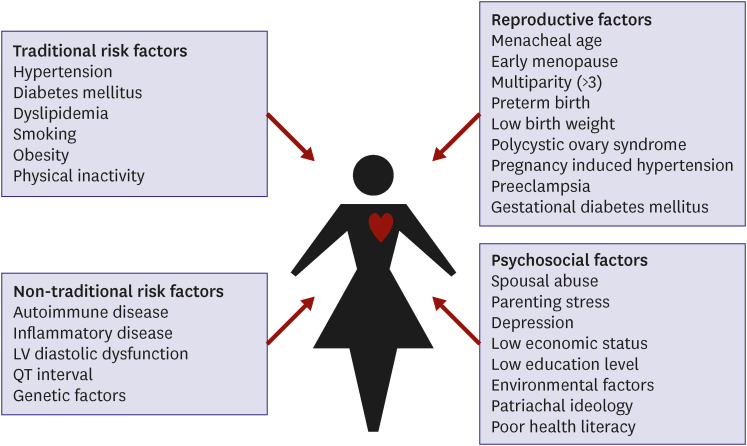
As the age of patients with CAD increases, the prevalence of cardiovascular risk factors also tends to increase. Therefore, it might be expected that women have more cardiovascular risk factors than men due to their older age. When looking at the prevalence of risk factors in CAD patients, it is notable that hypertension, diabetes and dyslipidemia are more common in women, while men tend to have higher rates of smoking and prior history of ischemic heart disease. This observation is consistent with both foreign and domestic data.23)24)25)26)27)28) Moving forward, we will provide a more detailed examination of the differences between men and women in terms of traditional and non-traditional risk factors.

Traditional risk factors

1) Hypertension

Hypertension is more prevalent in men under 65 years of age, whereas in women over 65 years of age, the prevalence is higher.29) In women, the increase in systolic blood pressure is more pronounced after menopause.30)31) This may be due to the loss of vasodilatory effect of endogenous estrogen, atherosclerosis, increased salt sensitivity, decreased endothelial nitric oxide production, and increased expression of angiotensin II receptors.30) As a result, the frequency of isolated systolic hypertension in women is higher than in men, which is a major risk factor for the development of cardiovascular complications. Several studies suggest that hypertension may have a stronger impact on CAD risk in women than in men, and may be associated with a higher prevalence of non-obstructive CAD and diffuse atherosclerosis in women.32) In a study of 1,254 patients with stable chest pain who had elective ICA in the suspicion of CAD from the KoROSE database, the prevalence of hypertension was similar between sexes (50.2% for men and 51.1% for women) but hypertension was one of risk factors for obstructive CAD only in women but not in men.33)

2) Diabetes mellitus

Sex differences in diabetes mellitus as a cardiovascular risk factor have been widely studied. For instance, a study comprising nondiabetic and type 2 diabetic subjects found that diabetes increased the risk for coronary heart disease more substantially in women than in men over a 13-year follow-up period.34) This was reinforced by a systematic review and meta-analysis showing that women with type 2 diabetes had a 46% higher risk of coronary heart disease mortality compared to men.35) Another comprehensive study found that diabetes roughly doubled the risk of occlusive vascular mortality in men and tripled the risk in women.36) Consistent with these findings, KoROSE data found that the presence of diabetes was an independent risk factor for obstructive CAD in women but not in men.33) These findings underline the need for more intensified management of cardiovascular risk in women with diabetes.

3) Dyslipidemia

Women tend to have higher levels of high-density lipoprotein (HDL) cholesterol, whereas, men tend to have higher levels of low-density lipoprotein (LDL) cholesterol.37)38)39) However, after menopause, women tend to have an increase in LDL cholesterol levels and a decrease in HDL cholesterol levels, which can increase their risk of CVD.40) The impact of dyslipidemia on CVD may differ between men and women, but the available data on sex differences in this regard is limited. It has been reported that the effect of low HDL cholesterol on cardiovascular risk is greater in women than in men.41) Additionally, a study of acute myocardial infarction demonstrated that the influence of lipoprotein (a) was more significant in women than in men in the young age group.37)

4) Cigarette smoking

Studies have consistently shown that smoking is a significant risk factor for CVD including CAD, stroke, and peripheral artery disease.6) Interestingly, women appear to be more susceptible to the detrimental effects of smoking, with female smokers showing a higher risk of developing CVD compared to their male counterparts, even after adjusting for other risk factors.42)43)44)45)46) In a prospective cohort study, female smokers had a relative risk of 2.24 for myocardial infarction, compared to 1.43 for male smokers.45) Similar findings were echoed in several meta-analyses.43)44) In a Korean study analyzing 12,565 patients with acute myocardial infarction, it was observed that the clinical outcomes were more unfavorable for women who are currently smoking, when compared to their male counterparts who are also smokers.46) Although a positive effect of smoking cessation on cardiovascular risk is observed in both men and women, the extent of sex differences remains inconsistent.47)48) The precise reason for this sex difference in smoking-related cardiovascular risk is unknown, but it underscores the urgency for smoking cessation education, particularly for women.

5) Obesity

The escalating global obesity epidemic, with its related complications, poses a significant threat to public health.49) Obesity is a significant risk factor for ischemic heart disease and stroke.4) In menopausal women, fat tends to accumulate in the abdominal area, similar to men, which heightens cardiovascular risk by inducing insulin resistance and inflammation.50) Abdominal obesity is well-known to increase cardiovascular risk by inducing insulin resistance and an inflammatory response.51) The KoROSE data showed that the association between abdominal obesity, as measured by waist circumference, and obstructive CAD in menopausal women was more pronounced than that measured by body mass index.52) Although obese men and women are at increased risk for CVD, a closure analysis of the data suggests that the relative risk of obesity is slightly higher in men than in women.4)53) However, most of this data uses body mass index as the measure of obesity, and further research using measures of abdominal obesity such as waist circumference or waist-to-hip ratio is needed.

Nontraditional risk factors

1) Chronic inflammation

Chronic inflammation is linked to an increased risk of CVD due to its destructive effects on blood vessels, such as promoting atherosclerosis, oxidative stress, and endothelial dysfunction.54)55) Inflammatory markers such as interleukin-6, tumor necrosis factor-alpha, and C-reactive protein (CRP) have been linked to an increased risk of CVD events such as heart attacks and strokes.54) Given that the prevalence of rheumatic diseases, which can cause chronic inflammation and increase cardiovascular risk,56) is higher in women than in men, it is essential to better understand this relationship when assessing cardiovascular risk, especially in women.57) The KoROSE data revealed that increased CRP levels predicted obstructive CAD in men, but not in women.58) This finding is consistent with other studies.59)60) While anti-inflammatory therapies have been used to reduce cardiovascular risk, their effectiveness remains uncertain.61)

2) Left ventricular diastolic dysfunction

Left ventricular (LV) diastolic dysfunction, identified as an independent predictor of poor cardiovascular outcomes,62) is associated with an increased risk of CAD.63)64) It results from myocardial ischemia, often caused by disturbed coronary blood flow,65) and shares risk factors with CAD.66) A study using KoROSE data found low septal e′ velocity to be an independent risk factor associated with obstructive CAD.67) Women are reported to be more susceptible to developing LV diastolic dysfunction in response to risk factors, although data on sex differences in its association with CAD is scarce.68)69)70)71) In a study with 1:1 matched samples from KoROSE data, the correlation between LV diastolic dysfunction and significant coronary artery stenosis was confirmed only in women.72) In another study that analyzed the KoROSE data, CRP was highly associated with CAD in men, while LV diastolic dysfunction was highly associated with CAD in women.58) Hence, more attention is warranted in evaluating CAD, especially in women with LV diastolic dysfunction.

3) Psychological stress

Psychological stress is one of the important risk factors for CVD.73)74)75) Chronic stress leads to increased release of stress hormones, such as cortisol and adrenaline, resulting in elevated blood pressure, heart rate, and inflammation. Over time, this response can damage blood vessel walls and contribute to the formation of blood clots and atherosclerotic plaques.74) Stressful condition can also induce lifestyle habits, such as overeating, excessive drinking, and smoking, which increase cardiovascular risk. Chronic job strain is associated with increased risk of recurrent coronary events.76) Stress impacts cardiovascular risk in both sexes, but the effect appears more pronounced in women.77)78)79) A study utilizing KoROSE data found a significant association between depression scores and obstructive CAD in women.80) However, some studies report inconsistent findings,81) highlighting the need for further research into the relationship between stress and cardiovascular health.

4) Pregnancy and reproductive factors

Pregnancy and reproductive factors may affect a woman’s cardiovascular risk.82) Studies suggest early or late menarche might increase CVD occurrence.83)84) A study using KoROSE data from 687 Korean women showed late menstrual age as an independent factor for significant coronary artery stenosis.85) Polycystic ovary syndrome, characterized by irregular menstrual cycles and hyperandrogenism, is linked with hypertension, dyslipidemia, and hyperglycemia, significantly escalating CVD incidence.86) Conditions such as hypertensive disorder of pregnancy87)88) and gestational diabetes89) are notable risk factors for later-life CVD development. Preterm birth and low birth weight also increase maternal cardiovascular risk, although the underlying reasons are unclear.88)90) A J-shaped correlation exists between the number of pregnancies and cardiovascular risk, with women experiencing over 5 pregnancies having higher rates of future cardiovascular events.91) In the analysis of KoROSE database, an increased number of pregnancies (odds ratio, 1.223, 95% confidence interval, 1.026–1.457, p=0.025, per pregnancy) were the independent predictors of obstructive CAD even after controlling for conventional risk factors.85) This connection could be due to repeated physical stress, metabolic abnormalities, and weight gain during pregnancies.92)

Clinical presentations

There are sex differences in the clinical presentation of CAD. Studies have shown that women may experience different types of chest pain compared to men, such as more dull or burning pain rather than the classic squeezing or pressure-like chest pain associated with CAD.93)94)95) Additionally, it has been reported that women with CAD were more likely to present with atypical symptoms such as fatigue, shortness of breath, and weakness, compared to men who are more likely to present with chest pain.93)94)95)96)97)98)99) Also, throat, jaw, shoulder and neck discomfort were more frequently reported by women than in men.94)95)97)99)100) The way chest pain is expressed, or the way language is used differs between men and women, and in particular, ambiguous expressions in women make diagnosis more difficult.101) KoROSE study investigating 1,549 patients found that men experienced more squeezing-type pain on the left side of the chest, while women demonstrated more dull quality pain in the retrosternal and epigastric area.93) Additionally, in the same study, palpitations and headaches were more frequently observed in women than in men as associated symptoms.
The sensation of chest pain is controlled by various complex factors. Although it is not fully understood why men and women exhibit different symptoms of myocardial ischemia, there are various suggested reasons, such as differences in coronary anatomy, sympathetic innervation, hemodynamic factors, and stress response thresholds. These complex factors may contribute to the observed differences in chest pain symptoms between men and women.102)
The first step for the accurate diagnosis of CAD depends on the proper recognition of chest pain.103) However, women often present with atypical symptoms, leading to potential delays or misdiagnosis of CAD. To address this challenge, it is essential for healthcare providers to acknowledge these sex differences in chest pain manifestations. By doing so, they can ensure accurate diagnosis and appropriate treatment for both men and women.

SEX DIFFERENCES IN DIAGNOSTIC TESTS FOR CORONARY ARTERY DISEASE

Cardiac biomarkers

Cardiac biomarkers can vary between males and females.104) For example, troponin is a protein released into the bloodstream when there is heart muscle damage. Cardiac troponin is an important biomarker used to diagnose and manage CAD. Studies have demonstrated that women usually have lower levels of troponin than men.105) Similarly, B-type natriuretic peptide (BNP) or N-terminal proBNP (NT-proBNP) are hormones released by the heart when it’s under stress. BNP or NT-proBNP play an important role in the diagnosis, management, and monitoring of CAD, particularly in relation to heart failure and myocardial ischemia.106) Women tend to have higher levels of BNP than men in heart failure and CAD.25)107)108)
It’s critical to consider sex differences when interpreting cardiac biomarkers since these differences can have clinical implications for the diagnosis and treatment of heart disease. For instance, using the same cutoff values for troponin in men and women could result in underdiagnosis of heart disease in women due to their typically lower troponin levels.109)

Noninvasive tests

Noninvasive tests play a crucial role in identifying patients who may require further evaluation with ICA.110) Sex differences in the noninvasive test results for the diagnosis of CAD have been well documented. Women tend to have a higher incidence of false-positive electrocardiogram (ECG) results,111)112) potentially due to differences in heart size and shape and interference from breast tissue. Research has indicated important differences in various ECG parameters between men and women, such as faster heart rate and longer QTc duration in women.113) It has been suggested that the QTc interval can be an important variable in diagnosing CAD.114)115) KoROSE data from 1,741 patients with suspected angina showed that a longer QTc interval was associated with more severe CAD.115) Furthermore, the authors indicated that the diagnostic value of QTc interval was stronger in women than in men.
Women may be less likely to achieve target heart rates during exercise stress testing, which can lead to false-negative results.116) KoROSE data analyzing 335 women who underwent ICA demonstrated that treadmill exercise test (TET) had a lower sensitivity (16.7–42.9%) and positive predictive value (37.0–61.8%), but it has high positive predictive value (85.4–95.1%) in the detection of obstructive CAD.117)
Studies have shown that in echocardiography, women usually have smaller heart and thinner LV walls,118) and a higher incidence of diastolic dysfunction than men.58) LV diastolic dysfunction in women diagnosed by echocardiography has been found to be a valuable tool in diagnosing CAD. In a study analyzing 1,307 patients with suspected CAD from KoROSE database, low septal e′ velocity was identified as an independent risk factor associated with obstructive CAD, and significantly increased diagnostic value of TET in the detection of obstructive CAD.67)
Another study using KoROSE data found that CRP was the most powerful CAD risk factor in men, while low e′ velocity was the most powerful risk factor in women.58) In a 1:1 matching analysis of 295 men and women from KoROSE data, the association between LV diastolic function and obstructive CAD was observed only in women, suggesting that LV diastolic function in women is valuable for diagnosing CAD.72) Various studies have suggested that stress echocardiography provides valuable prognostic and diagnostic information in patients with CAD or suspected CAD.119) A multi-center registry analysis has suggested that exercise stress echocardiography could be more cost-effective than TET.120) Analysis using KoROSE data (n=202) showed that dobutamine-stress echocardiography was more useful in diagnosing obstructive CAD than TET in women.121) Additionally, an analysis of 102 women who underwent dobutamine-stress echocardiography revealed that obstruction of the LV outflow tract that occurred during the stress test was associated with LV hypertrophy, LV diastolic dysfunction, decreased exercise capacity and dyspnea, but not associated with coronary artery stenosis.122) This finding suggests that labor dyspnea or chest discomfort during exercise in women can be caused by obstruction of the LV outflow tract, even if it is not caused by coronary artery stenosis.122) Although diagnostic accuracy or prognostic value of stress echocardiography were similar between men and women in general, it is still recommended that exercise stress echocardiography should be used with caution in women due to insufficient data.123)
Overall, these sex differences in noninvasive test results can have significant implications for the diagnosis and management of CAD in women. It is important for healthcare providers to be aware of these differences and to tailor their diagnostic and treatment strategies accordingly. For example, women may require different exercise stress test protocols or imaging techniques to achieve accurate results. Furthermore, studies have suggested that sex-specific diagnostic criteria may be necessary to improve the accuracy of noninvasive tests for the diagnosis of CAD in women.

Invasive coronary angiogram findings

Numerous studies have examined sex differences in ICA findings, identifying several notable differences. For instance, women have been found to be less likely to have obstructive CAD on ICA when compared to men. Analysis of the KoROSE database found men had a higher prevalence of obstructive CAD and high-risk angiographic findings like left main disease or 3-vessel disease.23) This could be attributed to the fact that men usually have a higher prevalence of CAD risk factors such as smoking, hypertension, and high cholesterol.23) Further, men develop CAD at a younger age than women, allowing more time for disease progression.23)24) Estrogen, higher in women, protects the cardiovascular system, possibly contributing to differences in CAD prevalence.124) Lifestyle behaviors, such as diet and exercise, could also affect the earlier onset of CAD in men, as men typically have higher rates of physical inactivity and unhealthy eating habits.125)
The pathophysiology of CAD may also differ between men and women.32) Women tend to have a higher prevalence of microvascular disease, characterized by abnormalities in small heart-supplying blood vessels, which traditional diagnostic methods like ICA may not easily detect. Women with CAD often exhibit non-obstructive disease, characterized by the presence of plaque without significant narrowing or occlusion, likely due to higher prevalence of diffuse and non-calcified plaque. Furthermore, women with CAD often have more diffuse disease, indicating the presence of plaque in multiple coronary artery segments, which can make diagnosing and treating CAD in women more challenging.

SEX DIFFERENCE IN CLINICAL OUTCOMES OF CORONARY ARTERY DISEASE

In-hospital outcomes

Many studies on in-hospital outcomes after PCI have been reported, but the results are inconsistent. While there is some evidence to suggest that women may have higher rates of in-hospital complications after PCI compared to men,24)28) but some studies have not observed this effect.126)127) For example, a Korean study24) found that women had 1.2 times higher rates of in-hospital composite events but not in-hospital mortality compared to men in multivariable analysis, while a Western study127) found no difference between sexes with respect to the composite efficacy endpoint in patients undergoing primary PCI due to ST-elevation myocardial infarction after adjusting for potential confounders. However, it is a relatively consistent finding that post-PCI bleeding complications are more common in women than in men.24)127) Further research is needed to fully understand the potential sex differences in outcomes in patients undergoing PCI.

Long-term outcomes

There is ongoing debate about whether sex differences exist in the long-term prognosis of patients with CAD. Several studies have explored this topic, providing varying findings. For instance, an analysis of 67,534 patients found worse long-term outcomes in women, especially among those undergoing PCI.128) A meta-analysis of over a million patients found males had significantly lower mortality rates at 1 and 2 years post-PCI.129) Women also displayed poorer clinical outcomes after PCI for left main disease130) and had higher rates of cardiovascular events post-coronary artery bypass grafting.131) However, contradictory results exist. A study involving 28,924 PCI patients showed women had a lower 5-year death risk than men.132) Similarly, a multicenter study with 23,604 patients indicated that although short-term or mid-term prognosis was worse for women, long-term prognosis was better.133) Other studies have reported no sex differences in long-term prognosis for CAD patients.134)135)136) Therefore, considering other variables, such as race, age, and risk factors, is crucial for understanding these differences. Further research is needed for comprehensive insight into the subject, with the results of the ongoing KoROSE study expected to add valuable data.

PATHOPHYSIOLOGY OF SEX DIFFERENCES

Genetic factors

Sex-specific gene expressions play an important role explaining the sex differences observed in CAD. It has been suggested that women tend to accumulate more subcutaneous fat, while men tend to accumulate more visceral fat, and these patterns are mainly determined by genetic factors.137) In addition, renin-angiotensin system associated genes in males compared to females may contribute to increased cardiac fibrosis in males.138) Researchers have identified several sex-specific single nucleotide polymorphisms associated with CAD or CAD risk factors.139)140) Variants on the Y chromosome have also been associated with elevated blood pressure,141) and it has been found that a father’s blood pressure status can influence blood pressure in male offspring.142) In an experimental study, the administration of androgens to macrophages derived from healthy males resulted in increased expression of genes associated with atherosclerosis compared to females.143) Recently, there have been attempts to utilize these genetic factors for predicting the occurrence of CVD.144) While the genetic predispositions associated with CAD are complex and involve multiple intertwined genes, understanding the expression and impact of sex-specific genetic factors can provide valuable insights into CAD.

Body size

Women typically have a smaller body size than men, which can be associated, at least partially, with a higher incidence of side effects from cardiovascular medications145) and complications related to procedures, such as bleeding.24) Due to their shorter stature and faster wave reflection, women may experience increased augmentation pressure.146) However, the relationship between height and CVD is still a topic of debate,147) and further research is needed to reach a consensus.

Psychosocial factors

Sex differences in CAD can be influenced by various psychosocial factors. Different stress levels according to the traditional sex role expectations may contribute to the development of CAD or its associated risk factors. For example, men may face work-related stress and pressures of raising a family, while women may experience stress related to caregiving and household responsibilities. In addition, it is known that women react sensitively to stress arising from relationships with people, and men react sensitively to stress in the workplace. Anxiety or depression, which are more common in women than in men, can also contribute to increased cardiovascular risk in women. Several studies have reported such a link.79)80)148)149)

Hormonal factors

Estrogen, a primary female hormone, exhibits cardiovascular effects by promoting angiogenesis, inhibiting fibrosis, preserving endothelial cell function, and inhibiting reactive oxygen species and inflammatory responses.124) Additionally, as stated above, estrogen depletion can lead to abdominal obesity50) and elevated blood pressure30) in older women. Cardiovascular risk escalates rapidly in postmenopausal women, which is closely related to a decline in blood estrogen levels.150)

Pregnancy and reproductive factors

The effects of pregnancy and reproductive factors on CAD have already been previously detailed in the section on “nontraditional risk factors.”

SEX DISPARITY

Delayed diagnosis or misdiagnosis of CAD is more common in women than in men.151)152) Truong et al.153) have reported that the improvement of cardiovascular outcome via intensive statin therapy in patients with acute coronary syndrome was comparable between men and women. In this study, the benefit of statins appeared to be numerically higher in women. However, in the actual clinical practice, several studies have noted that cardiovascular protective drugs, such as statins, aspirin and renin-angiotensin system blockers, are prescribed less frequently to women than to men.154)155)156) PCI and coronary artery bypass grafting were also found to be conducted less frequently in women.155)157) In clinical studies, it can be seen that the participation rate of women is much lower than that of men.14) This disparity in treatment eventually results in suboptimal control of risk factors and poor prognosis.158) Such sex disparity needs to be well acknowledged and corrected.

FUTURE DIRECTIONS

As detailed above, women have more CAD risk factors to consider than men (Figure 1). Particularly in women, many risk factors are not widely recognized in clinical practice, underscoring the importance of awareness. Furthermore, acknowledging the presence of sex disparities in CAD is critical. In the US, a campaign titled “Go Red for Women” has significantly increased awareness of women’s heart disease,15) concurrently contributing to a substantial reduction in women’s cardiovascular risk.16) In contrast, awareness of female heart disease in Korea may be notably lower among both the general public and medical professionals. The Women’s Heart Disease Research Working Group recently conducted a telephone survey with 1,050 middle-aged or elderly Korean women and found that awareness of women’s heart disease was strikingly low.159) Therefore, it is imperative to undertake systematic and multifaceted efforts to boost awareness of women’s heart disease. Additionally, current guidelines for CAD worldwide may not adequately account for sex differences. It’s crucial to increase women’s participation in clinical research to gather robust data. Based on this data, guidelines should underscore the distinctions between sexes. Furthermore, there could be a need for separate guidelines for men and women.160) Sex disparity and solutions for overcoming it is displayed in Figure 2.
Figure 1

CAD risk factors in Korean women.

CAD = coronary artery disease; LV = left ventricular.
kcj-53-655-g001
Figure 2

Sex disparity in CAD and solutions for overcoming it.

CABG = coronary artery bypass grafting; CAD = coronary artery disease; PCI = percutaneous coronary intervention.
kcj-53-655-g002

CONCLUSIONS

There are significant sex differences in the clinical characteristics and risk factors for CAD (Table 1), which were also confirmed in the KoROSE data for Korean population. These differences between men and women, which play a crucial role in the onset of CAD, often appear to be insufficiently recognized in clinical practice. Specifically, factors related to pregnancy, childbirth, and socio-psychological aspects are frequently overlooked as significant contributors to the occurrence of CAD in women. Instead, a sex disparity persists, where women tend to receive less CVD treatment than men. Medical professionals must first understand these sex differences and incorporate them into their diagnostic and treatment approaches. Additionally, clinical researchers should encourage more female participation in clinical research to generate robust evidence specific to women. This approach could potentially lead to improved patient outcomes, enhance the quality of life, and even save lives. Furthermore, an accurate recognition of these sex differences can assist in developing sex-specific diagnostic and treatment guidelines for CAD.
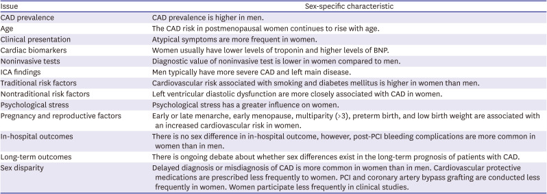
Table 1

Summary of major sex differences in CAD

kcj-53-655-i001
Issue Sex-specific characteristic
CAD prevalence CAD prevalence is higher in men.
Age The CAD risk in postmenopausal women continues to rise with age.
Clinical presentation Atypical symptoms are more frequent in women.
Cardiac biomarkers Women usually have lower levels of troponin and higher levels of BNP.
Noninvasive tests Diagnostic value of noninvasive test is lower in women compared to men.
ICA findings Men typically have more severe CAD and left main disease.
Traditional risk factors Cardiovascular risk associated with smoking and diabetes mellitus is higher in women than men.
Nontraditional risk factors Left ventricular diastolic dysfunction are more closely associated with CAD in women.
Psychological stress Psychological stress has a greater influence on women.
Pregnancy and reproductive factors Early or late menarche, early menopause, multiparity (>3), preterm birth, and low birth weight are associated with an increased cardiovascular risk in women.
In-hospital outcomes There is no sex difference in in-hospital outcome, however, post-PCI bleeding complications are more common in women than in men.
Long-term outcomes There is ongoing debate about whether sex differences exist in the long-term prognosis of patients with CAD.
Sex disparity Delayed diagnosis or misdiagnosis of CAD is more common in women than in men. Cardiovascular protective medications are prescribed less frequently to women. PCI and coronary artery bypass grafting are conducted less frequently in women. Women participate less frequently in clinical studies.
BNP = B-type natriuretic peptide; CAD = coronary artery disease; ICA = invasive coronary angiography; PCI = percutaneous coronary intervention.

Notes

Funding: The authors received no financial support for the research, authorship, and/or publication of this article.

Conflict of Interest: The authors have no financial conflicts of interest.

Data Sharing Statement: The data generated in this study is available from the corresponding author upon reasonable request.

Author Contributions:

  • Conceptualization: Kim HL, Kim MA.

  • Investigation: Kim HL.

  • Resources: Kim HL.

  • Supervision: Kim MA.

  • Writing - original draft: Kim HL.

  • Writing - review & editing: Kim MA.

References

1. Tsao CW, Aday AW, Almarzooq ZI, et al. Heart disease and stroke statistics-2023 update: a report from the American Heart Association. Circulation. 2023; 147:e93–621. PMID: 36695182.
2. Rao Kondapally Seshasai S, Kaptoge S, Thompson A, et al. Diabetes mellitus, fasting glucose, and risk of cause-specific death. N Engl J Med. 2011; 364:829–841. PMID: 21366474.
crossref
3. Lewington S, Clarke R, Qizilbash N, Peto R, Collins R. Prospective Studies Collaboration. Age-specific relevance of usual blood pressure to vascular mortality: a meta-analysis of individual data for one million adults in 61 prospective studies. Lancet. 2002; 360:1903–1913. PMID: 12493255.
crossref
4. Whitlock G, Lewington S, Sherliker P, et al. Body-mass index and cause-specific mortality in 900 000 adults: collaborative analyses of 57 prospective studies. Lancet. 2009; 373:1083–1096. PMID: 19299006.
5. Prospective Studies Collaboration. Lewington S, Whitlock G, et al. Blood cholesterol and vascular mortality by age, sex, and blood pressure: a meta-analysis of individual data from 61 prospective studies with 55,000 vascular deaths. Lancet. 2007; 370:1829–1839. PMID: 18061058.
crossref
6. Jha P, Ramasundarahettige C, Landsman V, et al. 21st-century hazards of smoking and benefits of cessation in the United States. N Engl J Med. 2013; 368:341–350. PMID: 23343063.
crossref
7. Arafa A, Lee HH, Eshak ES, et al. Modifiable risk factors for cardiovascular disease in Korea and Japan. Korean Circ J. 2021; 51:643–655. PMID: 34227266.
crossref
8. Arnett DK, Blumenthal RS, Albert MA, et al. 2019 ACC/AHA guideline on the primary prevention of cardiovascular disease: executive summary: a report of the American College of Cardiology/American Heart Association Task Force on Clinical Practice Guidelines. J Am Coll Cardiol. 2019; 74:1376–1414. PMID: 30894319.
9. Visseren FL, Mach F, Smulders YM, et al. 2021 ESC guidelines on cardiovascular disease prevention in clinical practice. Eur Heart J. 2021; 42:3227–3337. PMID: 34458905.
10. Kim HL, Chung J, Kim KJ, et al. Lifestyle modification in the management of metabolic syndrome: statement from Korean Society of CardioMetabolic Syndrome (KSCMS). Korean Circ J. 2022; 52:93–109. PMID: 35128848.
crossref
11. Baek J, Lee H, Lee HH, Heo JE, Cho SM, Kim HC. Thirty-six year trends in mortality from diseases of circulatory system in Korea. Korean Circ J. 2021; 51:320–332. PMID: 33821581.
crossref
12. Khot UN, Khot MB, Bajzer CT, et al. Prevalence of conventional risk factors in patients with coronary heart disease. JAMA. 2003; 290:898–904. PMID: 12928466.
crossref
13. Lee HH, Cho SM, Lee H, et al. Korea heart disease fact sheet 2020: analysis of nationwide data. Korean Circ J. 2021; 51:495–503. PMID: 34085422.
crossref
14. Agarwala A, Goldberg A. Special considerations for lipid-lowering therapy in women reflecting recent randomized trials. Curr Atheroscler Rep. 2021; 23:42. PMID: 34146167.
crossref
15. Mosca L, Hammond G, Mochari-Greenberger H, et al. Fifteen-year trends in awareness of heart disease in women: results of a 2012 American Heart Association national survey. Circulation. 2013; 127:1254–1263. e1–1229. PMID: 23429926.
crossref
16. Benjamin EJ, Virani SS, Callaway CW, et al. Heart disease and stroke statistics-2018 update: a report from the American Heart Association. Circulation. 2018; 137:e67–492. PMID: 29386200.
17. Women’s Heart Disease Research Working Group (KR). Women’s Heart Disease Research Working Group’s homepage [Internet]. Incheon: Women’s Heart Disease Research Working Group;2023. cited 2023 July 18. Available from: https://www.womensheart.or.kr/.
18. Vogel B, Acevedo M, Appelman Y, et al. The Lancet women and cardiovascular disease Commission: reducing the global burden by 2030. Lancet. 2021; 397:2385–2438. PMID: 34010613.
crossref
19. Statistics Korea. Causes of death statistics [Internet]. Daejeon: Statistics Korea;c2023. cited 2023 July 18. Available from: https://kostat.go.kr/ansk/.
20. North BJ, Sinclair DA. The intersection between aging and cardiovascular disease. Circ Res. 2012; 110:1097–1108. PMID: 22499900.
crossref
21. Yazdanyar A, Newman AB. The burden of cardiovascular disease in the elderly: morbidity, mortality, and costs. Clin Geriatr Med. 2009; 25:563–577. PMID: 19944261.
crossref
22. Centers for Disease Control and Prevention. National Center for Health Statistics. Summary health statistics: National Health Interview Survey, 2018: table A-1 [Internet]. Atlanta (GA): Centers for Disease Control and Prevention;2018. cited 2022 March 22. Available from: https://ftp.cdc.gov/pub/Health_Statistics/NCHS/NHIS/SHS/2018_SHS_Table_A-1.pdf.
23. Kim HL, Kim HJ, Kim M, et al. Sex differences in coronary angiographic findings in patients with stable chest pain: analysis of data from the KoRean wOmen’S chest pain rEgistry (KoROSE). Biol Sex Differ. 2022; 13:2. PMID: 34980251.
crossref
24. Kim HL, Jang JS, Kim MA, et al. Gender differences of in-hospital outcomes in patients undergoing percutaneous coronary intervention in the drug-eluting stent era. Medicine (Baltimore). 2019; 98:e15557. PMID: 31096458.
crossref
25. Korea Acute Myocardial Infarction Registry (KAMIR) Investigators. Lee KH, Jeong MH, et al. Gender differences of success rate of percutaneous coronary intervention and short term cardiac events in Korea Acute Myocardial Infarction Registry. Int J Cardiol. 2008; 130:227–234. PMID: 18077018.
crossref
26. Kunadian V, Qiu W, Lagerqvist B, et al. Gender differences in outcomes and predictors of all-cause mortality after percutaneous coronary intervention (data from United Kingdom and Sweden). Am J Cardiol. 2017; 119:210–216. PMID: 27816119.
crossref
27. Jacobs AK, Johnston JM, Haviland A, et al. Improved outcomes for women undergoing contemporary percutaneous coronary intervention: a report from the National Heart, Lung, and Blood Institute Dynamic registry. J Am Coll Cardiol. 2002; 39:1608–1614. PMID: 12020487.
crossref
28. Iyanoye A, Moreyra AE, Swerdel JN, et al. Gender disparity in the use of drug-eluting stents during percutaneous coronary intervention for acute myocardial infarction. Catheter Cardiovasc Interv. 2015; 86:221–228. PMID: 25645156.
crossref
29. Kim HC, Lee H, Lee HH, et al. Korea hypertension fact sheet 2021: analysis of nationwide population-based data with special focus on hypertension in women. Clin Hypertens. 2022; 28:1. PMID: 34980274.
crossref
30. Brahmbhatt Y, Gupta M, Hamrahian S. Hypertension in premenopausal and postmenopausal women. Curr Hypertens Rep. 2019; 21:74. PMID: 31451980.
crossref
31. Seo E, Jung S, Lee H, Kim HC. Sex-specific trends in the prevalence of hypertension and the number of people with hypertension: analysis of the Korea National Health and Nutrition Examination Survey (KNHANES) 1998-2018. Korean Circ J. 2022; 52:382–392. PMID: 35257521.
crossref
32. Shaw LJ, Bugiardini R, Merz CN. Women and ischemic heart disease: evolving knowledge. J Am Coll Cardiol. 2009; 54:1561–1575. PMID: 19833255.
33. Park J, Kim HL, Kim MA, et al. Traditional cardiovascular risk factors and obstructive coronary disease in patients with stable chest pain: gender-specific analysis. Cardiometab Syndr J. 2021; 1:101–110.
crossref
34. Juutilainen A, Kortelainen S, Lehto S, Rönnemaa T, Pyörälä K, Laakso M. Gender difference in the impact of type 2 diabetes on coronary heart disease risk. Diabetes Care. 2004; 27:2898–2904. PMID: 15562204.
crossref
35. Huxley R, Barzi F, Woodward M. Excess risk of fatal coronary heart disease associated with diabetes in men and women: meta-analysis of 37 prospective cohort studies. BMJ. 2006; 332:73–78. PMID: 16371403.
crossref
36. Prospective Studies Collaboration and Asia Pacific Cohort Studies Collaboration. Sex-specific relevance of diabetes to occlusive vascular and other mortality: a collaborative meta-analysis of individual data from 980 793 adults from 68 prospective studies. Lancet Diabetes Endocrinol. 2018; 6:538–546. PMID: 29752194.
37. Lu Y, Zhou S, Dreyer RP, et al. Sex differences in lipid profiles and treatment utilization among young adults with acute myocardial infarction: results from the VIRGO study. Am Heart J. 2017; 183:74–84. PMID: 27979045.
crossref
38. Rhee EJ. Prevalence and current management of cardiovascular risk factors in Korean adults based on fact sheets. Endocrinol Metab. 2020; 35:85–94.
crossref
39. Peters SA, Muntner P, Woodward M. Sex differences in the prevalence of, and trends in, cardiovascular risk factors, treatment, and control in the United States, 2001 to 2016. Circulation. 2019; 139:1025–1035. PMID: 30779652.
crossref
40. Abbey M, Owen A, Suzakawa M, Roach P, Nestel PJ. Effects of menopause and hormone replacement therapy on plasma lipids, lipoproteins and LDL-receptor activity. Maturitas. 1999; 33:259–269. PMID: 10656504.
crossref
41. Kannel WB, Castelli WP, Gordon T, McNamara PM. Serum cholesterol, lipoproteins, and the risk of coronary heart disease. The Framingham study. Ann Intern Med. 1971; 74:1–12. PMID: 5539274.
crossref
42. Grundtvig M, Hagen TP, German M, Reikvam A. Sex-based differences in premature first myocardial infarction caused by smoking: twice as many years lost by women as by men. Eur J Cardiovasc Prev Rehabil. 2009; 16:174–179. PMID: 19247184.
crossref
43. Huxley RR, Woodward M. Cigarette smoking as a risk factor for coronary heart disease in women compared with men: a systematic review and meta-analysis of prospective cohort studies. Lancet. 2011; 378:1297–1305. PMID: 21839503.
crossref
44. Peters SA, Huxley RR, Woodward M. Smoking as a risk factor for stroke in women compared with men: a systematic review and meta-analysis of 81 cohorts, including 3,980,359 individuals and 42,401 strokes. Stroke. 2013; 44:2821–2828. PMID: 23970792.
crossref
45. Prescott E, Hippe M, Schnohr P, Hein HO, Vestbo J. Smoking and risk of myocardial infarction in women and men: longitudinal population study. BMJ. 1998; 316:1043–1047. PMID: 9552903.
crossref
46. Kim YH, Her AY, Jeong MH, et al. Sex difference after acute myocardial infarction patients with a history of current smoking and long-term clinical outcomes: results of KAMIR registry. Cardiol J. 2022; 29:954–965. PMID: 33438183.
crossref
47. Pirie K, Peto R, Reeves GK, Green J, Beral V. Million Women Study Collaborators. The 21st century hazards of smoking and benefits of stopping: a prospective study of one million women in the UK. Lancet. 2013; 381:133–141. PMID: 23107252.
crossref
48. Woodward M, Lam TH, Barzi F, et al. Smoking, quitting, and the risk of cardiovascular disease among women and men in the Asia-Pacific region. Int J Epidemiol. 2005; 34:1036–1045. PMID: 15914503.
crossref
49. Ng M, Fleming T, Robinson M, et al. Global, regional, and national prevalence of overweight and obesity in children and adults during 1980-2013: a systematic analysis for the Global Burden of Disease Study 2013. Lancet. 2014; 384:766–781. PMID: 24880830.
50. Krotkiewski M, Björntorp P, Sjöström L, Smith U. Impact of obesity on metabolism in men and women. Importance of regional adipose tissue distribution. J Clin Invest. 1983; 72:1150–1162. PMID: 6350364.
crossref
51. Zhang C, Rexrode KM, van Dam RM, Li TY, Hu FB. Abdominal obesity and the risk of all-cause, cardiovascular, and cancer mortality: sixteen years of follow-up in US women. Circulation. 2008; 117:1658–1667. PMID: 18362231.
crossref
52. Cho JH, Kim HL, Kim MA, et al. Association between obesity type and obstructive coronary artery disease in stable symptomatic postmenopausal women: data from the KoRean wOmen’S chest pain rEgistry (KoROSE). Menopause. 2019; 26:1272–1276. PMID: 31688574.
crossref
53. Jee SH, Sull JW, Park J, et al. Body-mass index and mortality in Korean men and women. N Engl J Med. 2006; 355:779–787. PMID: 16926276.
crossref
54. Hansson GK. Inflammation, atherosclerosis, and coronary artery disease. N Engl J Med. 2005; 352:1685–1695. PMID: 15843671.
crossref
55. Chung J, Min KW, Son BK, Kim DH, Kim HL. Association between histological severity of Helicobacter pylori infection and cardiovascular risk scores in the Korean population. Atherosclerosis. 2021; 333:124–130. PMID: 34429194.
crossref
56. Mason JC, Libby P. Cardiovascular disease in patients with chronic inflammation: mechanisms underlying premature cardiovascular events in rheumatologic conditions. Eur Heart J. 2015; 36:482–489c. PMID: 25433021.
crossref
57. Di Florio DN, Sin J, Coronado MJ, Atwal PS, Fairweather D. Sex differences in inflammation, redox biology, mitochondria and autoimmunity. Redox Biol. 2020; 31:101482. PMID: 32197947.
crossref
58. Kim HL, Kim MA, Oh S, et al. Sex differences in traditional and nontraditional risk factors for obstructive coronary artery disease in stable symptomatic patients. J Womens Health (Larchmt). 2019; 28:212–219. PMID: 29958048.
crossref
59. Wang TJ, Larson MG, Levy D, et al. C-reactive protein is associated with subclinical epicardial coronary calcification in men and women: the Framingham Heart Study. Circulation. 2002; 106:1189–1191. PMID: 12208790.
crossref
60. Khera A, de Lemos JA, Peshock RM, et al. Relationship between C-reactive protein and subclinical atherosclerosis: the Dallas Heart Study. Circulation. 2006; 113:38–43. PMID: 16380546.
crossref
61. Yamashita T, Sasaki N, Kasahara K, Hirata K. Anti-inflammatory and immune-modulatory therapies for preventing atherosclerotic cardiovascular disease. J Cardiol. 2015; 66:1–8. PMID: 25744783.
crossref
62. Redfield MM, Jacobsen SJ, Burnett JC Jr, Mahoney DW, Bailey KR, Rodeheffer RJ. Burden of systolic and diastolic ventricular dysfunction in the community: appreciating the scope of the heart failure epidemic. JAMA. 2003; 289:194–202. PMID: 12517230.
crossref
63. Liu S, Moussa M, Wassef AW, Hiebert BM, Hussain F, Jassal DS. The utility of systolic and diastolic echocardiographic parameters for predicting coronary artery disease burden as defined by the SYNTAX score. Echocardiography. 2016; 33:14–22. PMID: 26114805.
crossref
64. Lin FY, Zemedkun M, Dunning A, et al. Extent and severity of coronary artery disease by coronary CT angiography is associated with elevated left ventricular diastolic pressures and worsening diastolic function. J Cardiovasc Comput Tomogr. 2013; 7:289–96.e1. PMID: 24268115.
crossref
65. Ohara T, Little WC. Evolving focus on diastolic dysfunction in patients with coronary artery disease. Curr Opin Cardiol. 2010; 25:613–621. PMID: 20827179.
crossref
66. Fischer M, Baessler A, Hense HW, et al. Prevalence of left ventricular diastolic dysfunction in the community. Results from a Doppler echocardiographic-based survey of a population sample. Eur Heart J. 2003; 24:320–328. PMID: 12581679.
crossref
67. Kim HM, Kim HL, Kim MA, et al. Additional roles of diastolic parameters in the diagnosis of obstructive coronary artery disease. Coron Artery Dis. 2021; 32:145–151. PMID: 33060532.
crossref
68. Kim HL, Kim MA, Oh S, et al. Sex difference in the association between metabolic syndrome and left ventricular diastolic dysfunction. Metab Syndr Relat Disord. 2016; 14:507–512. PMID: 27710153.
crossref
69. Kim HL, Lim WH, Seo JB, et al. Association between arterial stiffness and left ventricular diastolic function in relation to gender and age. Medicine (Baltimore). 2017; 96:e5783. PMID: 28072727.
crossref
70. Kim M, Kim HL, Lim WH, et al. Association between arterial stiffness and left ventricular diastolic function: a large population-based cross-sectional study. Front Cardiovasc Med. 2022; 9:1001248. PMID: 36312236.
crossref
71. Kim KJ, Kim HL, Kim MJ, et al. Gender difference in the association between aortic pulse pressure and left ventricular filling pressure in the elderly: an invasive hemodynamic study. J Card Fail. 2017; 23:224–230. PMID: 28087427.
crossref
72. Cho DH, Kim MA, Choi J, et al. Sex differences in the relationship between left ventricular diastolic dysfunction and coronary artery disease: from the Korean Women’s Chest Pain Registry. J Womens Health (Larchmt). 2018; 27:912–919. PMID: 29634453.
crossref
73. Schulz R, Beach SR, Ives DG, Martire LM, Ariyo AA, Kop WJ. Association between depression and mortality in older adults: the Cardiovascular Health Study. Arch Intern Med. 2000; 160:1761–1768. PMID: 10871968.
crossref
74. Steptoe A, Kivimäki M. Stress and cardiovascular disease. Nat Rev Cardiol. 2012; 9:360–370. PMID: 22473079.
crossref
75. Wei J, Rooks C, Ramadan R, et al. Meta-analysis of mental stress-induced myocardial ischemia and subsequent cardiac events in patients with coronary artery disease. Am J Cardiol. 2014; 114:187–192. PMID: 24856319.
crossref
76. Aboa-Eboulé C, Brisson C, Maunsell E, et al. Job strain and risk of acute recurrent coronary heart disease events. JAMA. 2007; 298:1652–1660. PMID: 17925517.
crossref
77. Vaccarino V, Sullivan S, Hammadah M, et al. Mental stress-induced-myocardial ischemia in young patients with recent myocardial infarction: sex differences and mechanisms. Circulation. 2018; 137:794–805. PMID: 29459465.
crossref
78. Samad Z, Boyle S, Ersboll M, et al. Sex differences in platelet reactivity and cardiovascular and psychological response to mental stress in patients with stable ischemic heart disease: insights from the REMIT study. J Am Coll Cardiol. 2014; 64:1669–1678. PMID: 25323254.
crossref
79. Kim NH, Lee YH, Kim M. Community-dwelling individuals with coronary artery disease have higher risk of depression than the general population in female, but not in male. Korean Circ J. 2021; 51:752–763. PMID: 34227268.
crossref
80. Cho KI, Shim WJ, Park SM, et al. Association of depression with coronary artery disease and QTc interval prolongation in women with chest pain: data from the KoRean wOmen’S chest pain rEgistry (KoROSE) study. Physiol Behav. 2015; 143:45–50. PMID: 25727022.
crossref
81. Almuwaqqat Z, Sullivan S, Hammadah M, et al. Sex-specific association between coronary artery disease severity and myocardial ischemia induced by mental stress. Psychosom Med. 2019; 81:57–66. PMID: 30571661.
crossref
82. O’Kelly AC, Michos ED, Shufelt CL, et al. Pregnancy and reproductive risk factors for cardiovascular disease in women. Circ Res. 2022; 130:652–672. PMID: 35175837.
crossref
83. Canoy D, Beral V, Balkwill A, et al. Age at menarche and risks of coronary heart and other vascular diseases in a large UK cohort. Circulation. 2015; 131:237–244. PMID: 25512444.
crossref
84. Lee JJ, Cook-Wiens G, Johnson BD, et al. Age at menarche and risk of cardiovascular disease outcomes: findings from the National Heart Lung and Blood Institute-sponsored women’s ischemia syndrome evaluation. J Am Heart Assoc. 2019; 8:e012406. PMID: 31165670.
crossref
85. Kim HL, Kim MA, Shim WJ, et al. Reproductive factors predicting angiographic obstructive coronary artery disease: the KoRean wOmen’S chest pain rEgistry (KoROSE). J Womens Health (Larchmt). 2016; 25:443–448. PMID: 26653708.
crossref
86. Glintborg D, Rubin KH, Nybo M, Abrahamsen B, Andersen M. Cardiovascular disease in a nationwide population of Danish women with polycystic ovary syndrome. Cardiovasc Diabetol. 2018; 17:37. PMID: 29519249.
crossref
87. Haug EB, Horn J, Markovitz AR, et al. Association of conventional cardiovascular risk factors with cardiovascular disease after hypertensive disorders of pregnancy: analysis of the Nord-Trøndelag Health Study. JAMA Cardiol. 2019; 4:628–635. PMID: 31188397.
crossref
88. Søndergaard MM, Hlatky MA, Stefanick ML, et al. Association of adverse pregnancy outcomes with risk of atherosclerotic cardiovascular disease in postmenopausal women. JAMA Cardiol. 2020; 5:1390–1398. PMID: 32936228.
crossref
89. Kramer CK, Campbell S, Retnakaran R. Gestational diabetes and the risk of cardiovascular disease in women: a systematic review and meta-analysis. Diabetologia. 2019; 62:905–914. PMID: 30843102.
crossref
90. Crump C, Sundquist J, Howell EA, McLaughlin MA, Stroustrup A, Sundquist K. Pre-term delivery and risk of ischemic heart disease in women. J Am Coll Cardiol. 2020; 76:57–67. PMID: 32616164.
crossref
91. Li W, Ruan W, Lu Z, Wang D. Parity and risk of maternal cardiovascular disease: a dose-response meta-analysis of cohort studies. Eur J Prev Cardiol. 2019; 26:592–602. PMID: 30567461.
crossref
92. Vladutiu CJ, Siega-Riz AM, Sotres-Alvarez D, et al. Parity and components of the metabolic syndrome among US Hispanic/Latina women: results from the Hispanic Community Health Study/Study of Latinos. Circ Cardiovasc Qual Outcomes. 2016; 9:S62–S69. PMID: 26908862.
93. Cho DH, Choi J, Kim MN, et al. Gender differences in the presentation of chest pain in obstructive coronary artery disease: results from the KoRean wOmen'S chest pain rEgistry. Korean J Intern Med. 2020; 35:582–592. PMID: 30879289.
crossref
94. Chen W, Woods SL, Puntillo KA. Gender differences in symptoms associated with acute myocardial infarction: a review of the research. Heart Lung. 2005; 34:240–247. PMID: 16027643.
crossref
95. Rubini Gimenez M, Reiter M, Twerenbold R, et al. Sex-specific chest pain characteristics in the early diagnosis of acute myocardial infarction. JAMA Intern Med. 2014; 174:241–249. PMID: 24275751.
crossref
96. D’Antono B, Dupuis G, Fortin C, Arsenault A, Burelle D. Angina symptoms in men and women with stable coronary artery disease and evidence of exercise-induced myocardial perfusion defects. Am Heart J. 2006; 151:813–819. PMID: 16569540.
crossref
97. Devon HA, Rosenfeld A, Steffen AD, Daya M. Sensitivity, specificity, and sex differences in symptoms reported on the 13-item acute coronary syndrome checklist. J Am Heart Assoc. 2014; 3:e000586. PMID: 24695650.
crossref
98. Bairey Merz CN, Shaw LJ, Reis SE, et al. Insights from the NHLBI-Sponsored Women’s Ischemia Syndrome Evaluation (WISE) study: part II: gender differences in presentation, diagnosis, and outcome with regard to gender-based pathophysiology of atherosclerosis and macrovascular and microvascular coronary disease. J Am Coll Cardiol. 2006; 47:S21–S29. PMID: 16458167.
99. van Oosterhout RE, de Boer AR, Maas AH, Rutten FH, Bots ML, Peters SA. Sex differences in symptom presentation in acute coronary syndromes: a systematic review and meta-analysis. J Am Heart Assoc. 2020; 9:e014733. PMID: 32363989.
crossref
100. Mackay MH, Ratner PA, Johnson JL, Humphries KH, Buller CE. Gender differences in symptoms of myocardial ischaemia. Eur Heart J. 2011; 32:3107–3114. PMID: 21920968.
crossref
101. Philpott S, Boynton PM, Feder G, Hemingway H. Gender differences in descriptions of angina symptoms and health problems immediately prior to angiography: the ACRE study. Appropriateness of Coronary Revascularisation study. Soc Sci Med. 2001; 52:1565–1575. PMID: 11314852.
crossref
102. Arora G, Bittner V. Chest pain characteristics and gender in the early diagnosis of acute myocardial infarction. Curr Cardiol Rep. 2015; 17:5. PMID: 25618302.
crossref
103. Montalescot G, Sechtem U, Achenbach S, et al. 2013 ESC guidelines on the management of stable coronary artery disease: the task force on the management of stable coronary artery disease of the European Society of Cardiology. Eur Heart J. 2013; 34:2949–3003. PMID: 23996286.
crossref
104. Mingels AM, Kimenai DM. Sex-related aspects of biomarkers in cardiac disease. Adv Exp Med Biol. 2018; 1065:545–564. PMID: 30051406.
crossref
105. Kimenai DM, Lindahl B, Jernberg T, Bekers O, Meex SJ, Eggers KM. Sex-specific effects of implementing a high-sensitivity troponin I assay in patients with suspected acute coronary syndrome: results from SWEDEHEART registry. Sci Rep. 2020; 10:15227. PMID: 32943674.
crossref
106. Jernberg T, James S, Lindahl B, et al. Natriuretic peptides in unstable coronary artery disease. Eur Heart J. 2004; 25:1486–1493. PMID: 15342167.
crossref
107. Chung J, Kim HL, Kim MA, et al. Sex differences in long-term clinical outcomes in patients hospitalized for acute heart failure: a report from the Korean Heart Failure Registry. J Womens Health (Larchmt). 2019; 28:1606–1613. PMID: 31216207.
crossref
108. Kim HL, Kim MA, Choi DJ, et al. Gender difference in the prognostic value of N-terminal pro-B type natriuretic peptide in patients with heart failure- a report from the Korean Heart Failure Registry (KorHF). Circ J. 2017; 81:1329–1336. PMID: 28442636.
crossref
109. Slagman A, Searle J, Vollert JO, et al. Sex differences of troponin test performance in chest pain patients. Int J Cardiol. 2015; 187:246–251. PMID: 25838225.
crossref
110. Mieres JH, Gulati M, Bairey Merz N, et al. Role of noninvasive testing in the clinical evaluation of women with suspected ischemic heart disease: a consensus statement from the American Heart Association. Circulation. 2014; 130:350–379. PMID: 25047587.
crossref
111. De Bacquer D, De Backer G, Kornitzer M, Blackburn H. Prognostic value of ECG findings for total, cardiovascular disease, and coronary heart disease death in men and women. Heart. 1998; 80:570–577. PMID: 10065025.
crossref
112. Greenland P, Xie X, Liu K, et al. Impact of minor electrocardiographic ST-segment and/or T-wave abnormalities on cardiovascular mortality during long-term follow-up. Am J Cardiol. 2003; 91:1068–1074. PMID: 12714148.
crossref
113. Mieszczanska H, Pietrasik G, Piotrowicz K, McNitt S, Moss AJ, Zareba W. Gender-related differences in electrocardiographic parameters and their association with cardiac events in patients after myocardial infarction. Am J Cardiol. 2008; 101:20–24. PMID: 18157959.
crossref
114. Nowinski K, Jensen S, Lundahl G, Bergfeldt L. Changes in ventricular repolarization during percutaneous transluminal coronary angioplasty in humans assessed by QT interval, QT dispersion and T vector loop morphology. J Intern Med. 2000; 248:126–136. PMID: 10947891.
crossref
115. Cho DH, Choi J, Kim MN, et al. Incremental value of QT interval for the prediction of obstructive coronary artery disease in patients with chest pain. Sci Rep. 2021; 11:10513. PMID: 34006974.
crossref
116. Alexander KP, Shaw LJ, Shaw LK, Delong ER, Mark DB, Peterson ED. Value of exercise treadmill testing in women. J Am Coll Cardiol. 1998; 32:1657–1664. PMID: 9822093.
crossref
117. Kim YH, Shim WJ, Kim MA, et al. Utility of pretest probability and exercise treadmill test in Korean women with suspected coronary artery disease. J Womens Health (Larchmt). 2016; 25:617–622. PMID: 26562494.
crossref
118. Choi JO, Shin MS, Kim MJ, et al. Normal echocardiographic measurements in a Korean population study: part I. cardiac chamber and great artery evaluation. J Cardiovasc Ultrasound. 2015; 23:158–172. PMID: 26448824.
crossref
119. Mieres JH, Shaw LJ, Arai A, et al. Role of noninvasive testing in the clinical evaluation of women with suspected coronary artery disease: consensus statement from the Cardiac Imaging Committee, Council on Clinical Cardiology, and the Cardiovascular Imaging and Intervention Committee, Council on Cardiovascular Radiology and Intervention, American Heart Association. Circulation. 2005; 111:682–696. PMID: 15687114.
crossref
120. Marwick TH, Shaw L, Case C, Vasey C, Thomas JD. Clinical and economic impact of exercise electrocardiography and exercise echocardiography in clinical practice. Eur Heart J. 2003; 24:1153–1163. PMID: 12804930.
crossref
121. Kim MN, Kim SA, Kim YH, et al. Head to head comparison of stress echocardiography with exercise electrocardiography for the detection of coronary artery stenosis in women. J Cardiovasc Ultrasound. 2016; 24:135–143. PMID: 27358706.
crossref
122. Park SM, Kim MN, Kim SA, et al. Clinical significance of dynamic left ventricular outflow tract obstruction during dobutamine stress echocardiography in women with suspected coronary artery disease. Circ J. 2015; 79:2255–2262. PMID: 26255612.
crossref
123. Nguyen PK, Nag D, Wu JC. Sex differences in the diagnostic evaluation of coronary artery disease. J Nucl Cardiol. 2011; 18:144–152. PMID: 21136229.
crossref
124. Iorga A, Cunningham CM, Moazeni S, Ruffenach G, Umar S, Eghbali M. The protective role of estrogen and estrogen receptors in cardiovascular disease and the controversial use of estrogen therapy. Biol Sex Differ. 2017; 8:33. PMID: 29065927.
crossref
125. Stamler J, Stamler R, Neaton JD, et al. Low risk-factor profile and long-term cardiovascular and noncardiovascular mortality and life expectancy: findings for 5 large cohorts of young adult and middle-aged men and women. JAMA. 1999; 282:2012–2018. PMID: 10591383.
crossref
126. Vasiljevic-Pokrajcic Z, Mickovski N, Davidovic G, et al. Sex and age differences and outcomes in acute coronary syndromes. Int J Cardiol. 2016; 217(Suppl):S27–S31. PMID: 27395070.
127. Mrdovic I, Savic L, Asanin M, et al. Sex-related analysis of short- and long-term clinical outcomes and bleeding among patients treated with primary percutaneous coronary intervention: an evaluation of the RISK-PCI data. Can J Cardiol. 2013; 29:1097–1103. PMID: 23462375.
crossref
128. Lin DS, Lin YS, Lee JK, Kao HL. Sex differences following percutaneous coronary intervention or coronary artery bypass surgery for acute myocardial infarction. Biol Sex Differ. 2022; 13:18. PMID: 35477482.
crossref
129. Guo Y, Yin F, Fan C, Wang Z. Gender difference in clinical outcomes of the patients with coronary artery disease after percutaneous coronary intervention: a systematic review and meta-analysis. Medicine (Baltimore). 2018; 97:e11644. PMID: 30045311.
130. Serruys PW, Cavalcante R, Collet C, et al. Outcomes after coronary stenting or bypass surgery for men and women with unprotected left main disease: the EXCEL trial. JACC Cardiovasc Interv. 2018; 11:1234–1243. PMID: 29976359.
crossref
131. Bryce Robinson N, Naik A, Rahouma M, et al. Sex differences in outcomes following coronary artery bypass grafting: a meta-analysis. Interact Cardiovasc Thorac Surg. 2021; 33:841–847. PMID: 34476494.
crossref
132. Lee SH, Choi J, Chang YJ, et al. Sex difference in long-term clinical outcomes after percutaneous coronary intervention: a propensity-matched analysis of National Health Insurance data in Republic of Korea. Catheter Cardiovasc Interv. 2021; 98:E171–E180. PMID: 33547718.
crossref
133. Park DW, Kim YH, Yun SC, et al. Sex difference in clinical outcomes after percutaneous coronary intervention in Korean population. Am Heart J. 2014; 167:743–752. PMID: 24766986.
crossref
134. Shin ES, Lee CW, Ahn JM, et al. Sex differences in left main coronary artery stenting: different characteristics but similar outcomes for women compared with men. Int J Cardiol. 2018; 253:50–54. PMID: 29306470.
crossref
135. Barthélémy O, Degrell P, Berman E, et al. Sex-related differences after contemporary primary percutaneous coronary intervention for ST-segment elevation myocardial infarction. Arch Cardiovasc Dis. 2015; 108:428–436. PMID: 25937358.
crossref
136. Takagi K, Chieffo A, Shannon J, et al. Impact of gender on long-term mortality in patients with unprotected left main disease: the Milan and New-Tokyo (MITO) Registry. Cardiovasc Revasc Med. 2016; 17:369–374. PMID: 27460302.
crossref
137. Shungin D, Winkler TW, Croteau-Chonka DC, et al. New genetic loci link adipose and insulin biology to body fat distribution. Nature. 2015; 518:187–196. PMID: 25673412.
138. Kararigas G, Bito V, Tinel H, et al. Transcriptome characterization of estrogen-treated human myocardium identifies myosin regulatory light chain interacting protein as a sex-specific element influencing contractile function. J Am Coll Cardiol. 2012; 59:410–417. PMID: 22261164.
crossref
139. Goodarzynejad H, Boroumand M, Behmanesh M, Ziaee S, Jalali A. The rs5888 single nucleotide polymorphism in scavenger receptor class B type 1 (SCARB1) gene and the risk of premature coronary artery disease: a case-control study. Lipids Health Dis. 2016; 15:7. PMID: 26754576.
crossref
140. Hartiala JA, Tang WH, Wang Z, et al. Genome-wide association study and targeted metabolomics identifies sex-specific association of CPS1 with coronary artery disease. Nat Commun. 2016; 7:10558. PMID: 26822151.
crossref
141. Ellis JA, Stebbing M, Harrap SB. Association of the human Y chromosome with high blood pressure in the general population. Hypertension. 2000; 36:731–733. PMID: 11082135.
crossref
142. Uehara Y, Shin WS, Watanabe T, et al. A hypertensive father, but not hypertensive mother, determines blood pressure in normotensive male offspring through body mass index. J Hum Hypertens. 1998; 12:441–445. PMID: 9702929.
crossref
143. Ng MK, Quinn CM, McCrohon JA, et al. Androgens up-regulate atherosclerosis-related genes in macrophages from males but not females: molecular insights into gender differences in atherosclerosis. J Am Coll Cardiol. 2003; 42:1306–1313. PMID: 14522500.
crossref
144. Abraham G, Havulinna AS, Bhalala OG, et al. Genomic prediction of coronary heart disease. Eur Heart J. 2016; 37:3267–3278. PMID: 27655226.
crossref
145. Karalis DG, Wild RA, Maki KC, et al. Gender differences in side effects and attitudes regarding statin use in the Understanding Statin Use in America and Gaps in Patient Education (USAGE) study. J Clin Lipidol. 2016; 10:833–841. PMID: 27578114.
crossref
146. Smulyan H, Marchais SJ, Pannier B, Guerin AP, Safar ME, London GM. Influence of body height on pulsatile arterial hemodynamic data. J Am Coll Cardiol. 1998; 31:1103–1109. PMID: 9562014.
147. Kim HL, Lee Y, Lee JH, Shin JH, Shin J, Sung KC. Lack of the association between height and cardiovascular prognosis in hypertensive men and women: analysis of national real-world database. Sci Rep. 2022; 12:18953. PMID: 36347912.
crossref
148. Guimarães PO, Granger CB, Stebbins A, et al. Sex differences in clinical characteristics, psychosocial factors, and outcomes among patients with stable coronary heart disease: insights from the STABILITY (Stabilization of Atherosclerotic Plaque by Initiation of Darapladib Therapy) trial. J Am Heart Assoc. 2017; 6:e006695. PMID: 28912210.
crossref
149. Yang JJ, Yoon HS, Lee SA, et al. Metabolic syndrome and sex-specific socio-economic disparities in childhood and adulthood: the Korea National Health and Nutrition Examination Surveys. Diabet Med. 2014; 31:1399–1409. PMID: 24925795.
crossref
150. Wenger NK. Coronary heart disease: an older woman’s major health risk. BMJ. 1997; 315:1085–1090. PMID: 9366743.
crossref
151. Kaul P, Armstrong PW, Sookram S, Leung BK, Brass N, Welsh RC. Temporal trends in patient and treatment delay among men and women presenting with ST-elevation myocardial infarction. Am Heart J. 2011; 161:91–97. PMID: 21167339.
crossref
152. Möllmann H, Liebetrau C, Nef HM, Hamm CW. The Swedish paradox: or is there really no gender difference in acute coronary syndromes? Eur Heart J. 2011; 32:3070–3072. PMID: 22003153.
crossref
153. Truong QA, Murphy SA, McCabe CH, Armani A, Cannon CP. TIMI Study Group. Benefit of intensive statin therapy in women: results from PROVE IT-TIMI 22. Circ Cardiovasc Qual Outcomes. 2011; 4:328–336. PMID: 21487089.
154. Blomkalns AL, Chen AY, Hochman JS, et al. Gender disparities in the diagnosis and treatment of non-ST-segment elevation acute coronary syndromes: large-scale observations from the CRUSADE (Can Rapid Risk Stratification of Unstable Angina Patients Suppress Adverse Outcomes With Early Implementation of the American College of Cardiology/American Heart Association Guidelines) National Quality Improvement Initiative. J Am Coll Cardiol. 2005; 45:832–837. PMID: 15766815.
crossref
155. Bugiardini R, Estrada JL, Nikus K, Hall AS, Manfrini O. Gender bias in acute coronary syndromes. Curr Vasc Pharmacol. 2010; 8:276–284. PMID: 19485901.
crossref
156. Zhao M, Woodward M, Vaartjes I, et al. Sex differences in cardiovascular medication prescription in primary care: a systematic review and meta-analysis. J Am Heart Assoc. 2020; 9:e014742. PMID: 32431190.
crossref
157. Ter Woorst JF, van Straten AH, Houterman S, Soliman-Hamad MA. Sex difference in coronary artery bypass grafting: preoperative profile and early outcome. J Cardiothorac Vasc Anesth. 2019; 33:2679–2684. PMID: 30928291.
crossref
158. Chen CY, Chuang SY, Fang CC, et al. Gender disparities in optimal lipid control among patients with coronary artery disease. J Atheroscler Thromb. 2014; 21(Suppl 1):S20–S28. PMID: 24452112.
crossref
159. Kim HJ, Kim HY, Kim HL, et al. Awareness of cardiovascular disease among Korean women: results from a nationwide survey. Prev Med Rep. 2022; 26:101698. PMID: 35111568.
crossref
160. Tannenbaum C, Norris CM, McMurtry MS. Sex-specific considerations in guidelines generation and application. Can J Cardiol. 2019; 35:598–605. PMID: 30910247.
crossref
TOOLS
Similar articles