Journal List > J Korean Med Sci > v.38(41) > 1516084257

Kim, Park, Cho, and Rhyu: Anatomical Structures to Be Concerned With During Peripherally Inserted Central Catheter Procedures

Abstract

Background

The central line has been frequently used for drug and nutrition supply and regular blood sampling of patients with chronic diseases. However, this procedure is performed in a highly sensitive area and has several potential complications. Therefore, peripherally inserted central catheters (PICC), which have various advantages, are being extensively used. Although the number of PICC procedures is increasing, the anatomy for safe procedures has not yet been properly established. Therefore, we studied basic anatomical information for safe procedures.

Methods

We used 20 fixed cadavers (40 arms) donated to the Korea University College of Medicine. The mean age was 76.75 years (range, 48–94 years). After dissection of each arm, the distribution pattern of the basilic vein and close structures was recorded, and some important parameters based on bony landmarks were measured. In addition, the number of vein branches (axillary region) and basilic vein diameter were also checked.

Results

The mean length from the insertion site to the right atrium was 38.39 ± 2.63 cm (left) and 34.66 ± 3.60 cm (right), and the basilic vein diameter was 4.93 ± 1.18 mm (left) and 4.08 ± 1.49 mm (right). The data showed significant differences between the left and right arms (P < 0.05). The mean distance from the basilic vein to brachial artery was 8.29 ± 2.78 mm in men and 7.81 ± 2.78 mm in women, while the distance to the ulnar nerve was 5.41 ± 1.67 mm in men and 5.52 ± 2.06 mm in women.

Conclusion

According to these results, the right arm has a shorter distance from the insertion site to the right atrium, and the left arm has a wider vein diameter, which is advantageous for the procedure. In addition, the ulnar nerve and brachial artery were located close to or behind the insertion site. Therefore, special attention is required during the procedure to avoid damaging these important structures.

Graphical Abstract

jkms-38-e329-abf001.jpg

INTRODUCTION

A central venous catheter (central line) inserted directly into the right atrium through large veins is one of the most fundamental techniques in modern medical procedures. Central lines are commonly used to inject medicines directly into the heart or for blood sampling. Various methods for this technique are widely used, depending on the patient’s condition or treatment methods and period, and type of medication. There are three cases in which a central venous catheter is needed during treatment: chronic patients who need consistent injection of medicines, such as patients with cancer; patients who patients who need complete nutritional supplementation due to unconsciousness or problems with the digestive system or food intake; and patients who need repeated checkups through blood tests.12 For these patients, it is highly inefficient to set a new line for every injection or blood sampling; therefore, a tube is inserted into the central vein for a certain period, and various important treatments are administered through this line.
For this, central lines are the most used conventional method. They have different advantages depending on the insertion site, and their use varies according to the type. Catheters are commonly inserted through the subclavian or internal jugular veins. The advantage of using the subclavian vein is that it is easier to dress and is stable even with a change in the patient’s posture. The advantage of using the internal jugular vein is that it has a high success rate in insertion and is easier to access the right atrium.34 There are various types of central lines, including non-tunneled infusion catheters, non-tunneled dialysis catheters, tunneled infusion catheters, tunneled dialysis catheters, and implantable ports. Currently, two representative central lines are most commonly used in the clinical field. The Hickman catheter, included in the tunneled infusion catheters, is connected outside of the body and is used for hematopoietic stem cell transplantation or blood sampling. The Chemo port, included in the implantable ports, plants the opening of the catheter hypodermically to lower the risk of infection. The central line is a clinically significant technique, but current studies have reported complications triggered during procedures and maintenance. Basically, the access region of the central line is the neck and shoulder, where large vessels, nerves, and pleura are complexly intertwined; thus, approaching this location is accompanied by high risks. In addition, the incidence of complications, such as pneumothorax, hemothorax, air embolism, phlebitis, and nerve and artery injury is high,5 and a high risk of infection makes long-term maintenance difficult,6 placing a burden on both practitioners and patients by having to repeat the procedure continuously.7
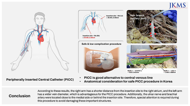
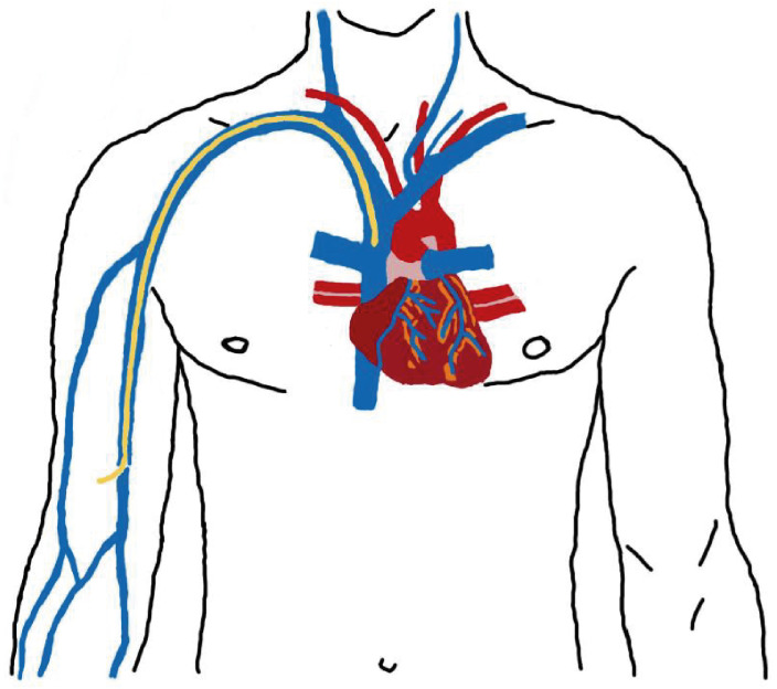
To utilize the advantages of the central line and overcome its limitations, a peripherally inserted central line (PICC) was introduced. This technique was first developed for chemotherapy in pediatric patients with cancer in 1960 and was first published by Hoshal8 in 1975; its usage was trivial before 1990, but is currently widely used in clinical practice.9 This is fundamentally the same procedure as the central line; however, the main difference is that it uses the peripheral veins of the arm instead of the large vein. Usually, the basilic vein, the largest deep vein of the arm, is used because the course is straight and the risk of arterial puncture is low. PICC has been known to have a higher possibility of causing deep vein thrombosis,1011121314 because of the long length the catheter travels, and its surface area exposure to blood is wider15; however, many studies have shown that this can be solved with plenty of practice and support from ultrasound or electrocardiogram.161718192021 Compared to the central line, PICC can be used safely even on neonates and children,22232425 does not require anesthesia or sedation, can be inserted without surgery,26 can be sustained for a long period of time, from 3 months to as long as an year,2728 is cost efficient,29 has low rates of complications and infection,303132 and can be performed by a professionally trained nurse, making it highly time efficient and cost effective for inpatients and patients cared for at home.3334 In addition, patients with a very high body mass index have a thick subcutaneous tissue in the neck, making it difficult to insert the central line. In this case, the PICC can be inserted through the peripheral vein of the arm using ultrasound guidance.35 PICC is a useful alternative for patients who have had renal transplants with neck vein thrombosis or multiple punctures that can cause chronic anemia or patients who are unable to be in a lying position due to severe pain or breathing difficulties.3637
Owing to these advantages, there has been a rising trend in the use of PICC in many hospitals. More than 2.5 million PICC procedures had been performed in the United States and more than 5 million around the world by 2008; by 2011, 15 million had been performed.6 Training programs for nurses started around 1970–80 and became pervasive during the 1990s; nowadays, 60% of the procedures are performed by professionally trained nurses.38 In Korea, several studies have been conducted on PICC, mainly summarizing basic procedures and precautions,39 or large-scale follow-up studies in hospitals, revealing the average duration and complication rates.404142 More specifically, there have been retrospective analyses of PICC procedures performed by well-trained nurse practitioners,43 safety studies of PICC in patients with burns, trauma, and chemotherapy,4445464748 and outcomes analyses of PICC performed at the bedside in critically ill patients who cannot be transported to an interventional radiology suite.4950 Other studies have devised formulas to optimize catheterization length and tip placement using patient height, weight, and ultrasound-determined anatomical features.5152
Despite this, there is a lack of research providing guidelines for PICC based on the position-relationship between anatomical structures, although guidelines for the central line have been mostly established.62053
Current studies have mainly focused on comparison analysis of complications and infections between central line and PICC that have been clinically proven, or established via medical imaging of anatomical structures56101112131617181920; therefore, this study was conducted to provide anatomic knowledge for safe PICC procedures through cadaver dissection.

METHODS

Study group

We used 40 arms from 20 fixed cadavers donated to Korea University College of Medicine (men: 10, women: 10, and mean age: 76.75 [48–94]).

Cadaveric study

Both arms of the cadavers were dissected to make the veins, arteries, and nerves clearly visible, and the parameters, which might be helpful when performing PICC procedures in the clinical field, were measured (Fig. 1). To determine the parameters, we consulted a radiologist with considerable practical experience.
Fig. 1

Vessel and nerve structures in the arm. Total length of the arm (greater tubercle–lateral epicondyle) (↔); Distance from the medial epicondyle to the splitting point of the brachial vein from the basilic vein (<--->); Insertion site (midpoint of total arm length, ▼); Distance from the basilic vein to the brachial artery and the ulnar nerve (◆); Vein branches in the axillary region (★).

LE = lateral epicondyle, ME = medial epicondyle, GT = greater tubercle.
jkms-38-e329-g001
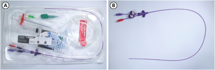
First, the total length of the arm and the distance from the greater tubercle to the lateral epicondyle of humerus were measured. The midpoint of the total arm length was designated as the insertion site. The length from the insertion site to the transition point from the superior vena cava to the right atrium was measured by inserting the catheter used in the actual PICC procedure into the basilic vein (Fig. 2). We used a Power PICC® Catheter (BARD Access Systems, Inc., Salt Lake City, UT, USA) for this procedure (Fig. 3). In addition, the distance from the basilic vein to the nearby structures such as median and ulnar nerves, and brachial artery was measured at the insertion site, and the diameter was measured using thinly sliced veins extracted at the same point. To set the upper limit of the insertion site, the location of brachial vein splitting from the basilic vein was confirmed, and the number of vein branches with diameters and paths that could prevent the catheter from progressing in the axillary region was also confirmed. Each parameter was measured after dissection, using a measuring tape and digital calipers, while trying to maintain the original position of every structure. To determine its diameter, a thin part of the basilic vein near the insertion was sampled, photographed, and measured using digital calipers.
Fig. 2

Route from the basilic vein in the arm to the right atrium. The yellow line is the inserted PICC, and this length was measured by directly inserting the PICC catheter.

PICC = peripherally inserted central catheters.
jkms-38-e329-g002
Fig. 3

PICC catheter. (A) Whole catheter kit (Power PICC® Catheter; BARD Access Systems, Inc., Salt Lake City, UT, USA), (B) Dual lumen PICC.

PICC = peripherally inserted central catheters.
jkms-38-e329-g003

Statistical analysis

Measured values were compared by sex and sides and then checked for significance using an unpaired t-test using mean values calculated in Microsoft Excel (Microsoft Office Excel 2007; Microsoft Inc., Redmond, WA, USA). Statistical significance was set at P < 0.05.

Ethics statement

This study was approved by "Cadaver Reserach Review Board", Korea University College of Medicine in 2017.

RESULTS

After determining the insertion site based on bony landmarks and the distance from the greater tubercle to the lateral epicondyle as the standard arm length, the anatomical relationship of the close structures was checked after dissection. Consequently, the nerve and vascular structures around the basilic vein were very close to each other. In particular, the brachial artery and ulnar nerve were located closer to each other than anticipated on both sides. When looking at the actual measured value, the brachial artery was situated on the deep lateral side of the basilic vein with a mean distance of 8.05 ± 2.42 mm, and the ulnar nerve was located on the medial side of the basilic vein with a mean distance of 5.46 ± 1.67 mm (Fig. 4).
Fig. 4

Dissected arm. Purple (BB) is the biceps brachii muscle, green (TB) is the triceps brachii muscle, yellow (UN) is the ulnar nerve, red (BA) is the brachial artery, blue (BV) is the basilic vein, and MN is median nerve. Based on the basilic vein, the brachial artery is located on the deep lateral side with a mean distance of 8.05 ± 2.43 mm, the ulnar nerve is located on the medial side with a mean distance of 5.46 ± 1.67 mm.

jkms-38-e329-g004
Mean values of all the measured parameters: the distance from the greater tubercle to the lateral epicondyle was 28.87 ± 1.94 cm, from the insertion site to the right atrium was 36.56 ± 3.56 cm, from the medial epicondyle to the meeting point of brachial and basilic veins was 192.81 ± 24.48 mm; the number of vein branches in the axillary region was 5.00 ± 1.21; and the diameter of the basilic vein was 4.55 ± 1.39 mm. (Fig. 5) When the results were analyzed considering sex and sides, the distance from the insertion site to the right atrium showed a significant difference with sex (P = 0.032); however, considering the natural difference based on sides due to the position of the heart, both sex and sides were considered in the calculation, thus showing insignificance (P = 0.059, left side and P = 0.198, right side). When comparing the distance from the insertion site to the right atrium based on the sides, there was a significant difference (P < 0.001) (Table 1).
Fig. 5

Vein branch observed in the axillary region and basilic vein extracted from the insertion site. (A) Vein branches in the axillary region that are wide enough for a catheter to pass through, which are divided by travel direction, upward or downward. (B) The thinly sliced basilic vein from the insertion site was placed under the measurement device, then a photo was taken and the precise diameter was measured using digital calipers.
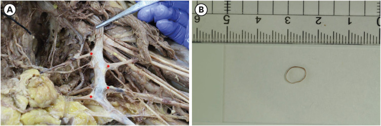
jkms-38-e329-g005
Table 1

Measured value by side and sex

jkms-38-e329-i001
Parameter Mean P value Mean P value
Left Right Men Women
Greater tubercle–Lat. Epicondyle, cm 28.63 29.05 0.196 29.45 28.23 0.065
Insertion site–RA, cm 38.39 34.66 0.001* 37.69 35.36 0.060/0.198a
Basilic v.–Brachial a., mm 8.19 7.91 0.560 8.29 7.81 0.571
Basilic v.–Ulnar n., mm 5.30 5.63 0.299 5.41 5.52 0.760
Med. Epicondyle–Basilic v. + Brachial v., mm 193.77 197.19 0.664 201.69 188.27 0.100
Number of v. branch, axilla part 4.90 5.10 0.592 4.90 5.10 0.650
Basilic v. diameter, mm 4.93 4.08 0.018* 4.48 4.53 0.908
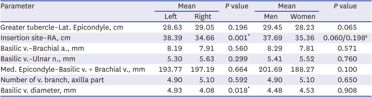
a. = artery, lat. = lateral, n. = nerve, v. = vein.
*P < 0.05.
aSignificance comes from the inevitable difference from the positioning of heart; therefore, re-analysis based on both sex and sides showed P ≥ 0.05, resulting in insignificance (men and women left side, P = 0.059 and men and women right side, P = 0.198). Compare with only sex, the P value was 0.032.

DISCUSSION

First, a parameter with significant difference was the distance from the insertion site to the right atrium, which was 3.73 cm longer on the left side than on the right side. This was hypothesized before the study, since the right atrium is located to the right.54 This result is expected to help determine the catheter length in clinical settings. In addition, the diameter of the basilic vein was not significantly different between the sexes, but it was larger on the left side than on the right side. The reason for this is unknown, but previous studies have speculated that doctors inserting central venous catheters prefer the right arm for convenience, and thus there is a possibility that the veins could have atrophied due to frequent injection of toxic drugs in the preferred right arm of patients with chronic diseases.55 This result could show the benefits of using the left arm for patients who cannot receive injections in the right arm for various reasons. Another advantage is that venous thrombosis, the most frequent side effect of inserting a PICC, has been reported to occur more frequently in thin blood vessels; therefore, this probability may be reduced when performed on the left arm.1556
In the axillary region, an average of five vein branches was thick enough for the catheter to pass through. Among these veins, the mean of 1.62 branches had an upward tendency, similar to the progress direction of the catheter, and the mean of 3.44 branches had a downward tendency (Fig. 5A).
Even though there was no significant correlation, these vein branches could interfere with the catheter pathway, and it would be helpful to know the number, location, and direction of these veins in advance.
It is important to note that the two closest structures, the brachial artery and ulnar nerve, were situated very close to the basilic vein, where the catheter was inserted. When performing the PICC procedure in the clinical setting, it is important to be careful not to damage major structures; thus, the distance and positional relationship found in this study will benefit in doing so.
The reason this study focused solely on the basilic vein is because the cephalic vein, an alternative, is too thin, has a high possibility of thrombosis, goes through the shoulder, causing movement of the catheter to increase, resulting in phlebitis56; axillary vein is an option with a large diameter but is close to the brachial artery and is covered with a thick subcutaneous fat layer57; the brachial vein, another alternative, has a higher possibility of damaging the median and anterior interosseous nerves or the brachial artery when injecting a needle,38585960 thus the basilic vein is most commonly used. However, depending on the state of the patient’s basilic vein, the cephalic or brachial veins are sometimes used; therefore, further research on these veins is needed.

Notes

Funding: This work was funded by the Brain Korea 21 Plus Project for Biomedical Science and a Korea University Grant (2018).

Disclosure: The authors have no potential conflicts of interest to disclose.

Author Contributions:

  • Conceptualization: Cho SB, Rhyu IJ.

  • Formal Analysis: Kim DS, Park JW.

  • Methodology: Rhyu IJ, Kim DS.

  • Writing - original draft: Kim DS.

  • Writing - review & editing: Park JW, Rhyu IJ.

References

1. Gabriel J. An intravenous alternative. Nurs Times. 1994; 90(31):39–41.
2. Walshe LJ, Malak SF, Eagan J, Sepkowitz KA. Complication rates among cancer patients with peripherally inserted central catheters. J Clin Oncol. 2002; 20(15):3276–3281. PMID: 12149302.
3. Ruesch S, Walder B, Tramèr MR. Complications of central venous catheters: internal jugular versus subclavian access--a systematic review. Crit Care Med. 2002; 30(2):454–460. PMID: 11889329.
4. Tan PL, Gibson M. Central venous catheters: the role of radiology. Clin Radiol. 2006; 61(1):13–22. PMID: 16356812.
5. Griffiths VR, Philpot P. Peripherally inserted central catheters (PICCs): do they have a role in the care of the critically ill patient? Intensive Crit Care Nurs. 2002; 18(1):37–47. PMID: 12008876.
6. O’Grady NP, Alexander M, Burns LA, Dellinger EP, Garland J, Heard SO, et al. Guidelines for the prevention of intravascular catheter-related infections. Am J Infect Control. 2011; 39(4):Suppl 1. S1–34. PMID: 21511081.
7. Bourgeois FC, Lamagna P, Chiang VW. Peripherally inserted central catheters. Pediatr Emerg Care. 2011; 27(6):556–561. PMID: 21642797.
8. Hoshal VL Jr. Total intravenous nutrition with peripherally inserted silicone elastomer central venous catheters. Arch Surg. 1975; 110(5):644–646. PMID: 805577.
9. Al Raiy B, Fakih MG, Bryan-Nomides N, Hopfner D, Riegel E, Nenninger T, et al. Peripherally inserted central venous catheters in the acute care setting: a safe alternative to high-risk short-term central venous catheters. Am J Infect Control. 2010; 38(2):149–153. PMID: 19836854.
10. Fearonce G, Faraklas I, Saffle JR, Cochran A. Peripherally inserted central venous catheters and central venous catheters in burn patients: a comparative review. J Burn Care Res. 2010; 31(1):31–35. PMID: 20061834.
11. Evans RS, Sharp JH, Linford LH, Lloyd JF, Woller SC, Stevens SM, et al. Reduction of peripherally inserted central catheter-associated DVT. Chest. 2013; 143(3):627–633. PMID: 22878346.
12. Chopra V, Ratz D, Kuhn L, Lopus T, Lee A, Krein S. Peripherally inserted central catheter-related deep vein thrombosis: contemporary patterns and predictors. J Thromb Haemost. 2014; 12(6):847–854. PMID: 24612469.
13. Parienti JJ, Mongardon N, Mégarbane B, Mira JP, Kalfon P, Gros A, et al. Intravascular complications of central venous catheterization by insertion site. N Engl J Med. 2015; 373(13):1220–1229. PMID: 26398070.
14. Puri A, Dai H, Giri M, Wu C, Huang H, Zhao Q. The incidence and risk of venous thromboembolism associated with peripherally inserted central venous catheters in hospitalized patients: a systematic review and meta-analysis. Front Cardiovasc Med. 2022; 9:917572. PMID: 35958406.
15. Patel GS, Jain K, Kumar R, Strickland AH, Pellegrini L, Slavotinek J, et al. Comparison of peripherally inserted central venous catheters (PICC) versus subcutaneously implanted port-chamber catheters by complication and cost for patients receiving chemotherapy for non-haematological malignancies. Support Care Cancer. 2014; 22(1):121–128. PMID: 24005884.
16. Sofocleous CT, Schur I, Cooper SG, Quintas JC, Brody L, Shelin R. Sonographically guided placement of peripherally inserted central venous catheters: review of 355 procedures. AJR Am J Roentgenol. 1998; 170(6):1613–1616. PMID: 9609183.
17. Fong NI, Holtzman SR, Bettmann MA, Bettis SJ. Peripherally inserted central catheters: outcome as a function of the operator. J Vasc Interv Radiol. 2001; 12(6):723–729. PMID: 11389224.
18. Barber JM, Booth DM, King JA, Chakraverty S. A nurse led peripherally inserted central catheter line insertion service is effective with radiological support. Clin Radiol. 2002; 57(5):352–354. PMID: 12014930.
19. Froehlich CD, Rigby MR, Rosenberg ES, Li R, Roerig PL, Easley KA, et al. Ultrasound-guided central venous catheter placement decreases complications and decreases placement attempts compared with the landmark technique in patients in a pediatric intensive care unit. Crit Care Med. 2009; 37(3):1090–1096. PMID: 19237922.
20. Bowdle A. Vascular complications of central venous catheter placement: evidence-based methods for prevention and treatment. J Cardiothorac Vasc Anesth. 2014; 28(2):358–368. PMID: 24008166.
21. Gullo G, Qanadli SD. ECG-based techniques to optimize peripherally inserted central catheters: rationale for tip positioning and practical use. Front Cardiovasc Med. 2022; 9:765935. PMID: 35600463.
22. Thiagarajan RR, Ramamoorthy C, Gettmann T, Bratton SL. Survey of the use of peripherally inserted central venous catheters in children. Pediatrics. 1997; 99(2):E4.
23. Jain A, Deshpande P, Shah P. Peripherally inserted central catheter tip position and risk of associated complications in neonates. J Perinatol. 2013; 33(4):307–312. PMID: 22955288.
24. Kleidon TM, Schults J, Rickard CM, Ullman AJ. Techniques and technologies to improve peripheral intravenous catheter outcomes in pediatric patients: systematic review and meta-analysis. J Hosp Med. 2021; 16(12):742–750. PMID: 34797998.
25. Zhu LB, Liu L, Zhang TS, Zheng YT, Lu CY, Lu K, et al. A clinical study on the tip localization of peripherally inserted central catheter (PICC) guided by intracavitary electrocardiography in newborns: a randomised trial. Transl Pediatr. 2021; 10(10):2409–2417. PMID: 34765464.
26. Yap YS, Karapetis C, Lerose S, Iyer S, Koczwara B. Reducing the risk of peripherally inserted central catheter line complications in the oncology setting. Eur J Cancer Care (Engl). 2006; 15(4):342–347. PMID: 16968315.
27. Kyle KS, Myers JS. Peripherally inserted central catheters. Development of a hospital-based program. J Intraven Nurs. 1990; 13(5):287–290. PMID: 2401933.
28. Cardella JF, Fox PS, Lawler JB. Interventional radiologic placement of peripherally inserted central catheters. J Vasc Interv Radiol. 1993; 4(5):653–660. PMID: 8219560.
29. Parkinson R, Gandhi M, Harper J, Archibald C. Establishing an ultrasound guided peripherally inserted central catheter (PICC) insertion service. Clin Radiol. 1998; 53(1):33–36. PMID: 9464433.
30. Duerksen DR, Papineau N, Siemens J, Yaffe C. Peripherally inserted central catheters for parenteral nutrition: a comparison with centrally inserted catheters. JPEN J Parenter Enteral Nutr. 1999; 23(2):85–89. PMID: 10081998.
31. Johansson E, Hammarskjöld F, Lundberg D, Arnlind MH. Advantages and disadvantages of peripherally inserted central venous catheters (PICC) compared to other central venous lines: a systematic review of the literature. Acta Oncol. 2013; 52(5):886–892. PMID: 23472835.
32. Pitiriga V, Bakalis J, Theodoridou K, Kanellopoulos P, Saroglou G, Tsakris A. Lower risk of bloodstream infections for peripherally inserted central catheters compared to central venous catheters in critically ill patients. Antimicrob Resist Infect Control. 2022; 11(1):137. PMID: 36352414.
33. Levy I, Bendet M, Samra Z, Shalit I, Katz J. Infectious complications of peripherally inserted central venous catheters in children. Pediatr Infect Dis J. 2010; 29(5):426–429. PMID: 20016395.
34. Chopra V, Flanders SA, Saint S. The problem with peripherally inserted central catheters. JAMA. 2012; 308(15):1527–1528. PMID: 23073947.
35. Uchida S, Takekawa D, Hori M, Hashiba E, Hirota K. Bedside insertion of a peripherally inserted central catheter into a patient with BMI of 84.8 kg/m2 using a magnetic tracking and electrocardiogram-based tip confirmation system: a case report. JA Clin Rep. 2022; 8(1):69. PMID: 36029359.
36. Delgado Márquez JC. Use of peripheral insertion central venous catheter in kidney transplant patients: an alternative. Exp Clin Transplant. 2022; 20:153–155. PMID: 35384828.
37. Narita A, Takehara Y, Maruchi Y, Matsunaga N, Ikeda S, Izumi Y, et al. Usefulness of peripherally inserted central catheter port system (PICC-PORT) implantation in the sitting position: a new technique for cases unsuitable for conventional implantation. Jpn J Radiol. 2023; 41(1):108–113. PMID: 35943686.
38. Moureau N. Vascular safety: it’s all about PICCs. Nurs Manage. 2006; 37(5):22–27.
39. Kim DH. Bedside peripherally inserted central catheter placement: focus on the procedure. Trauma Image Proced. 2022; 7(1):21–26.
40. Seo HK, Hwang JH, Shin MJ, Kim SY, Song KH, Kim ES, et al. Two-year hospital-wide surveillance of central line-associated bloodstream infections in a Korean hospital. J Korean Med Sci. 2018; 33(45):e280. PMID: 30402047.
41. Park JY, Kim HL. A comprehensive review of clinical nurse specialist-led peripherally inserted central catheter placement in Korea: 4101 cases in a tertiary hospital. J Infus Nurs. 2015; 38(2):122–128. PMID: 25723834.
42. Park S, Moon S, Pai H, Kim B. Appropriate duration of peripherally inserted central catheter maintenance to prevent central line-associated bloodstream infection. PLoS One. 2020; 15(6):e0234966. PMID: 32569313.
43. Park JY, Park KO, Baek MK, Kim SR, Kwon HL, Yang SJ. Analysis of PICC inserted patient data in a hospital by IV CNS-driven intervention. J Korean Biol Nurs Sci. 2004; 6(1):33–42.
44. Song C, Oh H. Burn patients’ experience of peripherally inserted central catheter insertion: analysis of focus group interviews from a South Korean burn center. Burns. 2016; 42(7):1439–1444. PMID: 27156805.
45. Kim HJ, Yun J, Kim HJ, Kim KH, Kim SH, Lee SC, et al. Safety and effectiveness of central venous catheterization in patients with cancer: prospective observational study. J Korean Med Sci. 2010; 25(12):1748–1753. PMID: 21165289.
46. Yun WS, Yang SS. Comparison of peripherally inserted central catheters and totally implanted venous access devices as chemotherapy delivery routes in oncology patients: a retrospective cohort study. Sci Prog. 2021; 104(2):368504211011871. PMID: 33950754.
47. Ryu DY, Lee SB, Kim GW, Kim JH. A peripherally inserted central catheter is a safe and reliable alternative to short-term central venous catheter for the treatment of trauma patients. J Trauma Inj. 2019; 32(3):150–156.
48. Lee Y, Ryu JA, Kim YO, Gil E, Song YM. Safety and feasibility of ultrasound-guided insertion of peripherally inserted central catheter performed by an intensive care trainee. J Neurocrit Care. 2020; 13(1):41–48.
49. Kim HJ, Jung CY, Min BJ. Clinical characteristics of peripherally inserted central catheter in critically ill patients. J Acute Care Surg. 2019; 9(1):18–24.
50. Yu B, Hong J. Safety and efficacy of peripherally inserted central catheter placement by surgical intensivist-led vascular access team. Vasc Spec Int. 2022; 38:41.
51. Cho HH, Jeon EY, Lee HJ, Lee H, Koh SH, Choi SY, et al. A new formula to estimate the length of right upper extremity vein from elbow crease to carina calculated by peripherally inserted central catheter insertion through right basilic vein puncture. J Korean Soc Radiol. 2012; 66(3):229–233.
52. Kang SS, Shin YS, Lee SY, Kim H. Simplified equation for determining proper depth of peripherally inserted central catheter in relation to anatomical landmarks. Korean J Anesthesiol. 2018; 71(4):300–304. PMID: 29684986.
53. Paterson RS, Schults JA, Slaughter E, Cooke M, Ullman A, Kleidon TM, et al. Review article: peripheral intravenous catheter insertion in adult patients with difficult intravenous access: a systematic review of assessment instruments, clinical practice guidelines and escalation pathways. Emerg Med Australas. 2022; 34(6):862–870. PMID: 36038953.
54. Forauer AR, Alonzo M. Change in peripherally inserted central catheter tip position with abduction and adduction of the upper extremity. J Vasc Interv Radiol. 2000; 11(10):1315–1318. PMID: 11099242.
55. Sperry BW, Roskos M, Oskoui R. The effect of laterality on venous thromboembolism formation after peripherally inserted central catheter placement. J Vasc Access. 2012; 13(1):91–95. PMID: 21948128.
56. Allen AW, Megargell JL, Brown DB, Lynch FC, Singh H, Singh Y, et al. Venous thrombosis associated with the placement of peripherally inserted central catheters. J Vasc Interv Radiol. 2000; 11(10):1309–1314. PMID: 11099241.
57. Lopez ON, Lorenc JM, Reading BD, Juang D, Aguayo P. Peripherally inserted central venous access. Semin Pediatr Surg. 2021; 30(6):151119. PMID: 34930595.
58. Puhaindran ME, Wong HP. A case of anterior interosseous nerve syndrome after peripherally inserted central catheter (PICC) line insertion. Singapore Med J. 2003; 44(12):653–655. PMID: 14770261.
59. Alomari A, Falk A. Median nerve bisection: a morbid complication of a peripherally inserted central catheter. J Vasc Access. 2006; 7(3):129–131. PMID: 17019665.
60. Kikuchi M, Sawada M, Nomura T, Mizuno Y, Goto T. Asymptomatic penetration of the median nerve by a peripherally inserted central catheter: a case report. A A Pract. 2022; 16(3):e01577. PMID: 35324514.
TOOLS
Similar articles