Journal List > Korean J Transplant > v.37(3) > 1516084162

Karaca, Işık, and Kemer: Optical quality of the cornea after Descemet membrane endothelial keratoplasty surgery: early results from Türkiye

Abstract

Background

Descemet membrane endothelial keratoplasty (DMEK) is increasingly favored in the treatment of endothelial dysfunction due to its benefits, which include swift visual rehabilitation and recovery, a relatively low rejection rate, and superior refractive stability. In this study, we examined alterations in corneal clarity among patients who underwent DMEK and correlated these densitometry findings with other optical parameters of corneal topography.

Methods

The study incorporated 35 eyes from 35 patients who had previously undergone DMEK surgery for pseudophakic bullous keratopathy at Ankara Bilkent City Hospital. The results from these patients were compared with those from a healthy control group. The preoperative and postoperative optical parameters of the patients were assessed using Pentacam Scheimpflug topography (Oculus).

Results

We observed significant decreases in corneal densitometry in the 0–2 mm and 2–6 mm zones of the anterior, posterior, central, and total layers at the sixth month postoperatively compared to the preoperative period (P<0.05). The corneal densitometry values at postoperative month 6 were elevated in all layers and zones relative to the healthy group (P<0.05). The root mean square of higher-order aberrations in postoperative period was elevated significantly (P<0.001).

Conclusions

Six months after operation, the optical quality of the cornea following DMEK surgery did not achieve the level of a healthy cornea.

HIGHLIGHTS
  • This paper discusses the disparity in optical quality between healthy individuals and patients after Descemet membrane (DMEK) endothelial keratoplasty.

  • Six months after operation, the optical quality of the cornea following DMEK surgery did not achieve the level of a healthy cornea.

INTRODUCTION

The corneal endothelium is crucial for maintaining the clarity of the cornea [1,2]. Descemet membrane endothelial keratoplasty (DMEK) is gaining popularity as a treatment for endothelial disorders due to its benefits, which include quick visual rehabilitation and recovery, a relatively low rejection rate, and enhanced refractive stability [3,4].
Corneal densitometry (CD) is a technique used to measure corneal haze, a condition characterized by clouding of the cornea due to injury, infection, or other eye conditions [5,6]. This measurement of backscatter and corneal transparency can be performed using the Pentacam HR (Oculus), a noncontact optical device that generates topographic maps of the anterior and posterior cornea [6,7]. CD provides a quantitative assessment of backlight scattering. The data obtained from this measurement can be utilized to diagnose and monitor corneal diseases, as well as to evaluate the effectiveness of various treatments. Following corneal surgery, CD can be employed to monitor the healing process of the cornea and assess the success of the surgical intervention. This technique allows clinicians to evaluate corneal transparency and identify any postoperative complications or issues. In certain cases, the goal of treatment for corneal diseases may be to decrease light scattering and improve corneal transparency. CD facilitates the objective evaluation of treatment efficacy by quantifying changes in corneal clarity after therapeutic interventions.
Following DMEK, the transplanted cells migrate and repopulate the recipient’s endothelial layer, thereby restoring its function and clarity. Research has demonstrated that DMEK can provide significant benefits in visual acuity, contrast sensitivity, and subjective visual quality when compared to other forms of corneal transplantation, such as penetrating keratoplasty [8]. Nevertheless, the success of surgery is contingent upon a variety of factors, including the patient’s preoperative visual acuity, the severity of the corneal disease, and the surgeon’s skill level [9]. Some patients may also report photophobia and glare despite the presence of a clear graft.
The aim of the current study was to identify changes in corneal clarity among participants who had undergone DMEK. The study was also designed to correlate the results of CD with other optical parameters found in corneal topography.

METHODS

The study was conducted in accordance with the ethical principles outlined in the Declaration of Helsinki and received approval from the Ethics Committee of Ankara City Hospital (No. E1-23-3519). Informed consent was obtained from all participants in the study.
The study incorporated 35 eyes from 35 patients who had previously undergone DMEK surgery for pseudophakic bullous keratopathy at Ankara Bilkent City Hospital and had completed regular follow-up visits. The data from these patients were compared with those from a healthy control group consisting of 30 individuals.
All participants underwent a detailed ophthalmological examination. This included measurements of best corrected visual acuity (BCVA), spherical equivalent values, anterior and posterior segment parameters, intraocular pressure (TX-20P noncontact tonometer; Canon Medical Systems), corneal topography (Pentacam), and specular microscopy (CEM-530; NIDEK). The exclusion criteria encompassed conditions such as cataract, uveitis, glaucoma, trauma, vitreoretinal disorders, dry eye, corneal infections, graft rejection, and the presence of any systemic or inflammatory diseases.
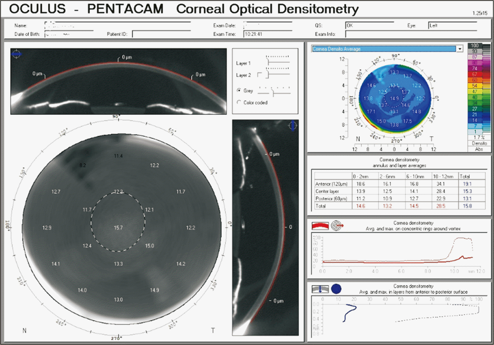
In this corneal topography study, we evaluated several parameters, including corneal topographic astigmatism, anterior K mean, anterior K maximum, anterior K minimum, posterior K mean, central corneal thickness (CCT), thinnest CCT, root mean square (RMS) total, and RMS of higher-order aberration (HOA) [10]. Additionally, we performed CD, which provides a measure of corneal transparency, with corneal backscattered light represented in grayscale units. These units range from 0, indicating complete transparency, to 100, indicating complete opacity. In this study, we measured densitometry across all annular zones (0–2 mm, 2–6 mm, 6–10 mm, and 10–12 mm). We conducted densitometry analysis by corneal depth, which included the anterior (the most anterior 120 µm), central, and posterior (the most posterior 60 µm) layers (Fig. 1). A trained technician captured all images in a darkened room prior to any other examination or drop instillation. We ensured the recording of high-quality images, as determined by Pentacam analysis. For this study, we analyzed backscattering values across all corneal zones, excluding the 6–10 and 10–12 mm areas; those peripheral sections lie outside the optical zone and may introduce artifacts due to corneal scarring between donor and host tissue. We compared the preoperative and 6-month postoperative topographic parameters of the patients.

Surgical Procedure

Each surgical procedure was performed by one of two expert cornea surgeons (EEK, ÖEK) under general anesthesia, following the same surgical procedure [11]. The DMEK grafts were prepared immediately prior to surgery. The eye was first cleaned with a 10% povidone-iodine solution and then covered with a surgical drape. Next, a lid speculum was inserted. An 8-mm descemetorhexis area was marked on the cornea, and three paracenteses were performed on the inferotemporal, superotemporal, and superonasal sides. Air was then injected, and descemetorhexis was performed using a reverse Sinskey hook under air. To minimize the risk of pupillary block, peripheral iridectomy at the 6 o'clock position was performed during surgery using an anterior vitrectomy probe (Centurion UltraVit 23 gauge; Alcon). A superior 2.4-mm clear corneal incision was made, and the Descemet membrane was removed. The 7.75-mm donor Descemet membrane was loaded into a glass pipet and injected through the clear corneal incision. A 10-0 nylon suture was immediately applied to the clear corneal incision. Techniques such as tapping, carpet unrolling while fixing one graft edge (the Dirisamer technique), or small air bubble-assisted unrolling (the Dapena maneuver) were used to unfold the graft [12,13]. Air was then used to attach the graft. Initially, the anterior chamber (AC) was left completely filled with air. Fifteen to 30 minutes after surgery, some of the tamponade was evacuated from the AC using a blunt cannula, leaving approximately a 50% air-fluid level. Patients were advised to maintain a supine position as much as possible on the first postoperative day.
The data were analyzed using IBM SPSS ver. 22 (IBM Corp). The Kolmogorov-Smirnov test was employed to assess the distribution of sample means. Continuous data were analyzed using the mean±standard deviation, while categorical variables were evaluated based on the number of cases and their respective percentages (%). The chi-square test was utilized for the comparison of categorical variables. To compare the groups, the Mann-Whitney U-test was implemented. Correlations between variables were examined using the Spearman correlation test. A P-value of less than 0.05 was considered to indicate statistical significance.

RESULTS

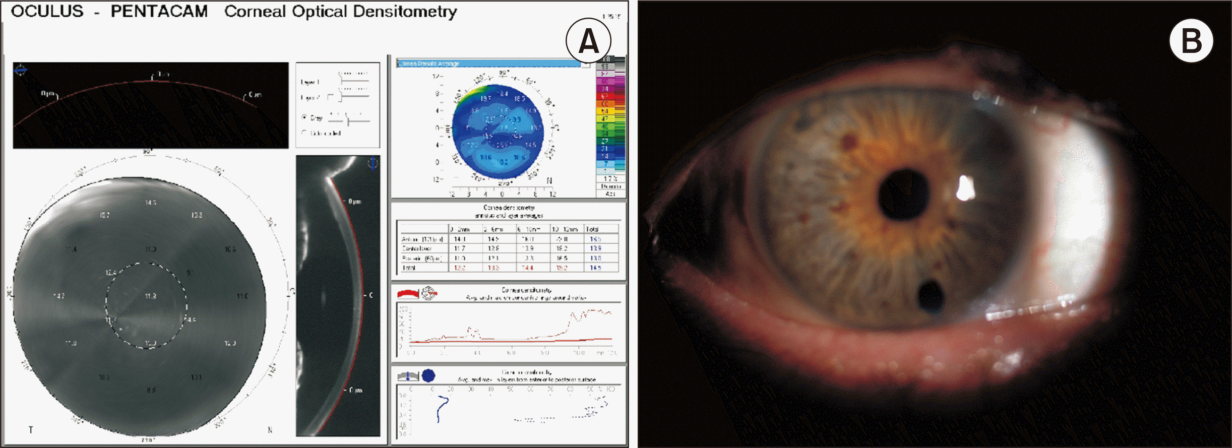
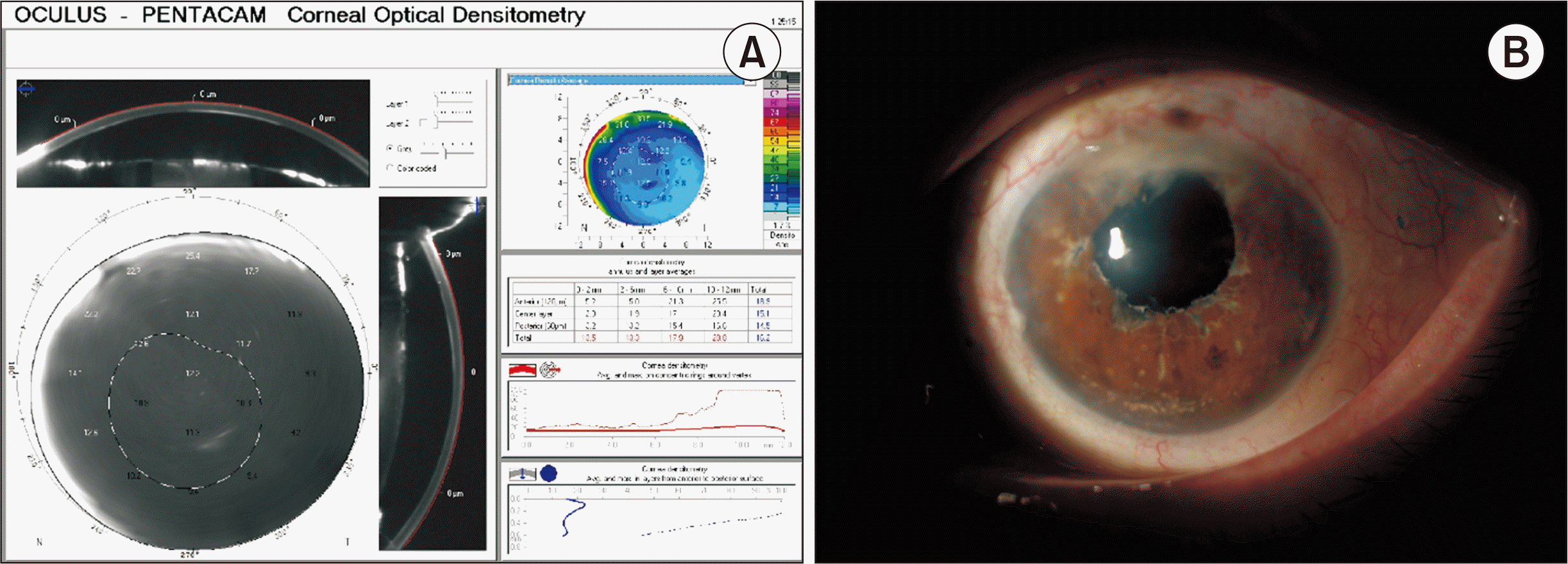
In this study, 42.8% of the patients (n=15) were male and 57.2% (n=20) were female. The mean age was 67.1±13.21 years. In the healthy control group, 40% (n=12) of participants were male and 60% (n=18) were female, with a mean age of 68.6±10.8 years (Table 1). We observed a significant decrease in CD in the 0–2 mm and 2–6 mm zones of the anterior, posterior, central and total layers at the sixth month compared to the preoperative period (P<0.001) (Table 2, Figs. 2 and 3). The CD values at postoperative month 6 were elevated in all layers and zones compared to the healthy group (P<0.001) (Table 3). No significant differences were observed in Kmax, K1, K2, or astigmatism values between the control group and the patients evaluated 6 months after DMEK (P>0.05). The endothelial cell count was 975.76±260.21 cells/mm2 (range, 630–2,130 cells/mm2) in the postoperative group at the 6-month visit, compared to 2,873±32.66 cells/mm2 (range, 2,077–3,435 cells/mm2) in the healthy group. The RMS HOA was higher in the postoperative group compared to preoperative period patients (P<0.001). Furthermore, a significant correlation was found between BCVA and total CD at the 0 to 2 mm and 2 to 6 mm zones at 6 months after DMEK (Table 4). A significant moderate negative correlation was found between CCT and total CD for the 0–2 mm and 2–6 mm zones after DMEK (P=0.035 and P<0.001, respectively).

DISCUSSION

DMEK is a surgical procedure that involves replacing the endothelial layer of the cornea with a thin, healthy donor graft. This procedure is known to result in rapid and improved visual acuity, with 50% of eyes achieving a BCVA of 0.0 logMAR or better [14,15]. However, vision quality is not solely dependent on BCVA; it is also influenced by other factors such as corneal clarity, thickness, and shape [16]. In this study, we not only investigated CD following DMEK, but also correlated these findings with other optical parameters derived from corneal topography. This correlation analysis enhanced the comprehensiveness of the study and aided in establishing relationships between corneal clarity and other topographic measures. In this study, we evaluated the early optical changes in the corneas of 35 eyes that were treated with DMEK surgery and compared these changes with those in healthy participants. This comparison can allow researchers to understand how the CD values in patients after DMEK differ from those in individuals with healthy corneas, thereby providing valuable insights into the impact of surgery on corneal clarity. We assessed the results of the patients 6 months after DMEK. Our findings suggest that the optical quality of healthy corneas is superior to that of DMEK patients at the 6-month mark.
The optical quality of the cornea plays an important role in overall visual function. As part of the eye’s optical system, the cornea refracts incoming light, focusing it onto the retina. Research indicates that the optical quality of a healthy cornea is generally superior to that of a cornea following DMEK surgery [17,18]. This is attributed to the DMEK procedure, which involves the removal of the host endothelium and its replacement with a donor graft. This process can induce changes in the cornea’s curvature along with irregularities at the interface between the donor graft and the host cornea. However, the extent of optical quality change after DMEK surgery varies among individuals and is influenced by several factors. These include the quality of the donor graft, the surgical technique employed, and the postoperative healing process. Numerous studies have explored the early changes in visual quality and corneal structure following DMEK surgery. Schoenberg et al. [19] discovered that patients who underwent DMEK surgery initially experienced a decrease in visual acuity and contrast sensitivity during the early postoperative period. However, these measurements significantly improved within the first 3 months following surgery. The study also revealed that corneal aberrations, or irregularities in the cornea’s shape, increased immediately postsurgery but gradually decreased over the first 3 months. Machalinska et al. [20] found that DMEK surgery led to a reduction in corneal thickness and an increase in curvature during the early postoperative period. However, these changes stabilized within the 6 months following surgery.
In this study, the indication for DMEK surgery was pseudophakic bullous keratopathy. Edema among these patients complicated the acquisition of accurate images of endothelial cells; thus, preoperative endothelial cell density (ECD) could not be measured. ECD measurements are crucial for the postoperative assessment of graft health. Although the endothelial cell count was 975.76±260.21 cells/mm2 (range, 630–2,130 cells/mm2) in the postoperative group at the 6-month visit, we observed positive outcomes in terms of corneal clarity and visual acuity. This could be attributed to the fact that corneal clarity is not solely dependent on ECD. Several other factors, such as intercellular connections and cell shape, may also contribute to this function.
Recent research on corneal aberrations following DMEK has yielded inconsistent results. Some studies suggest that DMEK can result in a decrease in HOAs, thereby improving visual acuity. In contrast, other studies indicate an increase in astigmatism and HOAs after DMEK [17,21,22]. In the present study, both anterior and posterior corneal aberrations were found to decrease following DMEK. However, it was observed that patients who underwent DMEK surgery exhibited higher aberrations postoperatively compared to healthy individuals.
CD is a widely used technique that offers an objective measurement of corneal transparency [23]. Patients with corneal endothelial disorders may experience an increase in light scattering, which leads to corneal haze. This is due to the disruption of the collagen matrix caused by corneal edema and scarring [24]. By monitoring changes in CD values of the anterior layer of the cornea, specifically in the second annular zone (2–6 mm), clinicians can gain valuable insights into the progression of the disease. This is particularly useful in patients who have reduced visual acuity following DMEK [23]. Increased backscatter in mid-peripheral corneal regions may be indicative of advanced stages of disease. Although the posterior layer of the cornea is typically the layer affected in endothelial disorders, the densitometry values in this layer do not always correlate well with visual acuity results [25]. This discrepancy may be due to the presence of stromal edema, which impacts visual acuity, in the central layer of the cornea. Furthermore, corneal edema can trigger subepithelial changes that result in a decrease in visual acuity. Consequently, correlations between visual acuity and CD values are more frequently observed in the central or anterior part of the cornea than in the posterior layer.
This study did have certain limitations. The most important limitations were the number of patients included, the lack of a long-term follow-up period after DMEK, and the absence of correlations of densitometry values with contrast sensitivity.
In conclusion, DMEK surgery may induce transient changes in visual quality and corneal structure during the early postoperative period. Although the optical quality of a healthy cornea typically exceeds that of a cornea after DMEK surgery, the extent of change in optical quality can fluctuate. The primary objective of DMEK surgery is to enhance the patient’s vision and quality of life. A substantial number of patients exhibit significant improvements in their visual acuity following the procedure.

ARTICLE INFORMATION

Conflict of Interest

No potential conflict of interest relevant to this article was reported.

Author Contributions

Conceptualization: EEK. Data curation: EEK. Formal Analysis: EEK. Investigation: FDI. Methodology: FDI. Supervision: ÖEK. Validation: ÖEK. Visualization: FDI. Writing–original draft: EEK, ÖEK. Writing–review & editing: EEK, ÖEK. All authors read and approved the final manuscript.

REFERENCES

1. DelMonte DW, Kim T. 2011; Anatomy and physiology of the cornea. J Cataract Refract Surg. 37:588–98. DOI: 10.1016/j.jcrs.2010.12.037. PMID: 21333881.
2. Schaub F, Enders P, Bluhm C, Bachmann BO, Cursiefen C, Heindl LM. 2017; Two-year course of corneal densitometry after Descemet membrane endothelial keratoplasty. Am J Ophthalmol. 175:60–7. DOI: 10.1016/j.ajo.2016.11.019. PMID: 27986425.
3. Melles GR, Ong TS, Ververs B, van der Wees J. 2008; Preliminary clinical results of Descemet membrane endothelial keratoplasty. Am J Ophthalmol. 145:222–7. DOI: 10.1016/j.ajo.2007.09.021. PMID: 18061137.
4. Laaser K, Bachmann BO, Horn FK, Cursiefen C, Kruse FE. 2012; Descemet membrane endothelial keratoplasty combined with phacoemulsification and intraocular lens implantation: advanced triple procedure. Am J Ophthalmol. 154:47–55. DOI: 10.1016/j.ajo.2012.01.020. PMID: 22465365.
5. Patel SV, McLaren JW, Hodge DO, Bourne WM. 2008; The effect of corneal light scatter on vision after penetrating keratoplasty. Am J Ophthalmol. 146:913–9. DOI: 10.1016/j.ajo.2008.07.018. PMID: 18774549. PMCID: PMC2612528.
6. Ní Dhubhghaill S, Rozema JJ, Jongenelen S, Ruiz Hidalgo I, Zakaria N, Tassignon MJ. 2014; Normative values for corneal densitometry analysis by Scheimpflug optical assessment. Invest Ophthalmol Vis Sci. 55:162–8. DOI: 10.1167/iovs.13-13236. PMID: 24327608.
7. Koh S, Maeda N, Nakagawa T, Nishida K. 2012; Quality of vision in eyes after selective lamellar keratoplasty. Cornea. 31 Suppl 1:S45–9. DOI: 10.1097/ICO.0b013e318269c9cd. PMID: 23038035.
8. Gellert A, Unterlauft JD, Rehak M, Girbardt C. 2022; Descemet membrane endothelial keratoplasty (DMEK) improves vision-related quality of life. Graefes Arch Clin Exp Ophthalmol. 260:3639–45. DOI: 10.1007/s00417-022-05711-9. PMID: 35612615. PMCID: PMC9581807.
9. Gurnani B, Kaur K, Lalgudi VG, Tripathy K. 2023; Risk factors for Descemet membrane endothelial keratoplasty rejection: current perspectives-systematic review. Clin Ophthalmol. 17:421–40. DOI: 10.2147/OPTH.S398418. PMID: 36755886. PMCID: PMC9899935.
10. Alio JL, Shabayek MH. 2006; Corneal higher order aberrations: a method to grade keratoconus. J Refract Surg. 22:539–45. DOI: 10.3928/1081-597X-20060601-05. PMID: 16805116.
11. Melles GR, Ong TS, Ververs B, van der Wees J. 2006; Descemet membrane endothelial keratoplasty (DMEK). Cornea. 25:987–90.
12. Yoeruek E, Bayyoud T, Hofmann J, Bartz-Schmidt KU. 2013; Novel maneuver facilitating Descemet membrane unfolding in the anterior chamber. Cornea. 32:370–3. DOI: 10.1097/ICO.0b013e318254fa06. PMID: 22820605.
13. Liarakos VS, Dapena I, Ham L, van Dijk K, Melles GR. 2013; Intraocular graft unfolding techniques in Descemet membrane endothelial keratoplasty. JAMA Ophthalmol. 131:29–35. DOI: 10.1001/2013.jamaophthalmol.4. PMID: 22965272.
14. Peraza-Nieves J, Baydoun L, Dapena I, Ilyas A, Frank LE, Luceri S, et al. 2017; Two-year clinical outcome of 500 consecutive cases undergoing Descemet membrane endothelial keratoplasty. Cornea. 36:655–60. DOI: 10.1097/ICO.0000000000001176. PMID: 28410548.
15. Monnereau C, Quilendrino R, Dapena I, Liarakos VS, Alfonso JF, Arnalich-Montiel F, et al. 2014; Multicenter study of Descemet membrane endothelial keratoplasty: first case series of 18 surgeons. JAMA Ophthalmol. 132:1192–8. DOI: 10.1001/jamaophthalmol.2014.1710. PMID: 24993643.
16. Turnbull AM, Tsatsos M, Hossain PN, Anderson DF. 2016; Determinants of visual quality after endothelial keratoplasty. Surv Ophthalmol. 61:257–71. DOI: 10.1016/j.survophthal.2015.12.006. PMID: 26708363.
17. van Dijk K, Droutsas K, Hou J, Sangsari S, Liarakos VS, Melles GR. 2014; Optical quality of the cornea after Descemet membrane endothelial keratoplasty. Am J Ophthalmol. 158:71–9. DOI: 10.1016/j.ajo.2014.04.008. PMID: 24784873.
18. Cabrerizo J, Livny E, Musa FU, Leeuwenburgh P, van Dijk K, Melles GR. 2014; Changes in color vision and contrast sensitivity after Descemet membrane endothelial keratoplasty for Fuchs endothelial dystrophy. Cornea. 33:1010–5. DOI: 10.1097/ICO.0000000000000216. PMID: 25119964.
19. Schoenberg ED, Price FW Jr, Miller J, McKee Y, Price MO. 2015; Refractive outcomes of Descemet membrane endothelial keratoplasty triple procedures (combined with cataract surgery). J Cataract Refract Surg. 41:1182–9. DOI: 10.1016/j.jcrs.2014.09.042. PMID: 26096520.
20. Machalinska A, Kuligowska A, Kaleta K, Kusmierz-Wojtasik M, Safranow K. 2021; Changes in corneal parameters after DMEK surgery: a swept-source imaging analysis at 12-month follow-up time. J Ophthalmol. 2021:3055722. DOI: 10.1155/2021/3055722. PMID: 34336256. PMCID: PMC8321767.
21. Satue M, Idoipe M, Gavin A, Romero-Sanz M, Liarakos VS, Mateo A, et al. 2018; Early changes in visual quality and corneal structure after DMEK: does DMEK approach optical quality of a healthy cornea? J Ophthalmol. 2018:2012560. DOI: 10.1155/2018/2012560. PMID: 30345110. PMCID: PMC6174748.
22. Rudolph M, Laaser K, Bachmann BO, Cursiefen C, Epstein D, Kruse FE. 2012; Corneal higher-order aberrations after Descemet's membrane endothelial keratoplasty. Ophthalmology. 119:528–35. DOI: 10.1016/j.ophtha.2011.08.034. PMID: 22197439.
23. Alnawaiseh M, Zumhagen L, Wirths G, Eveslage M, Eter N, Rosentreter A. 2016; corneal densitometry, central corneal thickness, and corneal central-to-peripheral thickness ratio in patients with Fuchs endothelial dystrophy. Cornea. 35:358–62. DOI: 10.1097/ICO.0000000000000711. PMID: 26655484.
24. Baratz KH, McLaren JW, Maguire LJ, Patel SV. 2012; Corneal haze determined by confocal microscopy 2 years after Descemet stripping with endothelial keratoplasty for Fuchs corneal dystrophy. Arch Ophthalmol. 130:868–74. DOI: 10.1001/archophthalmol.2012.73. PMID: 22410629.
25. Schaub F, Gerber F, Adler W, Enders P, Schrittenlocher S, Heindl LM, et al. 2019; Corneal densitometry as a predictive diagnostic tool for visual acuity results after Descemet membrane endothelial keratoplasty. Am J Ophthalmol. 198:124–9. DOI: 10.1016/j.ajo.2018.10.002. PMID: 30315754.

Fig. 1
Corneal densitometry.
kjt-37-3-203-f1.tif
Fig. 2
(A) Corneal densitometry with (B) increased corneal clarity.
kjt-37-3-203-f2.tif
Fig. 3
(A) Corneal densitometry with (B) decreased corneal clarity.
kjt-37-3-203-f3.tif
Table 1
Demographics of the patient and control groups
Parameter DMEK patients Control group P-value
Age (yr; mean±SD) 67.1±13.21 68.6±10.8 0.386
Sex (male:female) 15:20 12:18 0.783

DMEK, Descemet membrane endothelial keratoplasty; SD, standard deviation.

Table 2
Comparison of refractive and topographic outcomes between preoperative and 6-month postoperative patients
Parameter Preoperative Postoperative P-value
KmF (D) 43.90±2.84 43.42±1.86 <0.001
KmB (D) –5.69±0.71 –4.57±8.97 <0.001
TCRP (D) 43.2±2.1 42.02±1.70 <0.001
Central corneal thickness (µm) 646.7±78.2 525.41±25.20 <0.001
Thinnest corneal thickness (µm) 615.2±69.2 517.0±24.8 <0.001
BCVA (logMAR) 1.183±0.410 0.17±0.13 <0.001
Chord µ 0.30±0.06 0.31±0.08 >0.05
ACD (mm)
0–2 27.01±10.31 19.45±6.37 <0.001
2–6 28.72±9.15 18.42±7.10 <0.001
ATCD (GSU) 23.44±10.23 17.41±6.10 <0.001
CCD (0–2 mm) 27.01±10.31 15.10±3.95 <0.001
CCD (2–6 mm) 28.72±9.15 14.14±3.38 <0.001
CTCD (GSU) 22.38±6.49 16.52±5.45 <0.001
PCD (0–2 mm) 15.12±5.71 13.14±4.84 <0.001
PCD (2–6 mm) 16.63±5.04 12.73±4.52 <0.001
PTCD (GSU) 16.44±4.88 12.36±3.35 <0.001
RMS HOA (µm) 3.67±7.04 9.64±1.88 <0.001
RMS LOA (µm) 9.99±13.18 5.82±6.92 <0.001
RMS total (µm) 10.29±13.31 6.15±7.11 <0.001

Values are presented as mean±standard deviation.

KmF, mean keratometry front; D, diopters; KmB, mean keratometry back; TCRP, total cornea refractive power; BCVA, best corrected visual acuity; ACD, anterior cornea densitometry; ATCD, anterior total cornea densitometry; GSU, gray scale unit; CCD, central cornea densitometry; CTCD, central total cornea densitometry; PCD, posterior cornea densitometry; PTCD, posterior total cornea densitometry; RMS, root mean square; HOA, high-order aberration; LOA, low-order aberration.

Table 3
Comparison of refractive and topographic outcomes between healthy and 6-month postoperative DMEK patients
Parameter Control group DMEK P-value
KmF (D) 43.91±1.52 43.42±1.86 0.630
KmB (D) –6.30±1.24 –4.57±8.97 0.213
TCRP (D) 42.75±1.15 42.02±1.70 0.597
Central corneal thickness (µm) 544.6±30.7 525.41±25.20 0.148
Thinnest corneal thickness (µm) 540.01±32.10 517.0±24.8 0.194
BCVA 0 0.17±0.13 0.238
ECD 2,873±322.66 975.76±260.21 <0.001
Chord µ 0.32±0.74 0.31±0.08 0.845
ACD (0–2 mm) 11.10±1.11 19.45±6.37 <0.001
ACD (2–6 mm) 10.56±1.15 18.42±7.10 <0.001
ATCD (GSU) 11.35±1.79 17.41±6.10 <0.001
CCD (0–2 mm) 12.74±1.52 15.10±3.95 <0.001
CCD (2–6 mm) 9.21±0.78 14.14±3.38 <0.001
CTCD (GSU) 12.15±2.12 16.52±5.45 <0.001
PCD (0–2 mm) 8.64±0.67 13.14±4.84 <0.001
PCD (2–6 mm) 8.27±0.65 12.73±4.52 <0.001
PTCD (GSU) 8.24±1.27 12.36±3.35 <0.001
RMS HOA (µm) 0.10±0.22 9.64±1.88 <0.001
RMS LOA (µm) 2.38±0.75 5.82±6.92 <0.001
RMS total (µm) 2.91±1.15 6.15±7.11 <0.001

Values are presented as mean±standard deviation.

DMEK, Descemet membrane endothelial keratoplasty; KmF, mean keratometry front; D, diopters; KmB, mean keratometry back; TCRP, total cornea refractive power; BCVA, best corrected visual acuity; ECD, endothelial cell density; ACD, anterior cornea densitometry; ATCD, anterior total cornea densitometry; GSU, gray scale unit; CCD, central cornea densitometry; CTCD, central total cornea densitometry; PCD, posterior cornea densitometry; PTCD, posterior total cornea densitometry; RMS, root mean square; HOA, high-order aberration; LOA, low-order aberration.

Table 4
Correlations between total cornea densitometry and other clinical parameters following DMEK
Parameter R-value P-value
ECD (mm)
0–2 0.017 0.856
2–6 –0.125 0.426
BCVA (mm)
0–2 0.405 0.008
2–6 0.470 0.003
CCT (mm)
0–2 –0.325 0.035
2–6 –0.475 <0.001
KmF (mm)
0–2 0.220 0.270
2–6 0.204 0.308
KmB (mm)
0–2 –0.271 0.172
2–6 –0.226 0.257

DMEK, Descemet membrane endothelial keratoplasty; ECD, endothelial cell density; BCVA, best corrected visual acuity; CCT, central corneal thickness; KmF, mean keratometry front; KmB, mean keratometry back.

TOOLS
Similar articles