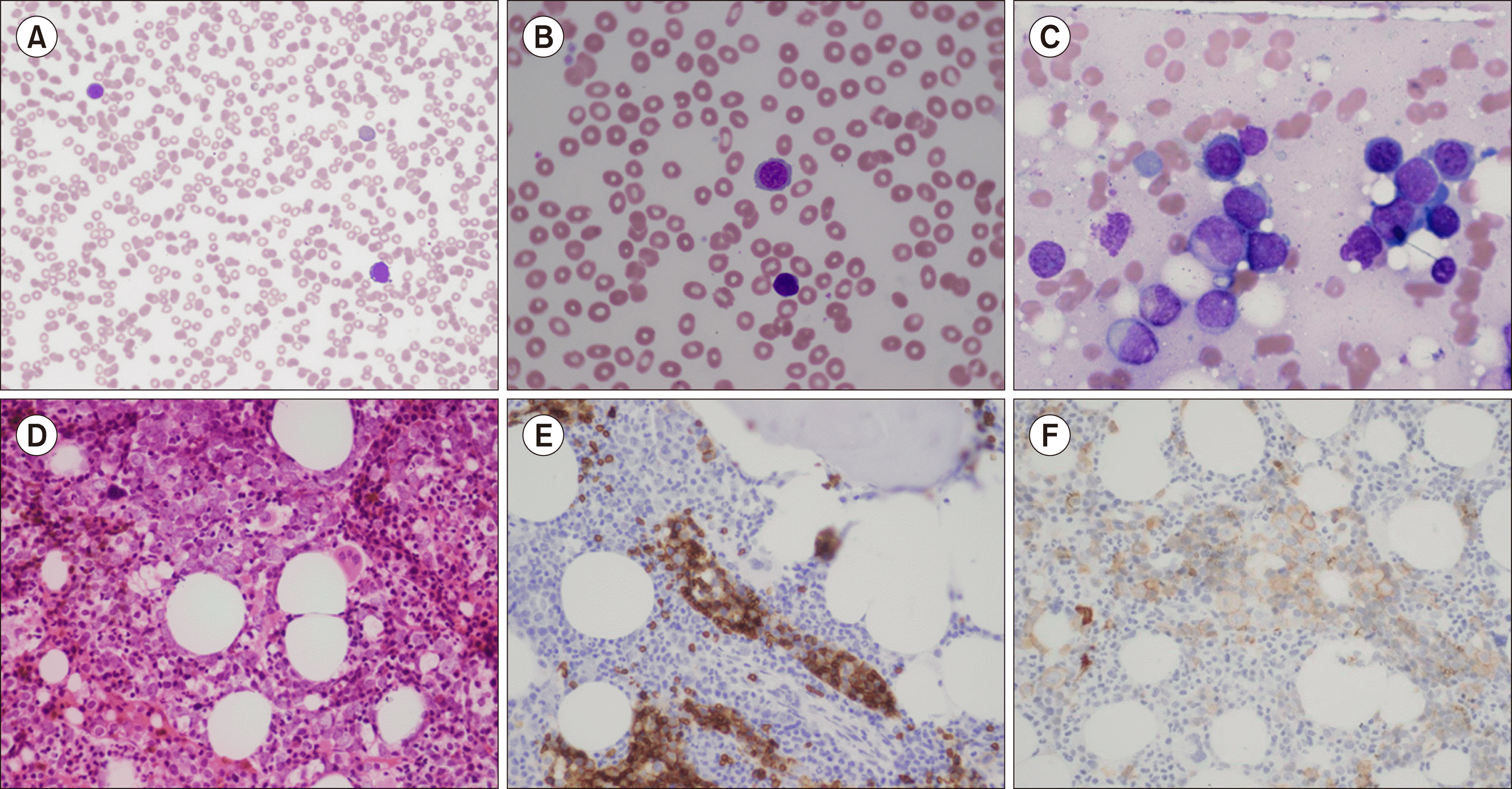
A 67-year-old patient received rituximab and bendamustine for follicular lymphoma and achieved complete response two years prior to developing moderate anaemia (94 g/L) and thrombocytopenia (44×109/L). Blood film showed circulating nucleated red cells and proerythroblasts with dysplasia (A, B).
Diagnostic bone marrow aspirate was a dry tap. Trephine imprints showed 42% nucleated red cells with significant dysplasia such as binucleation, nuclear irregularity and cytoplasmic vacuoles (C). Immature erythroblasts were increased but comprised <30% of nucleated cells. There was minimal maturation beyond the proerythroblast phase.
Trephine core was hypercellular with abnormal megakaryocytes and erythroblasts (D). The latter showed weak membranous staining with anti-CD117 (E) and variable patterns of glycophorin A (F). Intra-sinusoidal infiltration of proerythroblasts were highlighted. There was no evidence for increased myeloblasts, granulocytic dysplasia, or relapsed lymphoma.
Myeloid gene panel with next generation sequencing revealed somatic mutations in TP53, SRSF2, TET2 and KDM6A. Findings support a diagnosis of myeloid neoplasm post cytotoxic therapy according to the World Health Organisation; and myelodysplastic syndrome/acute myeloid leukaemia with mutated TP53, therapy-related according to the International Consensus Classification.


 PDF
PDF Citation
 Print
Print


 XML Download