Abstract
The first extensor compartment of the wrist is a distinctly variable anatomical area. Anatomical variations in this region contribute to the pathophysiology and treatment failure of de Quervain’s disease, which is a kind of tenosynovitis that develops in the first extensor compartment of the wrist. We aim to describe the first extensor compartment morphology, to evaluate the septum frequency, location of the septum, and the number of tendons of abductor pollicis longus (APL) and extensor pollicis brevis muscles (EPB). First extensor compartment of 87 wrists of 45 cadavers were dissected. The presence or absence of septum and number of tendon slips of APL and EPB revealed. The proximal and distal widths of the compartments were measured. Septums were detected in 60.9% (n=53) of the wrists. Incomplete (distal) and complete (proximal) septa were present in 35.6% (n=31) and 25.3% (n=22) of the cases. Only 26.4% of the wrists had a single slip of APL tendon. The Remaining had multiple slips. The median inner width of the proximal and distal compartments in all wrists were calculated as in the order of 9.11±1.14 mm and 8.55±1.12 mm. We believe that understanding the anatomy of the first extensor compartment in the Turkish population would be helpful to surgeons, radiologists, and physiotherapists to diagnose and manage de Quervain’s disease.
The wrist has six extensor compartments. A synovial sheath surrounds tendons in each compartment. The normal anatomy of the first extensor compartment was originally described as containing the tendons of the extensor pollicis brevis (EPB) muscle and abductor pollicis longus (APL) muscle and the tendons reaching out in a unified fibro-osseous tunnel compartment formed by extensor retinaculum to the first metacarpal and proximal phalanx in the 1960s [1]. These illustrations still have been accepted as the standard anatomy of the first compartment [2]. However subsequent cadaver and radiological studies have proved that the compartment is a greatly variable space [3, 4]. Two important variations have been identified regarding the compartment and persistently emphasized in ongoing studies. These main variations are multiple slips of tendons of APL muscle and the division of the compartment into two subcompartments by a fibro-osseous septum [4]. Contrary to the historical standard anatomic description, instead of 2 tendons lying in a single tunnel in the compartment, a septum could be detected which creates subcompartment areas in the tunnel. Septums extending the proximal end of the first extensor compartment (complete type) or terminated distally (incomplete type) have also been reported in various studies [5, 6].
de Quervain’s disease is a kind of stenosing tenosynovitis of the APL and EPB. Overuse of the wrist through ulnar deviation increases the friction between APL and EPB tendon and tendon sheath. The disease is also known as radial styloid tenosynovitis and is characterized by mucosal degeneration, fibrocartilage degeneration, and mucopolysaccharide deposition [7]. It has been reported that it is more common in adults between the ages of 30–50 and especially women. The presenting complaint is mostly tenderness, swelling, and pain in the radial styloid region. A positive Finkelstein test is defined as severe pain in the radial styloid when the patient's thumb is grasped and the hand is brought to a sudden ulnar deviation [8]. Finkelstein test is more indicative of APL muscle pathology than EPB muscle pathology. Eichhoff maneuver is another test expected to be positive in this disease: while the patient clenches her thumb in her fist, the wrist comes to ulnar deviation, then when the thumb is extended, the pain is relieved even if the wrist remains in the ulnar deviation [9]. The logic of both tests is based on providing a slip between the thickened tendons of APL and EPB and the floor of the first compartment. The thickened fibrous sheath may be palpated during the examination. As a differential diagnosis, superficial radial nerve compression or neuroma in the trapeziometacarpal scaphotrapeziotrapezoid and radiocarpal joint, tenosynovitis occurring where the tendons of EPB and APL overlap with the extensor carpi radialis longus and brevis may cause the same symptoms. Splint use and steroid injection are among the conservative and first-stage treatments. Surgical treatment is considered in patients whose pain persists despite conservative treatment [8].
The anatomical variations shown may be the cause of de Quervain’s disease and may also affect the treatment option [10, 11]. The aim of this study is to describe the first extensor compartment morphology, to evaluate the septum frequency, location of the septum, and the number of APL-EPB tendon slips of cadavers in a Turkish population.
All cadavers studied were obtained from the Department of Anatomy. The cadavers were either donated or procured according to Turkish law and regulations [12]. IRB approval for the study was obtained from the Istanbul University Ethical Committee (Date: 10/09/2021; Number: 16).
The wrists of 18 female and 27 male cadavers were dissected which were fixed by formalin-ethanol-glycerin-phenol solution and kept in cold storage (5°C–8°C) after embalming. Since they were used in educational activities, 3 cadavers were evaluated unilaterally. Wrist dissection was performed bilaterally in the remaining cadavers. Female 41.4% (n=36) and male cadaver 58.6% (n=51) of 87 wrists were dissected totally. Right 50.6% (n=44) and left 49.4% (n=43) wrists were dissected.
None of the cadavers had any clinical history of de Quervain’s disease or visible prior wrist surgery.
The skin incision was made as a transverse incision connecting both styloid processes of radius and ulna, another incision was made longitudinally extending from the middle of this line to the elbow. After the skin was removed, the superficial radial nerve, cephalic vein, and subcutaneous tissue were dissected. The compartment was opened by making an incision along the entire length of the dorsal part of it. Osseo-fibrous septum presence and type of the septum as complete or incomplete according to the radial styloid process were noted. Osseo-fibrous septum position had the determinative role of septum type. If the septum was positioned distal to the radial styloid process we named this type as the incomplete type. If the septum was reached and passed the radial styloid process we named this type as the complete type.Thenumber of tendon slips of APLand EPB in the compartment were examined and recorded. Proximal and distal internal widths of the compartment were measured by a digital caliper.
While evaluating the findings obtained in the study, Number Cruncher Statistical System (NCSS) 2020 Statistical Software (NCSS LLC) program was used for statistical analysis. While evaluating the study data, quantitative variables were shown with mean, standard deviation, median, min and max values, and qualitative variables were shown with descriptive statistical methods such as frequency and percentage. Shapiro Wilks test and Box Plot graphics were used to evaluate the conformity of the data to the normal distribution. Student’s t-test was used for quantitative evaluation of two groups with normal distribution.
Pearson Chi-Square test, Fisher’s Exact test and Fisher Freeman Halton Test were used to compare qualitative data. The results were evaluated at the 95% confidence interval and the significance level of P<0.05.
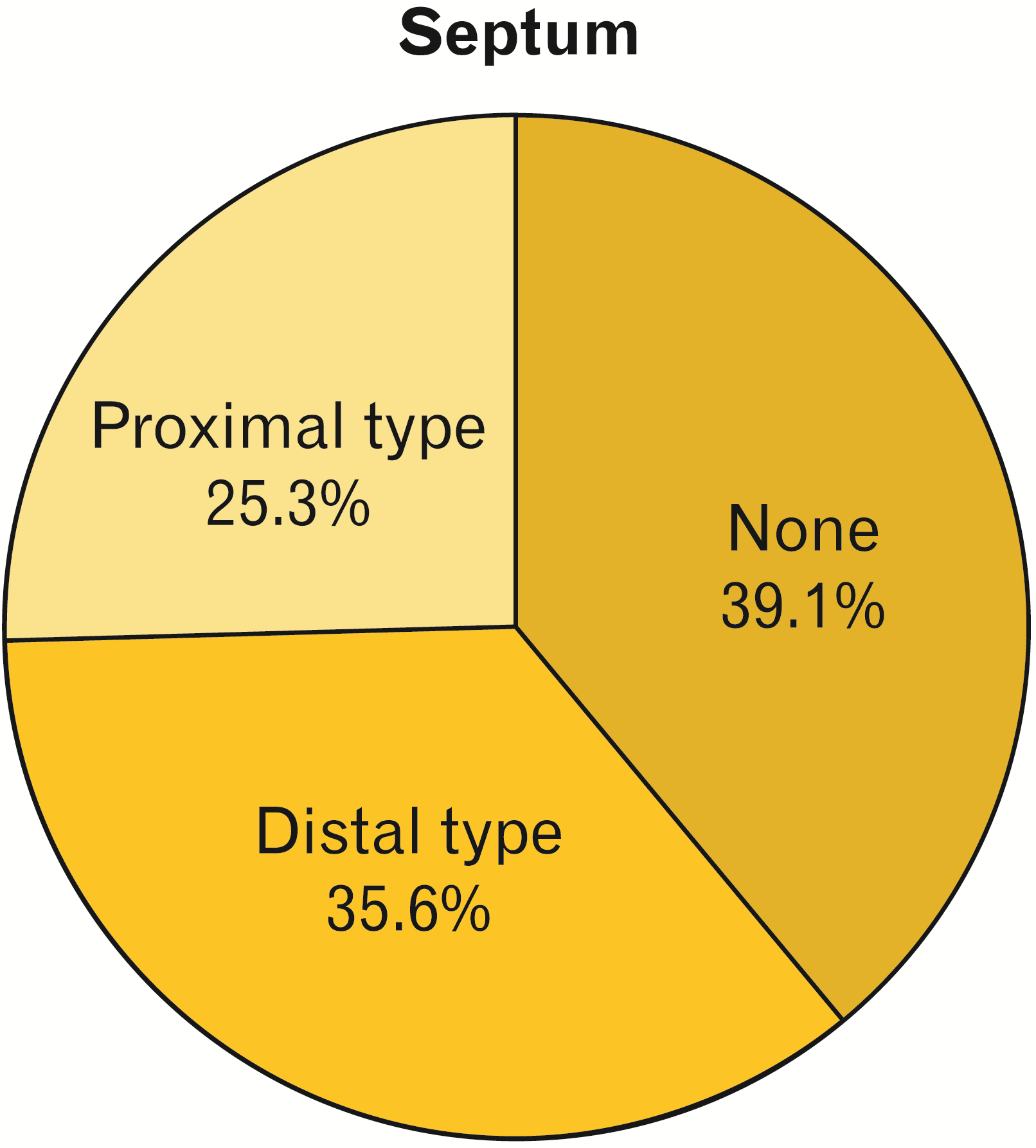
Septum was detected in 60.8% (n=53). The wrists of 35.6% (n=31) had incomplete and 25.3% (n=22) of the wrists had complete type septum (Figs. 1, 2).
The incidence of the septum in female cadavers was 63.9% (23 wrists). Wrists of 41.7% (15 wrists) had incomplete and 22.2% (8 wrists) had complete type septum. The septum was also detected in 58.8% (30 wrists) of the male cadavers. Results revealed 31.4% (16 wrists) had incomplete and 27.5% (14 wrists) had complete type septum (Tables 1 and 2).
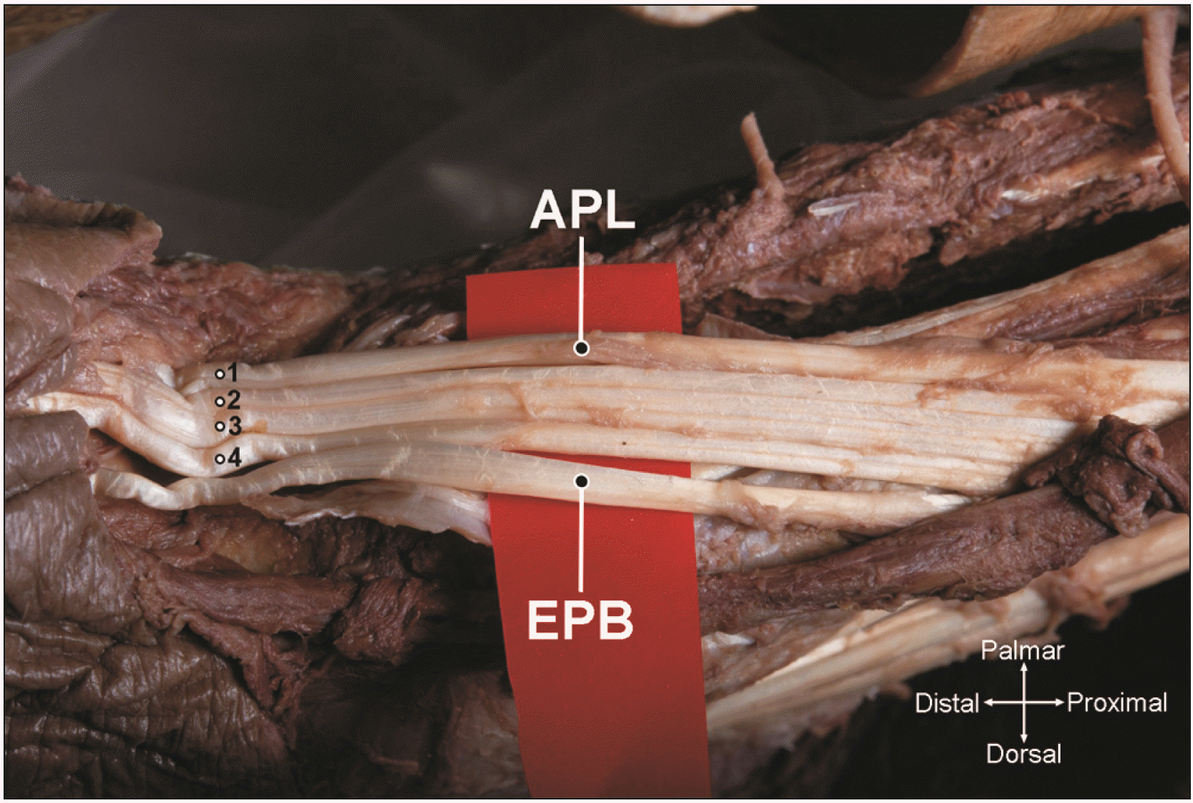
When the tendons of APL were examined, only 26.4% (23 wrists) had a single tendon (Fig. 3). The remaining 73.6% (64 wrists) tendons of APL had multiple slips. The highest number of tendon slips detected was 4 which occurred in the left wrist of a male cadaver (Fig. 4). The most frequent anatomical finding was two tendon slips in the first compartment. This finding was present in 56.3% of the cadavers (49 wrists). All the tendons of the EPB had a single slip.
The proximal width of the compartment varies between 6.43 and 11.20, with a mean of 9.11±1.14 mm. The distal width of the compartment varies between 6.03 and 12.40, with a mean of 8.55±1.12 mm.
The 1st extensor compartment of the wrist is a highly variable anatomical region. However, the most depicted 1st extensor compartment anatomy is with a single tunnel and a tendon slip of each APL and EPB. A previous cadaveric study in the Turkish population reported the incidence of a dividing septum in the first dorsal compartment as 28% [13]. In different cadaveric studies, septum incidences were also reported as 33.3% and 54% in the Turkish population [6, 14]. The septum rate was detected in the present study as 60.9% (Fig. 5). High rates of septum incidence were also obtained in the previous studies also from the different countr’s populations [15-17]. Diverse studies verified compartment variations correlate with de Quervain’s disease frequency [6, 18]. In one of them, Yuasa and Kiyoshige [18], reported septum in the compartment increases the friction between APLand EPB. These studies and the results of the present study specify that septum presence should be considered as prevalent by physicians dealing with the treatment of de Quervain’s disease.
The presence of the septum contributes to the development of de Quervain’s disease and it’s also important to know its presence in the treatment of the disease [10, 11]. Corticosteroid injection is the first-stage treatment of the disease. It has been shown that two-point injection instead of one point by considering the septum in the compartment was more effective. The same study also reported that subcompartment morphology could be the reason for unsuccessful injection treatment [19]. Incomplete injection for both subcompartments may result in treatment failure. Zingas et al. [20], stated that if the injection was chosen as the treatment option, injection into both subcompartments would not be possible in compartments with a septum, and this procedure should be done under the guidance of ultrasonography. The percutaneous release is a minimally invasive approach and an alternative surgical treatment for de Quervain’s disease. Güleç et al. [14], reported a statistically significant relationship between incomplete release and septation in the wrists to which percutaneous release was performed. In another study, it was reported that the failure in the surgical treatment of the disease was due to the insufficient recognition of the compartment and the inability to decompress all the subcompartments [16]. These studies indicate that the definition of a classical and unidirectional 1st extensor compartment anatomy may mislead physicians dealing with the treatment of this region.
Considering the variations of APL tendon is important in clinical assessment. Mansur et al. [21], suggested that multiple APL tendons may contribute to the development of de Quervain’s disease. Additionally, the number of multiple APL tendons that are not taken into account when surgical treatment is applied to the disease may make the intervention of surgeons insufficient. Knowing the number of multiple tendons may also be important for surgeons who intervene in the dorsolateral compartment for graft surgeries. Öztürk et al. [22], encountered multiple APL tendon numbers as 97.6% in 83 extremities. In the present study, the presence of multiple tendons of APL was found similarly to 73.5% of all wrists. The number of multiple APL tendons, together with the presence of a septum, should be another important variation that should be considered by clinicians. Variation of EPB tendon was not encountered in the present study. Ethnicity difference was noted as an important point in EPB tendon variation in a review [23]. A study reported the mean size of thefirst extensor compartrment as 8.65±0.67 mm [24]. In our study we measured the width of the first external compartment proximally and distally as 9.11±1.14 mm and 8.55±1.12 mm, respectively.
The first limitation of our study is the relatively small number of cadavers in our sample. The second limitation of the study is unknown medical history of the cadavers. Therefore we could not manage to compare the correlation between de Quervain’s disease and septum types.
Our study and previous studies have shown that physicians approaching de Quervain’s disease should consider the anatomical variation in this region as a prevailing rather than an exception [15, 19]. Considering the variations in this region will reduce treatment failure and recurrence of the disease.
Notes
References
1. Anson BJ. 1963. An atlas of human anatomy. 2nd ed. Saunders;180e1. DOI: 10.7326/0003-4819-59-1-129_1.
2. Netter FH.
M Cumhur
. 2015. Human anatomy atlas. 6th ed. Nobel Tıp Kitabevleri;p. 457. Turkish.
3. Pollack HJ. 1978; Variations of the first extensor tendon compartment and their significance in the surgical therapy of Quervain's disease. Beitr Orthop Traumatol. 25:148–50. German. PMID: 656035.
4. Jackson WT, Viegas SF, Coon TM, Stimpson KD, Frogameni AD, Simpson JM. 1986; Anatomical variations in the first extensor compartment of the wrist. A clinical and anatomical study. J Bone Joint Surg Am. 68:923–6. DOI: 10.2106/00004623-198668060-00016. PMID: 3733780.

5. Gao ZY, Tao H, Xu H, Xue JQ, Ou-Yang Y, Wu JX. 2017; A novel classification of the anatomical variations of the first extensor compartment. Medicine (Baltimore). 96:e7875. DOI: 10.1097/MD.0000000000007875. PMID: 28858099. PMCID: PMC5585493.

6. Gurses IA, Coskun O, Gayretli O, Kale A, Ozturk A. 2015; The anatomy of the fibrous and osseous components of the first extensor compartment of the wrist: a cadaveric study. Surg Radiol Anat. 37:773–7. DOI: 10.1007/s00276-015-1439-2. PMID: 25645546.

7. Clarke MT, Lyall HA, Grant JW, Matthewson MH. 1998; The histopathology of de Quervain's disease. J Hand Surg Br. 23:732–4. DOI: 10.1016/S0266-7681(98)80085-5. PMID: 9888670.

8. Canale ST, Azar FM, Beaty JH, Campbell WC. 2017. Campbell's operative orthopaedics. 13th ed. Elsevier;p. 765–7.
9. Gokkus K, Atik T, Hughes M. 2022. De Quervain's tenosynovitis [Internet]. Orthobullets;Available from: https://www.orthobullets.com/hand/6026/de-quervains-tenosynovitis. cited 2022 Jun 15.
10. Harvey FJ, Harvey PM, Horsley MW. 1990; De Quervain's disease: surgical or nonsurgical treatment. J Hand Surg Am. 15:83–7. DOI: 10.1016/S0363-5023(09)91110-8. PMID: 2299173.

11. Bahm J, Szabo Z, Foucher G. 1995; The anatomy of de Quervain's disease. A study of operative findings. Int Orthop. 19:209–11. DOI: 10.1007/BF00185223. PMID: 8557414.
12. Gürses İA, Coşkun O, Öztürk A. 2018; Current status of cadaver sources in Turkey and a wake-up call for Turkish anatomists. Anat Sci Educ. 11:155–65. DOI: 10.1002/ase.1713. PMID: 28657659.
13. Mirzanli C, Ozturk K, Esenyel CZ, Ayanoglu S, Imren Y, Aliustaoglu S. 2012; Accuracy of intrasheath injection techniques for de Quervain's disease: a cadaveric study. J Hand Surg Eur Vol. 37:155–60. DOI: 10.1177/1753193411409126. PMID: 21593074.

14. Güleç A, Türkmen F, Toker S, Acar MA. 2016; Percutaneous release of the first dorsal extensor compartment: a cadaver study. Plast Reconstr Surg Glob Open. 4:e1022. DOI: 10.1097/GOX.0000000000001022. PMID: 27826460. PMCID: PMC5096515.

15. Lee ZH, Stranix JT, Anzai L, Sharma S. 2017; Surgical anatomy of the first extensor compartment: a systematic review and comparison of normal cadavers vs. De Quervain syndrome patients. J Plast Reconstr Aesthet Surg. 70:127–31. DOI: 10.1016/j.bjps.2016.08.020. PMID: 27693273.

16. Nayak SR, Hussein M, Krishnamurthy A, Mansur DI, Prabhu LV, D'Souza P, Potu BK, Chettiar GK. 2009; Variation and clinical significance of extensor pollicis brevis: a study in South Indian cadavers. Chang Gung Med J. 32:600–4. DOI: 10.1007/s00276-009-0526-7. PMID: 19554250.
17. Roy AJ, Roy AN, De C, Banerji D, Das S, Chatterjee B, Halder TC. 2012; A cadaveric study of the first dorsal compartment of the wrist and its content tendons: anatomical variations in the Indian population. J Hand Microsurg. 4:55–9. DOI: 10.1007/s12593-012-0073-z. PMID: 24293951. PMCID: PMC3509286.

18. Yuasa K, Kiyoshige Y. 1998; Limited surgical treatment of de Quervain's disease: decompression of only the extensor pollicis brevis subcompartment. J Hand Surg Am. 23:840–3. DOI: 10.1016/S0363-5023(98)80160-3. PMID: 9763259.

19. Sawaizumi T, Nanno M, Ito H. 2007; De Quervain's disease: efficacy of intra-sheath triamcinolone injection. Int Orthop. 31:265–8. DOI: 10.1007/s00264-006-0165-0. PMID: 16761148. PMCID: PMC2267566.

20. Zingas C, Failla JM, Van Holsbeeck M. 1998; Injection accuracy and clinical relief of de Quervain's tendinitis. J Hand Surg Am. 23:89–96. DOI: 10.1016/S0363-5023(98)80095-6. PMID: 9523961.

21. Mansur DI, Krishnamurthy A, Nayak SR, Kumar CG, Rai R, D'Costa S, Prabhu LV. 2010; Multiple tendons of abductor pollicis longus. Int J Anat Var. 3:25–6. DOI: 10.32388/qkgn8q.
22. Öztürk K, Dursun A, Kastamoni Y, Albay S. 2021; Anatomical variations of the extensor tendons of the fetal thumb. Surg Radiol Anat. 43:755–62. DOI: 10.1007/s00276-020-02611-7. PMID: 33170332.

23. Jabir S, Lyall H, Iwuagwu FC. 2013; The extensor pollicis brevis: a review of its anatomy and variations. Eplasty. 13:e35. PMID: 23882301. PMCID: PMC3701420.
24. Nam YS, Doh G, Hong KY, Lim S, Eo S. 2018; Anatomical study of the first dorsal extensor compartment for the treatment of de Quervain's disease. Ann Anat. 218:250–5. DOI: 10.1016/j.aanat.2018.04.007. PMID: 29746921.

Fig. 1
Incomplete (distal) type septum: Septum terminated distally of the first extensor compartment. APL, abductor pollicis longus; EPB, extensor pollicis brevis.
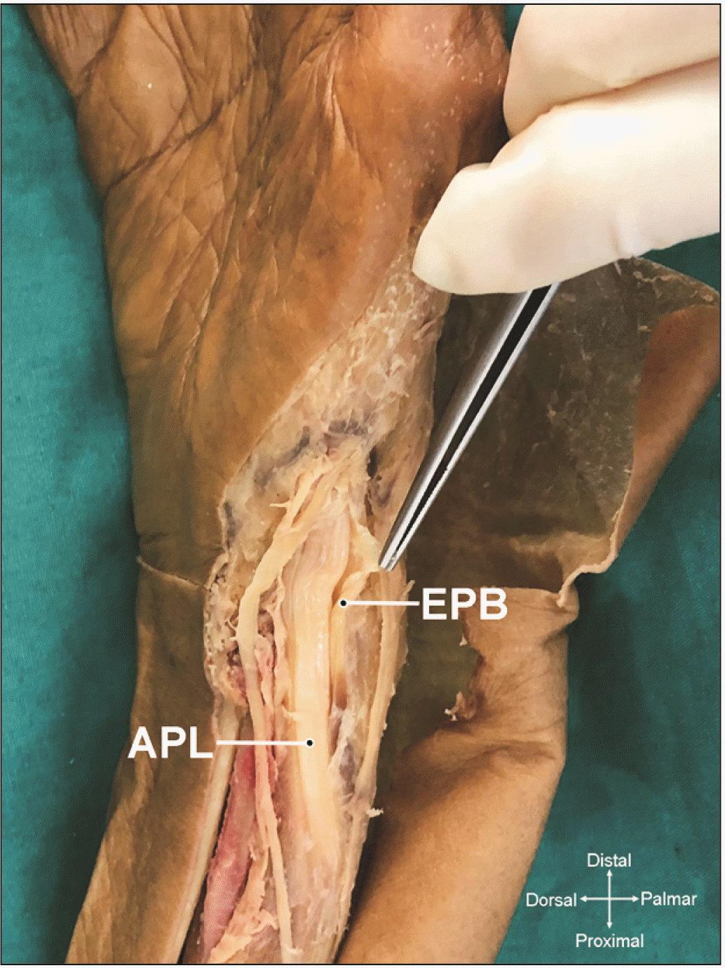
Fig. 2
Complete (proximal) type septum: Septum extending the proximal end of the first extensor compartment. APL, abductor pollicis longus; EPB, extensor pollicis brevis.
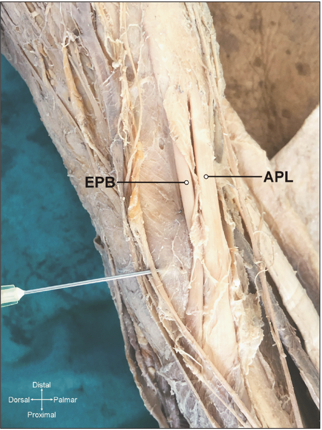
Fig. 3
First extensor compartment with no septum. APL, abductor pollicis longus; EPB, extensor pollicis brevis.
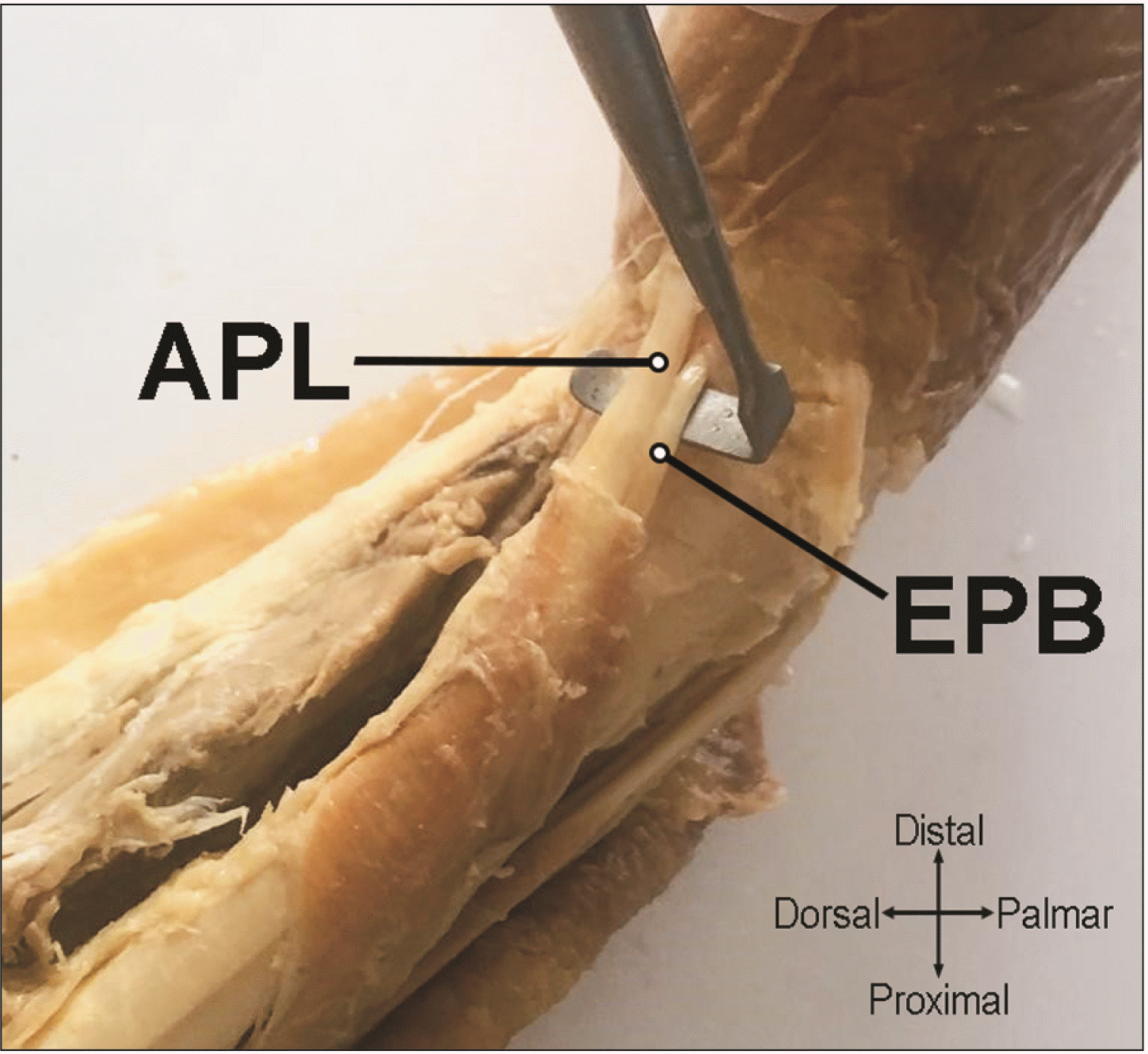
Table 1
Results classified by gender and septum type
| Septum type | No septum | Complete septum | Incomplete septum |
|---|---|---|---|
| Female wrists | 11 (55.0) | 4 (20.0) | 5 (25.0) |
| Male wrists | 21 (41.2) | 14 (27.5) | 16 (31.4) |
| Total | 33 (45.8) | 18 (25.0) | 21 (29.2) |
Table 2
Gender differences
| Gender | P-value | ||
|---|---|---|---|
| Woman (n=36) | Man (n=51) | ||
| Proximal total | 0.088a | ||
| Ort±Ss | 8.86±1.25 | 9.28±1.04 | |
| Median (range) | 8.75 (6.43–11.1) | 9.4 (6.99–11.2) | |
| Distal total | 0.009a,* | ||
| Ort±Ss | 8.18±0.91 | 8.82±1.19 | |
| Median (range) | 8.2 (6.03–9.63) | 8.78 (6.4–12.4) | |
| Septum | 0.608b | ||
| None | 13 (36.1) | 21 (41.2) | |
| Distal type | 15 (41.7) | 16 (31.4) | |
| Proximal type | 8 (22.2) | 14 (27.5) | |
| APL-T | 0.541c | ||
| 1 tendon | 8 (22.2) | 15 (29.4) | |
| 2 tendon | 20 (55.6) | 29 (56.9) | |
| 3 tendon | 5 (13.9) | 6 (11.8) | |
| 4 tendon | 3 (8.3) | 1 (2.0) | |


 PDF
PDF Citation
 Print
Print


 XML Download