Journal List > Ann Lab Med > v.44(1) > 1516083892

Han, Min, Jo, Lee, Kim, Han, Kang, Ahn, Oh, Song, and Ha: Association Between Low Anti-spike Antibody Levels After the Third Dose of SARS-CoV-2 Vaccination and Hospitalization due to Symptomatic Breakthrough Infection in Kidney Transplant Recipients

Abstract

Background

Whether anti-severe acute respiratory syndrome coronavirus 2 (SARS-CoV-2) antibody levels post-third coronavirus disease (COVID-19) vaccination correlate with worse outcomes due to breakthrough infection is unclear. We evaluated the association between anti-SARS-CoV-2 antibody levels and symptomatic breakthrough infection or hospitalization during the Omicron surge in kidney transplant recipients.

Methods

In total, 287 kidney transplant recipients expected to receive a third vaccination were enrolled between November 2021 and February 2022. The Abbott SARS-CoV-2 IgG II Quant test (Abbott, Chicago, IL, USA) was performed within three weeks before and four weeks after the third vaccination. The incidence of symptomatic breakthrough infection and hospitalization from two weeks to four months post-third vaccination was recorded.

Results

After the third vaccination, the seropositive rate and median antibody titer of the 287 patients increased from 57.1% to 82.2% and from 71.7 (interquartile range [IQR] 7.2–402.8) to 1,612.1 (IQR 153.9–5,489.1) AU/mL, respectively. Sixty-four (22.3%) patients had symptomatic breakthrough infections, of whom 12 required hospitalization. Lower anti-receptor-binding domain (RBD) IgG levels (<400 AU/mL) post-third vaccination were a risk factor for symptomatic breakthrough infection (hazard ratio [HR]=3.46, P<0.001). Anti-RBD IgG levels <200 AU/mL were a critical risk factor for hospitalization (HR=36.4, P=0.007).

Conclusions

Low anti-spike IgG levels after third vaccination in kidney transplant recipients were associated with symptomatic breakthrough infection and, particularly, with hospitalization during the Omicron surge. These data can be used to identify patients requiring additional protective measures, such as passive immunization using monoclonal antibodies.

INTRODUCTION

Severe acute respiratory syndrome coronavirus 2 (SARS-CoV-2) Omicron has rapidly become the dominant variant since its emergence in November 2021. Two doses of the primary vaccine series have shown waning effectiveness in patients, and extra doses of vaccinations were recommended worldwide [1]. Additional doses of vaccinations are prioritized in immunocompromised patients, including solid organ transplant patients, considering their increased risk of morbidity and mortality reported during the early years of the pandemic [2]. Hence, for the transplant population, a third vaccination dose is highly recommended [3].
While enhanced immunogenicity in the transplant population after the third vaccine dose has been reported [4-11], whether the elicited immunity can protect against Omicron infection remains unclear. Considering that Omicron, with multiple variants, can partially escape immunity acquired from coronavirus disease (COVID-19) vaccines targeted towards the wild-type strain [12-14], immunogenicity elicited through vaccination may not correlate with the actual clinical infection outcome. Recent studies on the association between anti-SARS-CoV-2 antibody levels and breakthrough infections have shown variable results and variable cutoff values of antibodies for predicting breakthrough infection [6, 15-19]. This is partially because many studies were conducted retrospectively, and in prospective studies, the number of subjects was not sufficient. We prospectively evaluated the association between anti-spike antibody levels after completing a three-dose COVID-19 vaccine series and the incidence and severity of subsequent breakthrough infection in 287 kidney transplant recipients (KTRs) during the Omicron surge.

MATERIALS AND METHODS

This was a noninterventional prospective study that examined the efficacy of a third dose of COVID-19 vaccination in KTRs. The study has been registered at clinicaltrials.gov (NCT05156086). All participants provided written consent, and the study was approved by the Institutional Review Board of the Seoul National University Hospital (IRB number: H-2110-175-1266).

Study population

Between November 15, 2021 and February 10, 2022, we enrolled 287 patients who had received a kidney transplant at least six months before the first dose of vaccination and two doses of a COVID-19 vaccine (61 patients with two doses of a viral vector vaccine [ChAdOx1 of Astra Zeneca], 143 with two doses of an mRNA vaccine [126 with BNT162b2 of Pfizer-BioNTech and 17 with mRNA-1273 of Moderna], and 83 with a heterologous combination of ChAdOx1/BNT162b2) and who planned to receive a third mRNA monovalent vaccination (Supplemental Data Fig. S1). The exclusion criteria were as follows: patients with a prior history of SARS-CoV-2 infection, those who had received B cell-depleting therapy (e.g., rituximab and bortezomib) or T cell-depleting therapy (anti-thymocyte globulin) within the last six months, and those who restarted dialysis due to graft failure. All patients were followed up every one to two months after the third dose of vaccination.

Serologic measurements of anti-SARS-CoV-2 antibody

Vaccine-induced SARS-CoV-2-specific antibodies were quantified using an Abbott SARS-CoV-2 IgG II Quant test (Abbott, Chicago, IL, USA), which is a two-step chemiluminescent microparticle immunoassay that detects IgG antibodies against the receptor-binding domain (RBD) of the S1 subunit of the spike protein of SARS-CoV-2. Measurements were between 6.8 and 40,000 arbitrary units (AU)/mL, and the seropositivity cutoff, as per the manufacturer’s instruction, was ≥50 AU/mL. Serum samples were collected from all patients within three weeks before the planned date of the third vaccine dose and at four weeks (± one week) post-third vaccination. Additionally, 126 patients who were seropositive post-third vaccination and consented to additional tests underwent additional antibody tests three months (± one week) post-third vaccination.

Assessment of symptomatic breakthrough infections

Symptomatic SARS-CoV-2 breakthrough infection cases were identified through self-reporting or relevant symptoms with proof of a positive real-time reverse transcription–PCR test or rapid antigen test [20], performed by medical personnel between two weeks and four months post-third vaccination. Asymptomatic infections could not be detected because periodic SARS-CoV-2 confirmation tests were not performed in all subjects. Data on the types and duration of symptoms, types of treatments received, and treatment outcomes were collected through a questionnaire and electronic medical records.

Statistical analysis

Descriptive statistics are expressed as means (SDs) or medians (interquartile range, IQR), as appropriate. Between-group differences in dichotomous variables were assessed using the chi-square test or Fisher’s exact test, and those in continuous variables were assessed using an independent sample t-test or the Mann–Whitney U test. Anti-RBD titers before the third dose of vaccination were compared with those assessed four weeks after the third dose using Wilcoxon’s signed-rank tests. Stepwise multivariable logistic regression was used to identify factors potentially associated with breakthrough infection and hospitalization during infection. Age, sex, and factors associated with P< 0.2 in univariable logistic tests were included in the multivariable model. Cutoff values of anti-RBD IgG, used to group patients according to infection risk, were determined through ROC tests with the Youden index. The difference in the cumulative incidence of breakthrough infection was demonstrated using a Kaplan–Meier curve and analyzed using the log-rank test. A two-tailed P<0.05 was considered significant. Statistical analyses were performed using SPSS (version 21.0; IBM Corp., Armonk, NY, USA) and R 4.1.1 (http://www.r-project.org).

RESULTS

The enrolled patients had a median age of 49 yrs (IQR 38.5–60.0), and 51.9% were male. The median time after kidney transplantation was 5.6 yrs (IQR 1.9–10.7) (Table 1). The median intervals from the second dose of vaccination to the third dose were 111 (IQR 92–128) days for ChAdOX1-S/ChAdOX1-S primary vaccination, 94 (IQR 74–118) days for mRNA/mRNA vaccination, and 147 (IQR 133–159) days for ChAdOX1-S/BNT162b2 vaccination. Approximately three quarters of the patients were being administered a triple regimen comprising tacrolimus, antimetabolite, and steroids.

Anti-RBD IgG levels after third vaccination

All patients received an mRNA vaccine as the third dose of vaccination (BNT162b2 [N=247] or mRNA-1273 [N=40]). The seropositive rate of anti-RBD IgG increased from 57.1% (164/287) to 82.2% (236/287) after the third dose, with seroconversion in 58.5% (72/123) of seronegative patients after two doses of vaccination. The antibody levels also significantly increased from a median of 71.7 (IQR 7.2–402.8) AU/mL to 1,612.1 (IQR 153.9–5,489.1) AU/mL (P<0.001). The anti-RBD IgG levels after the third vaccination were 546.8 (IQR 25.4–1,832.5) AU/mL for ChAdOX1-S/ChAdOX1-S primary vaccination, 2,730 (229.5–7,847.1) AU/mL for mRNA/mRNA vaccination, and 1,689 (227.8–3,843.9) AU/mL for ChAdOX1-S/BNT162b2 vaccination (Fig. 1).
Through multivariable logistic regression, pre-third dose anti-RBD IgG seronegativity was identified as a risk factor for seronegative anti-RBD IgG after the third vaccination dose (hazard ratio [HR]=158.56) (Supplemental Data Table S1), even when including the type of primary vaccination instead of pre-third dose anti-RBD IgG seronegativity (because these two variables showed collinearity; data not shown). The use of mycophenolic acid or mycophenolate mofetil (HR=5.98 for a dose ≤500 mg/day and 19.01 for a dose <500 mg/day), the tacrolimus trough level (HR= 1.16), and primary vaccination with ChAdOX1-S/ChAdOx1-2 (HR=3.34) were significant risk factors for anti-RBD IgG seronegativity after the third vaccination dose (Supplemental Data Table S1). At three months after the third vaccination, the median anti-spike IgG titers of 124 patients (seropositive at four weeks after the third dose) were significantly decreased from 2,245.0 (IQR 657.8–6,292.3) AU/mL to 914.2 (IQR 148.9–3,751.2) AU/mL (P<0.001). Among them, 11/126 (8.9%) had become seronegative (<50 AU/mL) and 35/126 (23.2%) had anti-spike levels <200 AU/mL.

Symptomatic breakthrough infections and anti-spike IgG titers

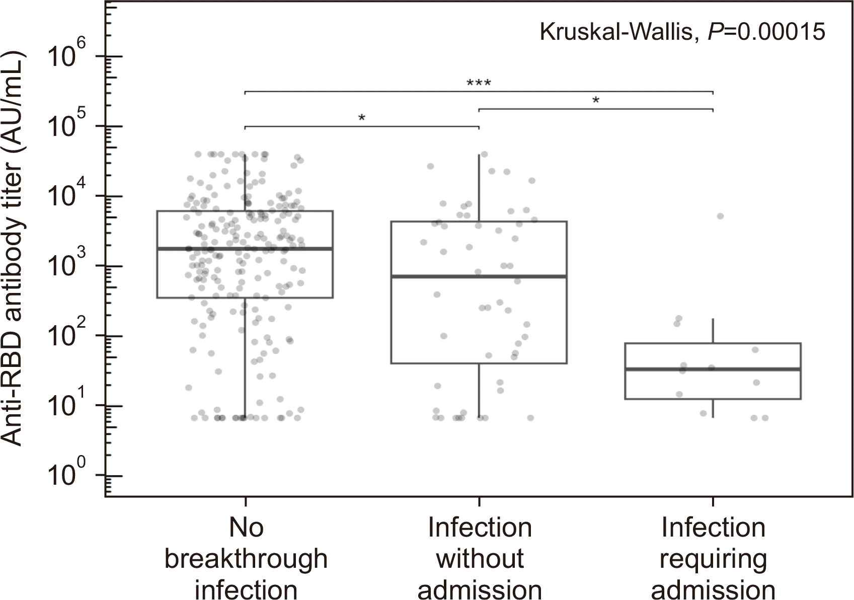
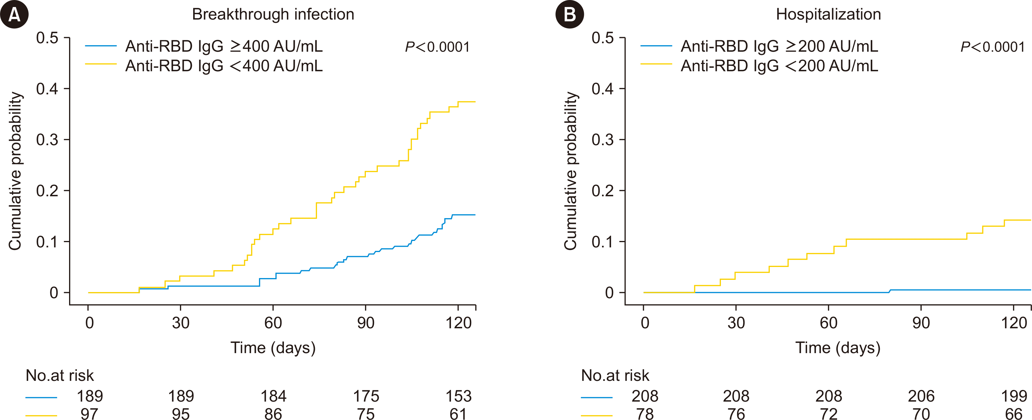
Symptomatic SARS-CoV-2 breakthrough infection occurred in 65 patients within four months after the third vaccination dose. Among them, excluding one patient who was infected within two weeks post-third vaccination, 64 patients had symptomatic breakthrough infection. The median time to breakthrough infection was 83.5 (IQR 58.0–105.5) days. Post-third vaccination anti-RBD IgG levels significantly differed among the patients with and without symptomatic breakthrough infections or hospitalization (median±quartile, 1,779.6±5,936.7 for the no infection group, 722.2±4,509.4 for the symptomatic breakthrough infection without hospitalization group, and 33.5±119.5 for the symptomatic breakthrough infection with hospitalization group; P=0.00015) (Fig. 2). When the patients were regrouped according to an anti-spike IgG level of 400 AU/mL (determined based on an ROC curve; data not shown), the risk of developing symptomatic breakthrough infection differed significantly between the two groups (log-rank test, P<0.0001; Fig. 3A). In multivariable logistic regression tests, a post-third vaccination anti-spike IgG level <400 AU/mL was the only risk factor for symptomatic breakthrough infection (HR=3.46, P<0.001; Table 2). When the patients were grouped into four groups according to quartile values of anti-spike IgG after the third vaccination (Q1, 6.8–152.3; Q2, 152.3–1,624.5; Q3, 1,624.5–5,495.6; Q4, >5,495.6 AU/mL), the risk of symptomatic breakthrough infection did not differ among the Q2, Q3, and Q4 groups (P<0.0001) (Supplemental Data Fig. S2A).

Association between anti-spike IgG titer and hospitalization

Twelve patients (18.8%) with symptomatic breakthrough infections required hospital admission. Reasons for in-hospital care included oxygen therapy for pneumonia (N=3), severe gastrointestinal symptoms (e.g., diarrhea and vomiting; N=6), fever and myalgia for more than four days (N=2), and acute thrombotic occlusion of previous arteriovenous access (N=1). Among the nine patients who were admitted for non-pulmonary symptoms, seven showed poor oral intake and evidence of dehydration, of whom five showed acute kidney injury on admission. One patient with pneumonia required intensive care unit care, but the symptoms alleviated without mechanical ventilatory support.
The 52 patients who did not require admission had mild symptoms, which mostly alleviated with oral medication. Among the patients with symptomatic breakthrough infections, those who required hospital admission had lower post-third anti-RBD IgG levels than those who did not require admission (33.5± 119.5 vs. 722.2±4,509.4 AU/mL, P=0.016; Fig. 2). When the patients were regrouped according to an anti-RBD IgG level of 200 AU/mL (determined based on an ROC curve; data not shown), the risk of hospitalization was higher in patients with anti-RBD IgG <200 AU/mL than in those with anti-RBD IgG ≥200 AU/mL (log-rank test, P<0.0001; Fig. 3B). In multivariable models, a low post-V3 antibody level (<200 AU/mL) was a significant risk factor for hospitalization (HR=36.4, P=0.007; Table 3). When the patients were grouped into Q1, Q2, Q3, and Q4 groups, the risk of symptomatic breakthrough infection requiring hospitalization did not differ among the Q2, Q3, and Q4 groups (P<0.0001; Supplemental Data Fig. S2B).

DISCUSSION

The third COVID-19 vaccination dose significantly increased the seropositive rate and anti-spike antibody levels, in line with the results of previous studies [4-11]. However, 17.8% (51/287) of patients were seronegative despite being administered the third vaccination dose. As has been suggested by Manothummetha, et al. [21], mycophenolic acid use, tacrolimus levels, and the type of priming vaccination were risk factors for seronegativity post-third vaccination. However, these factors were unrelated to the incidence of breakthrough infection in both the univariable and multivariable models. Clinical risk factors for seronegativity were not risk factors for symptomatic breakthrough infection or hospitalization. Therefore, to reduce the infection risk, patients should be more accurately stratified using serological tests rather than known clinical factors of a poor vaccine response.
The anti-spike antibody level after the third dose of COVID-19 vaccination was associated with the rate of symptomatic breakthrough infections and hospitalization. Studies on the correlation between anti-spike antibody levels after the third vaccination dose and outcomes of SARS-CoV-2 breakthrough infections in KTRs are still limited and have shown variable results [6, 16-19, 22]. In contrast to the results of the present study, Lammert, et al. [6] and Kemlin, et al. [17] did not observe a significant association between post-third vaccination anti-SARS-CoV-2 antibody levels and breakthrough infections. However, the number of patients analyzed in these studies was relatively small (103 and 65, respectively), limiting the ability to reach statistical significance, and the use of different modeling methods can also have an influence. We considered antibody levels as a dichotomous variable using cutoff values from ROC curves because of the assumed nonlinear relationship between antibody levels and infection or hospitalization (Fig. 2 and Supplemental Data Fig. S2). Patients with antibody levels in the second (Q2), third (Q3), and fourth (Q4) quartiles did not show differences in the risk of symptomatic breakthrough infection or hospitalization. Moreover, when we analyzed antibody levels as a continuous variable, the results did not reach statistical significance.
In a US multicenter observational cohort study of 666 solid organ transplant recipients (including 351 KTRs) who had three or more vaccinations, lower anti-RBD antibody levels of 0.8–250 IU/mL (based on the Roche kit, similar to the WHO standard unit) were associated with an increased risk of breakthrough infection during the Omicron surge [16]. In France, Bertrand, et al. [18] reported that lower anti-spike antibody levels (<264 binding antibody units/mL, WHO standard) were associated with an increased risk of breakthrough infection, hospitalization, and death. A prospective cohort study in Italy, including 614 solid organ transplantations (including 275 KTRs), reported that antibody levels <817 IU/mL were associated with breakthrough infection [19]. These three studies presented results similar to ours, i.e., that anti-RBD antibody levels <400 IU/mL (based on the Abbott anti-SARS-CoV-2 IgG II Quant) were associated with an increased risk of symptomatic breakthrough infection. The antibody levels associated with an increased risk of breakthrough infection varied between the studies, which is thought to be due to differences in the test kits used (antibody units are still not standardized), patient immunosuppression status based on different drug regimens among hospitals, exposure to and amount of virus from the person of care, facilities, and the prevalence of Omicron infection in the community.
An anti-RBD antibody level <200 IU/mL (based on Abbott anti-SARS-CoV-2 IgG II Quant) was associated with an increased risk of hospitalization. In a Spanish study that included 965 KTRs [22], patients with an anti-RBD antibody level <100 AU/mL (based on Abbott anti-SARS-CoV-2 IgG II Quant) were at increased risk of breakthrough infection and pneumonia, and patients with an antibody level <20 AU/mL had an increased risk of death during the Delta or Omicron surge after two or three vaccinations. The severity of Omicron infection is markedly attenuated compared with that of the previous variant, and deaths from COVID-19 are decreasing. However, immunosuppressed patients, such as KTRs, have a higher chance of acquiring severe disease than healthy people.
Identifying patients with severe outcomes, such as hospitalization and death, and taking additional preventive measures will be essential for the management of COVID-19 in the transplant population. The modulation of immunosuppressive regimens during vaccination [23], vaccinations using novel vaccines of different platforms [24] or vaccines targeting multiple antigens, and pre- or post-exposure passive immunization using monoclonal antibodies or convalescent plasma [25, 26] have been suggested as additional measures to enhance immunogenicity in KTRs. Multiple studies [27], including our own, have demonstrated a positive association between mycophenolic acid use or tacrolimus levels and the vaccine response in KTRs. While reducing immunosuppression, e.g., with mycophenolic acid or tacrolimus, appears to be a potential strategy to enhance the vaccine response [28], its efficacy and safety must be critically evaluated. Specifically, recent investigations have shown that mycophenolic acid withdrawal does not significantly improve the vaccine response or only yields a short-lived response with an increase in alloimmunity, as indicated by elevated levels of donor-specific antibodies [29]. Therefore, although reducing immunosuppression may be a promising approach to enhance the vaccine response, its potential benefits and risks should be carefully assessed in future studies. The optimal method for preventing symptomatic infection in KTRs with low anti-SARS-CoV-2 antibody levels is yet to be determined. Although additional booster shots are effective in inducing seroconversion [28], the current evidence does not strongly support bivalent vaccines over monovalent vaccines for protection against the Omicron variant in the transplant population. Pre-exposure prophylaxis with monoclonal antibodies, such as tixagevimab and cilgavimab, has demonstrated neutralizing activity against Omicron, but recent evidence suggests that a higher dose is required to prevent breakthrough infection, and neutralizing antibody activity commonly wanes by three months post-injection [30, 31]. Given the complex nature of immunosuppression in KTRs and the evolving nature of the COVID-19 pandemic, continued studies are crucial for developing effective prevention and treatment strategies.
There are some limitations when stratifying KTRs using widely used automated binding anti-RBD antibody tests, such as the Abbott anti-SARS-CoV-2 IgG II Quant. First, as tests have been usually developed against the ancestral strain, they may not accurately reflect the neutralizing capacity against the Omicron variant. However, studies have demonstrated that such tests exhibit a good correlation with the results of live virus neutralization tests against both the ancestral strain [32] and the Omicron variant [33, 34]. It should be noted that T cell responses also play a role in preventing SARS-CoV-2 infection, and weak correlations between T and B cell responses have been reported, particularly in cases of weak immune responses [35-37]. Nevertheless, automated binding antibody reagents are useful in identifying immunocompromised patients who require additional measures at a low cost. In some countries, antibody testing in transplant patients is used as a criterion for recommending monoclonal antibody therapy in cases of low antibody titers [18].
There are some precautions in applying the results of this study to the current COVID-19 situation or healthy people. First, most of the population worldwide has had a prior history of SARS-CoV-2 infection and probably has sufficiently high antibody levels. The association between anti-RBD IgG levels and symptomatic breakthrough infection was weak in patients with higher anti-RBD IgG levels. Second, immune responses to new SARS-CoV-2 variants that continue to emerge will differ from those to Omicron. Third, social and behavioral factors, which are essential in explaining the risk of infection, should be considered. Breakthrough infections can even occur in healthy individuals with very high antibody titers to SARS-CoV-2 [38]. The extent of virus exposure likely plays a critical role in breakthrough infections. Nevertheless, this study is meaningful in that it prospectively confirmed the association between anti-RBD binding antibody levels and the rate of symptomatic breakthrough infections and particularly very low antibody levels with hospitalization, in KTRs who are vulnerable to severe complications from COVID-19. These results can be useful as a basis for identifying patients who could benefit from additional protective measures among KTRs.

SUPPLEMENTARY MATERIALS

Supplementary materials can be found via https://doi.org/10.3343/alm.2024.44.1.64

ACKNOWLEDGEMENTS

None.

Notes

AUTHOR CONTRIBUTIONS

Han A, Ha J, and Song EY designed the study. Min S, Han A, Jo E, Lee H, Kim YC, Han SS, Kang HG, Ahn YH, Oh I, Ha J, and Song EY participated in the investigation and data collection. Han A and Oh I curated the data. Han A performed the formal analysis and wrote the original draft. Min S, Song EY, and Ha J reviewed and edited the manuscript. All authors approved the final version for submission.

CONFLICTS OF INTEREST

None declared.

REFERENCES

1. Levin EG, Lustig Y, Cohen C, Fluss R, Indenbaum V, Amit S, et al. 2021; Waning immune humoral response to BNT162b2 Covid-19 vaccine over 6 months. N Engl J Med. 385:e84. DOI: 10.1056/NEJMoa2114583. PMID: 34614326. PMCID: PMC8522797.
crossref
2. Coates PT, Wong G, Drueke T, Rovin B, Ronco P. Associate Editors, for the Entire Editorial Team. 2020; Early experience with COVID-19 in kidney transplantation. Kidney Int. 97:1074–5. DOI: 10.1016/j.kint.2020.04.001. PMID: 32354635. PMCID: PMC7142690.
crossref
3. Centers for Disease Control and Prevention. COVID-19: stay up to date with COVID-19 vaccines including boosters 2022. https://www.cdc.gov/coronavirus/2019-ncov/vaccines/stay-up-to-date.html. Updated on Dec 2022.
4. Benotmane I, Gautier G, Perrin P, Olagne J, Cognard N, Fafi-Kremer S, et al. 2021; Antibody response after a third dose of the mRNA-1273 SARS-CoV-2 vaccine in kidney transplant recipients with minimal serologic response to 2 doses. JAMA. 326:1063–5. DOI: 10.1001/jama.2021.12339. PMID: 34297036. PMCID: PMC8456389.
crossref
5. Cassaniti I, Gregorini M, Bergami F, Arena F, Sammartino JC, Percivalle E, et al. 2022; Effect of a third dose of SARS-CoV-2 mRNA BNT162b2 vaccine on humoral and cellular responses and serum anti-HLA antibodies in kidney transplant recipients. Vaccines (Basel). 10:921. DOI: 10.3390/vaccines10060921. PMID: 35746528. PMCID: PMC9227063.
6. Lammert A, Schnuelle P, Rabenau HF, Ciesek S, Krämer BK, Göttmann U, et al. 2022; SARS-CoV-2 vaccination in kidney transplant recipients-stratified analysis of the humoral immune response. Transplant Direct. 8:e1384. DOI: 10.1097/TXD.0000000000001384. PMID: 36259077. PMCID: PMC9575732.
crossref
7. Massa F, Cremoni M, Gérard A, Grabsi H, Rogier L, Blois M, et al. 2021; Safety and cross-variant immunogenicity of a three-dose COVID-19 mRNA vaccine regimen in kidney transplant recipients. EBioMedicine. 73:103679. DOI: 10.1016/j.ebiom.2021.103679. PMID: 34763205. PMCID: PMC8573385.
crossref
8. Stumpf J, Tonnus W, Paliege A, Rettig R, Steglich A, Gembardt F, et al. 2021; Cellular and humoral immune responses after 3 doses of BNT162b2 mRNA SARS-CoV-2 vaccine in kidney transplant. Transplantation. 105:e267–9. DOI: 10.1097/TP.0000000000003903. PMID: 34342963. PMCID: PMC8549130.
crossref
9. Tylicki L, Dębska-Ślizień A, Muchlado M, Ślizień Z, Gołębiewska J, Dąbrowska M, et al. 2021; Boosting humoral immunity from mRNA COVID-19 vaccines in kidney transplant recipients. Vaccines (Basel). 10:56. DOI: 10.3390/vaccines10010056. PMID: 35062717. PMCID: PMC8779302.
crossref
10. Werbel WA, Boyarsky BJ, Ou MT, Massie AB, Tobian AAR, Garonzik-Wang JM, et al. 2021; Safety and immunogenicity of a third dose of SARS-CoV-2 vaccine in solid organ transplant recipients: a case series. Ann Intern Med. 174:1330–2. DOI: 10.7326/L21-0282. PMID: 34125572. PMCID: PMC8252023.
crossref
11. Kim J, Bang HI, Shin JW, Park Y, Kim S, Kim M, et al. 2022; Immunogenicity of Third-dose BNT162b2 mRNA Vaccine Following Two Doses of ChAdOx1 in Health Care Workers: A Prospective Longitudinal Study. Ann Lab Med. 42:688–92. DOI: 10.3343/alm.2022.42.6.688. PMID: 35765878. PMCID: PMC9277035.
crossref
12. Altarawneh HN, Chemaitelly H, Hasan MR, Ayoub HH, Qassim S, AlMukdad S, et al. 2022; Protection against the Omicron variant from previous SARS-CoV-2 infection. N Engl J Med. 386:1288–90. DOI: 10.1056/NEJMc2200133. PMID: 35139269. PMCID: PMC8849180.
crossref
13. Chaguza C, Coppi A, Earnest R, Ferguson D, Kerantzas N, Warner F, et al. 2022; Rapid emergence of SARS-CoV-2 Omicron variant is associated with an infection advantage over Delta in vaccinated persons. Med. 3:325–34.e4. DOI: 10.1016/j.medj.2022.03.010. PMID: 35399324. PMCID: PMC8983481.
crossref
14. Tseng HF, Ackerson BK, Luo Y, Sy LS, Talarico CA, Tian Y, et al. 2022; Effectiveness of mRNA-1273 against SARS-CoV-2 Omicron and Delta variants. Nat Med. 28:1063–71. DOI: 10.1038/s41591-022-01753-y. PMID: 35189624. PMCID: PMC9117141.
crossref
15. Werbel WA, Segev DL. 2022; SARS-CoV-2 antibody testing for transplant recipients: a tool to personalize protection versus COVID-19. Am J Transplant. 22:1316–20. DOI: 10.1111/ajt.16993. PMID: 35119179. PMCID: PMC9111420.
16. Alejo JL, Chiang TPY, Bowles Zeiser L, Kim JD, Mitchell J, Avery RK, et al. 2022; Incidence and severity of COVID-19 among vaccinated solid organ transplant recipients during the Omicron wave. Transplantation. 106:e413–5. DOI: 10.1097/TP.0000000000004226. PMID: 35655363.
17. Kemlin D, Gemander N, Depickère S, Olislagers V, Georges D, Waegemans A, et al. 2023; Humoral and cellular immune correlates of protection against COVID-19 in kidney transplant recipients. Am J Transplant. 23:649–58. DOI: 10.1016/j.ajt.2023.02.015. PMID: 36773936. PMCID: PMC9911984.
crossref
18. Bertrand D, Laurent C, Lemée V, Lebourg L, Hanoy M, Le Roy F, et al. 2022; Efficacy of anti-SARS-CoV-2 monoclonal antibody prophylaxis and vaccination on the Omicron variant of COVID-19 in kidney transplant recipients. Kidney Int. 102:440–2. DOI: 10.1016/j.kint.2022.05.007. PMID: 35618097. PMCID: PMC9125992.
crossref
19. Bonazzetti C, Tazza B, Gibertoni D, Pasquini Z, Caroccia N, Fanì F, et al. Relationship between immune response to severe acute respiratory syndrome coronavirus 2 vaccines and development of breakthrough infection in solid organ transplant recipients: the CONTRAST cohort. Clin Infect Dis. 2023; 76:1761–7. DOI: 10.1093/cid/ciad016. PMID: 36636955. PMCID: PMC10209432.
crossref
20. Hong KH, Kim GJ, Roh KH, Sung H, Lee J, Kim SY, et al. 2022; Update of guidelines for laboratory diagnosis of COVID-19 in Korea. Ann Lab Med. 42:391–7. DOI: 10.3343/alm.2022.42.4.391. PMID: 35177559. PMCID: PMC8859556.
crossref
21. Manothummetha K, Chuleerarux N, Sanguankeo A, Kates OS, Hirankarn N, Thongkam A, et al. 2022; Immunogenicity and risk factors associated with poor humoral immune response of SARS-CoV-2 vaccines in recipients of solid organ transplant: a systematic review and meta-analysis. JAMA Netw Open. 5:e226822. DOI: 10.1001/jamanetworkopen.2022.6822. PMID: 35412626. PMCID: PMC9006106.
22. Rodríguez-Cubillo B, Moreno de la Higuera MA, Pérez-Flores I, Calvo Romero N, Aiffil AS, Arribi Vilela A, et al. 2022; Clinical effectiveness of SARS-CoV-2 vaccination in renal transplant recipients. Antibody levels impact in pneumonia and death. Transplantation. 106:e476–87. DOI: 10.1097/TP.0000000000004261. PMID: 35859270.
crossref
23. Yahav D, Rozen-Zvi B, Mashraki T, Atamna A, Ben-Zvi H, Bar-Haim E, et al. 2021; Immunosuppression reduction when administering a booster dose of the BNT162b2 mRNA SARS-CoV-2 vaccine in kidney transplant recipients without adequate humoral response following two vaccine doses: protocol for a randomised controlled trial (BECAME study). BMJ Open. 11:e055611. DOI: 10.1136/bmjopen-2021-055611. PMID: 34635537. PMCID: PMC8506046.
crossref
24. Kumari M, Lu RM, Li MC, Huang JL, Hsu FF, Ko SH, et al. 2022; A critical overview of current progress for COVID-19: development of vaccines, antiviral drugs, and therapeutic antibodies. J Biomed Sci. 29:68. DOI: 10.1186/s12929-022-00852-9. PMID: 36096815. PMCID: PMC9465653.
25. Al Jurdi A, Morena L, Cote M, Bethea E, Azzi J, Riella LV. 2022; Tixagevimab/cilgavimab pre-exposure prophylaxis is associated with lower breakthrough infection risk in vaccinated solid organ transplant recipients during the Omicron wave. Am J Transplant. 22:3130–6. DOI: 10.1111/ajt.17128. PMID: 35727916. PMCID: PMC9906353.
crossref
26. Avery RK. 2022; Update on COVID-19 therapeutics for solid organ transplant recipients, including the Omicron surge. Transplantation. 106:1528–37. DOI: 10.1097/TP.0000000000004200. PMID: 35700481. PMCID: PMC9311293.
crossref
27. Li J, Ayada I, Wang Y, den Hoed CM, Kamar N, Peppelenbosch MP, et al. 2022; Factors associated with COVID-19 vaccine response in transplant recipients: a systematic review and meta-analysis. Transplantation. 106:2068–75. DOI: 10.1097/TP.0000000000004256. PMID: 35761439. PMCID: PMC9521391.
crossref
28. Kho MML, Messchendorp AL, Frölke SC, Imhof C, Koomen VJ, Malahe SRK, et al. 2023; Alternative strategies to increase the immunogenicity of COVID-19 vaccines in kidney transplant recipients not responding to two or three doses of an mRNA vaccine (RECOVAC): a randomised clinical trial. Lancet Infect Dis. 23:307–19. DOI: 10.1016/S1473-3099(22)00650-8. PMID: 36354032.
crossref
29. Kühn T, Speer C, Morath C, Bartenschlager M, Kim H, Beimler J, et al. 2023; Immune response to COVID-19 mRNA vaccination in previous nonresponder kidney transplant recipients after short-term withdrawal of mycophenolic acid 1 and 3 months after an additional vaccine dose. Transplantation. 107:1139–50. DOI: 10.1097/TP.0000000000004516. PMID: 36617671. PMCID: PMC10125015.
crossref
30. Benotmane I, Velay A, Gautier-Vargas G, Olagne J, Obrecht A, Cognard N, et al. 2022; Breakthrough COVID-19 cases despite prophylaxis with 150 mg of tixagevimab and 150 mg of cilgavimab in kidney transplant recipients. Am J Transplant. 22:2675–81. DOI: 10.1111/ajt.17121. PMID: 35713984. PMCID: PMC9350296.
crossref
31. Karaba AH, Kim JD, Chiang TP, Alejo JL, Sitaras I, Abedon AT, et al. 2023; Neutralizing activity and 3-month durability of tixagevimab and cilgavimab prophylaxis against Omicron sublineages in transplant recipients. Am J Transplant. 23:423–8. DOI: 10.1016/j.ajt.2022.11.002. PMID: 36906295. PMCID: PMC9835002.
crossref
32. Gilbert PB, Montefiori DC, McDermott AB, Fong Y, Benkeser D, Deng W, et al. 2022; Immune correlates analysis of the mRNA-1273 COVID-19 vaccine efficacy clinical trial. Science. 375:43–50. DOI: 10.1126/science.abm3425. PMID: 34812653. PMCID: PMC9017870.
crossref
33. Simon G, Favresse J, Gillot C, Closset M, Catry É, Dogné JM, et al. Kinetics and ability of binding antibody and surrogate virus neutralization tests to predict neutralizing antibodies against the SARS-CoV-2 Omicron variant following BNT162b2 booster administration. Clin Chem Lab Med. 2023; DOI: 10.1515/cclm-2022-1258. PMID: 37078220.
crossref
34. Suntronwong N, Assawakosri S, Kanokudom S, Yorsaeng R, Auphimai C, Thongmee T, et al. 2022; Strong correlations between the binding antibodies against wild-type and neutralizing antibodies against Omicron BA.1 and BA.2 variants of SARS-CoV-2 in individuals following booster (third-dose) vaccination. Diagnostics (Basel). 12:1781. DOI: 10.3390/diagnostics12081781. PMID: 35892491. PMCID: PMC9394243.
crossref
35. Le Bert N, Chia WN, Wan WY, Teo AKJ, Chong SZ-R, Tan N, et al. 2021; Widely heterogeneous humoral and cellular immunity after mild SARS-CoV-2 infection in a homogeneous population of healthy young men. Emerg Microbes Infect. 10:2141–50. DOI: 10.1080/22221751.2021.1999777. PMID: 34709140. PMCID: PMC8604544.
crossref
36. Malipiero G, Moratto A, Infantino M, D'Agaro P, Piscianz E, Manfredi M, et al. 2021; Assessment of humoral and cellular immunity induced by the BNT162b2 SARS-CoV-2 vaccine in healthcare workers, elderly people, and immunosuppressed patients with autoimmune disease. Immunol Res. 69:576–83. DOI: 10.1007/s12026-021-09226-z. PMID: 34417958. PMCID: PMC8379062.
crossref
37. Seo YJ, Oh I, Nam M, Shin S, Roh EY, Song EY. 2023; Comparison of Four T-cell Assays and Two Binding Antibody Assays in SARS-CoV-2 Vaccinees With or Without Omicron Breakthrough Infection. Ann Lab Med. 43:596–604. DOI: 10.3343/alm.2023.43.6.596. PMID: 37387492. PMCID: PMC10345181.
crossref
38. Kuhlmann C, Mayer CK, Claassen M, Maponga T, Burgers WA, Keeton R, et al. 2022; Breakthrough infections with SARS-CoV-2 omicron despite mRNA vaccine booster dose. Lancet. 399:625–6. DOI: 10.1016/S0140-6736(22)00090-3. PMID: 35063123.
crossref

Fig. 1
Anti-RBD IgG levels according to the primary vaccine type before and after four weeks and three months following the third vaccination.
Abbreviation: RBD, receptor-binding domain.
alm-44-1-64-f1.tif
Fig. 2
Post-third vaccination anti-spike IgG antibody levels according to the rate of symptomatic breakthrough infections or hospitalization. The P-values for all pairwise comparisons by pairwise Wilcoxon tests are as follows: no infection vs. infection without hospitalization, P=0.040; infection without hospitalization vs. infection with hospitalization, P=0.016; no infection vs. infection with hospitalization, P=0.0001. *P<0.05, **P<0.005, ***P<0.001.
Abbreviation: RBD, receptor-binding domain.
alm-44-1-64-f2.tif
Fig. 3
Kaplan–Meier survival curves of risk of symptomatic breakthrough infection or hospitalization according to post-third vaccination anti-spike IgG levels. (A) When the patients were regrouped according to an anti-spike IgG level of 400 AU/mL (determined based on a ROC curve; data not shown), the risk of developing symptomatic breakthrough infection differed significantly between the groups (log-rank test, P< 0.0001). (B) When the patients were regrouped according to an anti-RBD IgG level of 200 AU/mL (determined based on a ROC curve; data not shown), the risk of hospitalization was higher in patients with anti-RBD IgG <200 AU/mL than in patients with anti-RBD IgG ≥200 AU/mL (log-rank test, P<0.0001).
Abbreviation: RBD, receptor-binding domain.
alm-44-1-64-f3.tif
Table 1
Patient demographics and baseline characteristics
Baseline characteristics Study population (N=287)
Age, median (IQR), yrs 49.0 (38.5–60.0)
Male sex, N (%) 149 (51.9)
Body mass index, median (IQR), kg/m2 23.0 (20.6–25.5)
Time since transplantation, median (IQR), yrs 5.6 (1.9–10.7)
Multiple kidney transplantations, N (%) 10 (3.5)
Original kidney disease, N (%)
Glomerulonephritis 90 (31.4)
Diabetes 38 (13.2)
ADPKD 21 (7.3)
Hypertension 9 (3.1)
Others 45 (15.7)
Unknown 84 (29.3)
Donor type, N (%)
Living donor 186 (64.8)
Deceased donor 101 (35.2)
Comorbidities, N (%)
Hypertension 166 (57.8)
Diabetes 77 (26.8)
Chronic liver disease 9 (3.1)
Chronic pulmonary disease 1 (0.3)
Immunosuppressive regimen, N (%)
CNI+antimetabolite+steroid 216 (75.3)
CNI+steroid 34 (11.8)
CNI+antimetabolite 15 (5.2)
CNI+mTORi+antimetabolite+steroid 6 (2.1)
mTORi+antimetabolite+steroid 3 (1.0)
Other regimens 13 (4.4)
Laboratory values, median (IQR) or mean±SD
WBCs, ×109/L 7.0 (5.8–8.4)
Hb, g/dL 13.4±1.8
ANC, /µL 3,994 (3,154–5,216)
MDRD eGFR, mL/min/1.73 m2 59.7±18.2
Tacrolimus trough level, ng/mL* 5.7 (4.6–6.7)
Type of previous COVID-19 vaccination (first/second), N (%)
ChAdOX1-S/ChAdOX1-S 61 (21.2)
BNT162b2/BNT162b2 126 (43.9)
mRNA-1273/mRNA-1273 17 (5.9)
ChAdOX1-S/BNT162b2 83 (28.9)

*Tacrolimus trough levels were available for 274 patients on tacrolimus.

Abbreviations: ADPKD, autosomal dominant polycystic kidney disease; ANC, absolute neutrophil count; CNI, calcineurin inhibitor; COVID-19, coronavirus disease; eGFR, estimated glomerular filtration rate; IQR, interquartile range; MDRD, modification of diet in renal disease; mTORi, mammalian target of rapamycin inhibitor; WBCs, white blood cells; SARS-CoV-2, severe acute respiratory syndrome coronavirus 2.

Table 2
Factors associated with symptomatic breakthrough infection after a third vaccination dose
Variables Breakthrough infection (N=64) No breakthrough infection (N=222) Univariable Multivariable*
HR 95% CI P HR 95% CI P
Age, median (IQR), yrs 50.0 (40.5–59.5) 49.0 (38.0–60.0) 1.00 0.99–1.02 0.644
Male sex, N (%) 34 (53.1) 114 (51.4) 1.07 0.62–1.88 0.802
Time since transplantation, median (IQR), yrs 4.0 (1.6–9.6) 6.1 (2.2–11.3) 0.96 0.91–1.00 0.075
Dialysis duration, median (IQR), month 8.3 (0–48.3) 14.6 (1.9–64.8) 0.99 0.99–1.00 0.089 0.99 0.99–1.00 0.066
Donor type–living donor, N (%) 48 (75.0) 137 (61.7) 1.86 1.01–3.57 0.052
Hypertension, N (%) 42 (65.6) 124 (55.9) 1.51 0.85–2.73 0.164
Steroid dose, median (IQR) 5.0 (2.5–5.0) 5.0 (2.5–5.0) 1.08 0.99–1.19 0.068 1.08 0.99–1.18 0.069
PLR 121.6 (89.5–162.0) 111.1 (86.4–146.8) 1.00 1.00–1.01 0.070
Tacrolimus trough level, ng/mL 6.0 (5.2–7.0) 5.4 (4.5–6.6) 0.91 0.35–2.40 0.171
Type of primary vaccination, N (%)
mRNA/mRNA vaccine 21 (41.2) 30 (41.7) Ref
ChAdOX1-S/ChAdOX1-S 17 (33.3) 23 (31.9) 1.01 0.48–2.02 0.988
ChAdOX1-S/BNT162b2 13 (25.5) 18 (25.0) 0.85 0.43–1.63 0.631
Janssen 0 1 (1.4) 0.00 0.988
Type of third dose vaccine, N (%)
mRNA-1273 6 (9.4) 34 (15.3) Ref
BNT162b2 58 (90.6) 188 (84.7) 1.75 0.75–4.81 0.232
Post-third dose anti-spike antibody titer <400 AU/mL, N (%) 36 (56.2) 61 (27.5) 3.39 1.92–6.07 <0.001 3.46 1.93–6.20 <0.001

*The multivariable logistic regression model included sex, age, and factors with P≤0.2 in univariable tests (i.e., time since transplantation, dialysis duration, donor type, hypertension, PLR, tacrolimus trough level, steroid dose, and post-booster anti-RBD IgG titer <400 AU/mL).

Post-booster anti-RBD IgG titer was <400 AU/mL.

Abbreviations: IQR, interquartile range; PLR, platelet:lymphocyte ratio; Ref, reference; RBD, receptor-binding domain.

Table 3
Factors associated with the hospitalization during symptomatic breakthrough infection after a third vaccination dose
Variables BI needing hospitalization (N=12) BI without hospitalization (N=52) Univariable Multivariable*
HR 95% CI P HR 95% CI P
Age, mean±SD, yrs 49.7±18.0 48.3±13.6 1.01 0.96–1.06 0.766
Male sex, N (%) 8 (66.7) 26 (50.0) 2.00 0.56–8.26 0.303 5.42 0.71–41.37 0.103
Body mass index, mean±SD, kg/m2 21.4±2.5 23.6±3.8 0.83 0.67–1.00 0.070 0.71 0.50–1.01 0.056
Dialysis duration, median (IQR), month 50.5 (7.2–76.3) 5.8 (0–29.5) 1.01 1.00–1.03 0.057
Time from third dose to infection, mean±SD, days 62.8±33.9 85.4±24.9 0.97 0.95–0.99 0.017 0.97 0.94–1.00 0.076
Post-third dose anti-spike IgG titer <200 AU/mL, N (%) 11 (91.7) 20 (38.5) 17.6 3.07–334.00 0.008 36.38 2.69–492.60 0.007

*The multivariable logistic regression model included sex, age, and factors with P≤0.2 in univariable tests (i.e., body mass index, dialysis duration, platelet count, Hb level, time from booster vaccination to infection, and post-booster anti-RBD IgG titer <200 AU/mL).

Post-booster anti-RBD IgG titer was <200 AU/mL.

Abbreviations: BI, breakthrough infection; IQR, interquartile range; RBD, receptor-binding domain.

TOOLS
Similar articles