

 PDF
PDF Citation
 Print
Print


INTRODUCTION
Disease-specific autoantibodies serve a critical role in diagnosing central nervous system (CNS) inflammatory disorders [1]. With the discovery of aquaporin-4 antibodies (AQP4-Abs) in neuromyelitis optica spectrum disorders (NMOSDs), the disease spectrum has expanded, and the presence of these antibodies constitutes a key component of the current diagnostic criteria for NMOSDs [2, 3]. Autoantibodies against myelin oligodendrocyte glycoprotein (MOG-Abs) such as AQP4-Abs are clinically significant in patients with CNS inflammatory disorders. MOG-Ab-associated disease (MOGAD) is considered a distinct CNS inflammatory disease from NMOSD or multiple sclerosis (MS) [4].
MOG-Abs can be detected in various clinical syndromes; MOGAD has multiple clinical phenotypes, including optic neuritis (ON), acute disseminated encephalomyelitis (ADEM), AQP4-Ab negative NMOSD, encephalitis, myelitis, and brainstem encephalitis [4-6]. MOG-Abs are occasionally identified only in the cerebrospinal fluid (CSF), and some MOG-Ab-positive patients with myelitis show no spinal cord lesions upon magnetic resonance imaging [6-8]. MOG-Abs have been associated with the risk of relapse; MOGAD cases with a monophasic clinical course become MOG-Ab-negative earlier than those with a relapsing course [9]. Nevertheless, the clinical implications of MOG-Abs have yet to be thoroughly evaluated. Thus, efforts to improve related assays and assess clinical associations with antibodies are essential, as such information could increase our understanding of the spectrum of MOGAD and change clinical practice.
We established an in-house cell-based assay (CBA) for detecting MOG-Abs and evaluated the clinical characteristics of patients with CNS inflammatory disorders according to the MOG-Ab serostatus.
MATERIALS AND METHODS
Patients and samples
To establish an in-house MOG-Ab CBA, we obtained samples from the Korean CNS inflammatory disorder registry, a nationwide multicenter network registry, between February 2020 and December 2022, including clinical information and serum samples from patients with CNS inflammatory disorders [10]. We stored all serum samples in 300-µL aliquots in microfuge tubes at –80°C. After evaluating samples from 172 patients, we included samples from 166 patients and excluded samples from six patients because of a lack of clinical information. Demographic-feature, clinical-diagnostic, and relapse data were collected. The clinical diagnoses of the registered samples included transverse myelitis (TM), ON, MS, seropositive NMOSD, seronegative NMOSD, other demyelinating diseases (ODDs), and optic neuropathy. We based our NMOSD diagnoses on the 2015 international consensus diagnostic criteria for NMOSDs [2] and used the 2010 or 2017 McDonald criteria for diagnosing MS [11, 12]. We recruited patients with MOG-Abs from our hospital (Samsung Medical Center [SMC], Seoul, Korea) to evaluate the clinical characteristics of patients with MOGAD and to compare them with those with AQP4-Abs. We retrospectively reviewed the clinical, laboratory, and radiological data of the patients. The SMC institutional review board approved this study (SMC 2020-04-030).
In-house CBA for MOG-Ab and AQP4-Ab detection
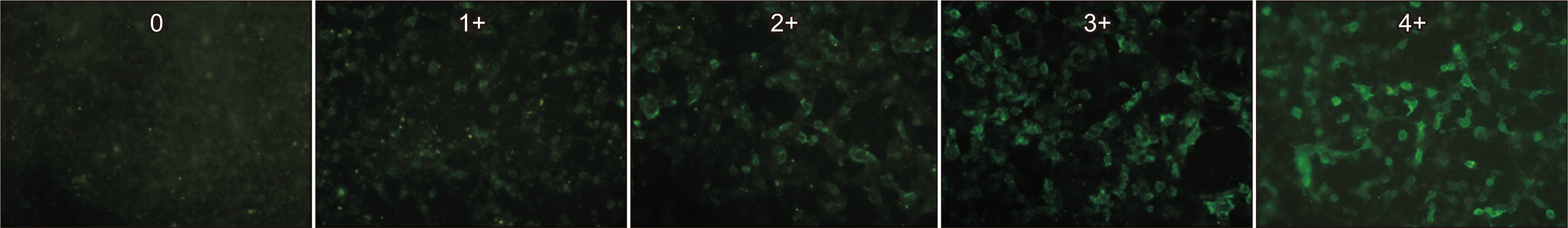
For the live CBA, we transfected HEK 293 cells overnight with a plasmid encoding full-length human MOG using Lipofectamine 3000 (Thermo Fisher Scientific Inc., Waltham, MA, USA), according to the manufacturer’s specifications. We washed the transfected cells with DMEM, incubated them with serum samples diluted with 1% bovine serum albumin in DMEM (1:20 dilution) at room temperature (20°C–25°C) for 60 mins, fixed them with 4% paraformaldehyde for 1 min, washed them three times with 10 mM HEPES-buffered DMEM, and incubated them with a goat anti-human IgG1-specific antibody conjugated with Alexa Fluor 488. After antibody labeling, we washed the cells thrice with phosphate-buffered saline and evaluated them under a fluorescence microscope (Eclipse 80i; Nikon, Tokyo, Japan). We confirmed that the full-length human MOG protein was expressed in HEK 293 cells by western blotting using a commercial MOG-Ab (Santa Cruz Technology, Santa Cruz, CA, USA). When scoring MOG-Ab seropositivity, two investigators were blinded to the clinical and laboratory information of the patients as well as to each other’s data. Any discrepancies were resolved by repeating the experiment and seeking a third opinion. A description of the intensity scores of the surface immunofluorescence is presented in Supplemental Data Table S1. The presence of MOG-Ab was confirmed if the staining intensity was 1+ or stronger, and the final score was calculated as the mean score of the readings from two or three investigators.
To conduct validation and concordance analyses of our in-house MOG-Ab CBA, we tested all included samples from the registry for the presence of MOG-Abs in our laboratory (SMC) and the Oxford laboratory (University of Oxford, United Kingdom). The Oxford laboratory is a referral neuroimmunology center for diagnostic testing that established the current MOG-Ab CBA method involving the use of an IgG1 antibody and live cells transiently expressing the full-length MOG protein [13]. All tested samples were stored at –80°C and delivered frozen to the Oxford laboratory. We also compared the results of our in-house MOG-Ab CBA to those of a commercial MOG-Ab CBA kit (FA 1156-1005-50; Euroimmun, Lübeck, Germany), which is a fixed cell-based indirect immunofluorescence assay for MOG-IgG involving HEK 293 cells transiently expressing the full-length human MOG protein that were fixed with formaldehyde [14]. The assay was performed according to the manufacturer’s instructions, with results classified as positive or negative at a 1:10 dilution, although we did not measure the antibody titer by serial dilution.
We included patients who tested positive for MOG-Abs and those with AQP4-Abs in our hospital to compare their clinical characteristics. We used our in-house MOG-Ab CBA to determine the MOG-Ab serostatus and determined the AQP4-Ab serostatus using a fixed cell-based indirect immunofluorescence assay kit for AQP4-Ab detection (FA 1128-1010-50; Euroimmun) according to the manufacturer’s instructions, yielding reliable results for AQP4-Ab detection in our laboratory [15].
Statistical analysis
We analyzed the clinical characteristics of the enrolled patients and determined appropriate summary statistics. For continuous data, we calculated the mean and SD or the median and interquartile range (IQR). For categorical variables, we determined the absolute and relative frequencies. For the comparison of MOG-Ab CBA results between the in-house CBA and Oxford laboratory CBA, Cohen’s kappa statistics were used for the concordance of the presence of MOG-Ab and then Spearman’s correlation analysis was used to assess the correlation between the CBA scores from both laboratories.
We also analyzed differences between the characteristics of patients with MOG-Abs and those with AQP4-Abs. We used the chi-square or Fisher’s exact test for categorical variables and Student’s t-test, the Mann–Whitney U-test, or the Kruskal–Wallis test for continuous variables. All statistical analyses and graph plotting were performed using SPSS for Windows (IBM Corp., version 25.0 Armonk, NY, USA) or the R software (version 4.0.2) with the ggplot2 package. Statistical significance was defined as P<0.05 in a two-tailed test.
RESULTS
Baseline patient characteristics
We collected a total of 166 samples from 166 patients, including 113 (68.1%) from female patients. The mean patient age at sampling was 43.3±15.2 yrs, and 95 samples (42.2%) were collected at remission. The most common clinical diagnosis was seropositive NMOSD (76 samples, 45.8%), followed by TM (29 samples), MS (26 samples), ON (15 samples), seronegative NMOSD (11 samples), ODDs (eight samples), and optic neuropathy (one sample). Patient characteristics and MOG-Ab CBA results are presented in Table 1 according to the diagnosis.
Establishment and results of the in-house MOG-Ab CBA
We transfected HEK 293 cells with a plasmid encoding the full-length human MOG protein and confirmed expression of the MOG protein by western blotting. Representative results of the in-house MOG-Ab CBA with intensities of 0–4+ are presented in Fig. 1. Supplemental Data Fig. S1 shows an example of MOG-Ab positivity.
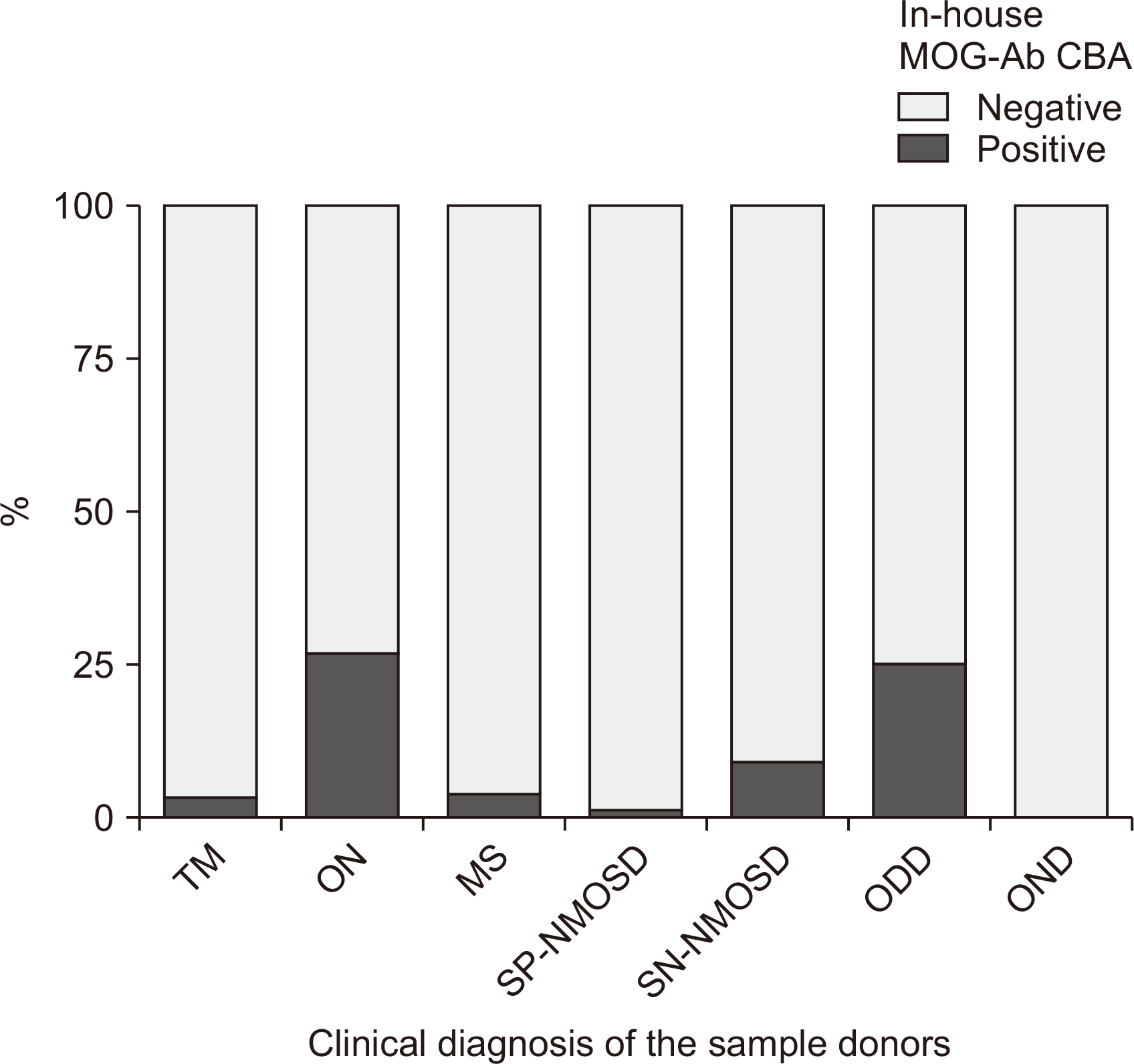
Of the 166 patients, 10 were positive for MOG-Abs. The most common clinical syndrome for which MOG-Abs were detected was ON (4/15, 26.7%). In addition, 2/8 (25.0%) patients with ODDs had MOG-Abs in their serum, and one patient each with MOG-Abs was found in the TM (1/29, 3.5%), seronegative NMOSD (1/76, 1.3%), and MS (1/26, 3.9%) groups (Fig. 2). In one case of recurrent ON, both MOG-Abs and AQP4-Abs tested positive, as confirmed by the Oxford laboratory. The other nine patients positive for MOG-Abs had known clinical phenotypes of MOGAD.
Comparison of the results of MOG-Ab CBAs
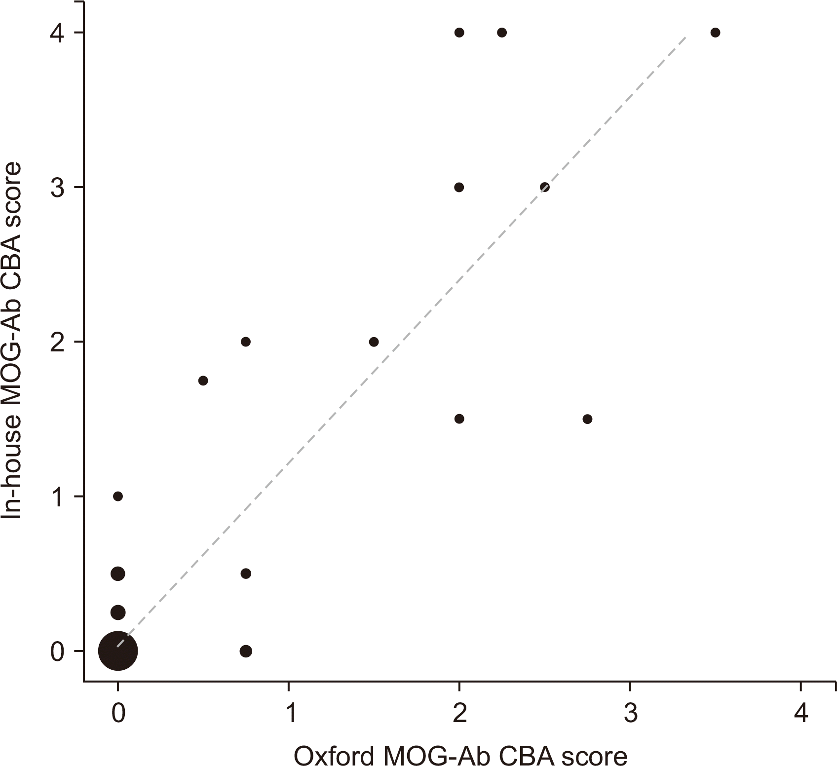
All samples were examined at both our laboratory and the Oxford laboratory for the presence of MOG-Abs. We also used a commercial fixed-cell-based MOG-Ab kit in our laboratory after blinding the investigators to the clinical information and results of the other CBAs. Both laboratories showed consistent MOG-Ab test results in 164/166 (98.8%) samples (κ=0.883, P<0.001); two patients (2/166, 1.2%) were only positive using our in-house CBA (Supplemental Data Table S2). The in-house MOG-Ab CBA scores correlated well with those of the Oxford CBA (r=0.663, P<0.001; Fig. 3). However, the results of the commercial MOG-Ab CBA kit differed from those of the live CBAs in both laboratories. Despite the high concordances between the results obtained using the commercial kit and the Oxford and in-house MOG-Ab CBAs (96.4% and 97.6%, respectively), three false-positive findings were solely observed with the commercial MOG-Ab CBA (two cases of seropositive NMOSD and one case of seronegative NMOSD). Additionally, in one case, the commercial MOG-Ab CBA showed a negative finding for ON, whereas the in-house and Oxford assays showed positive results (CBA scores of 3+ and 2+, respectively).
Clinical characteristics of patients with MOG-Abs
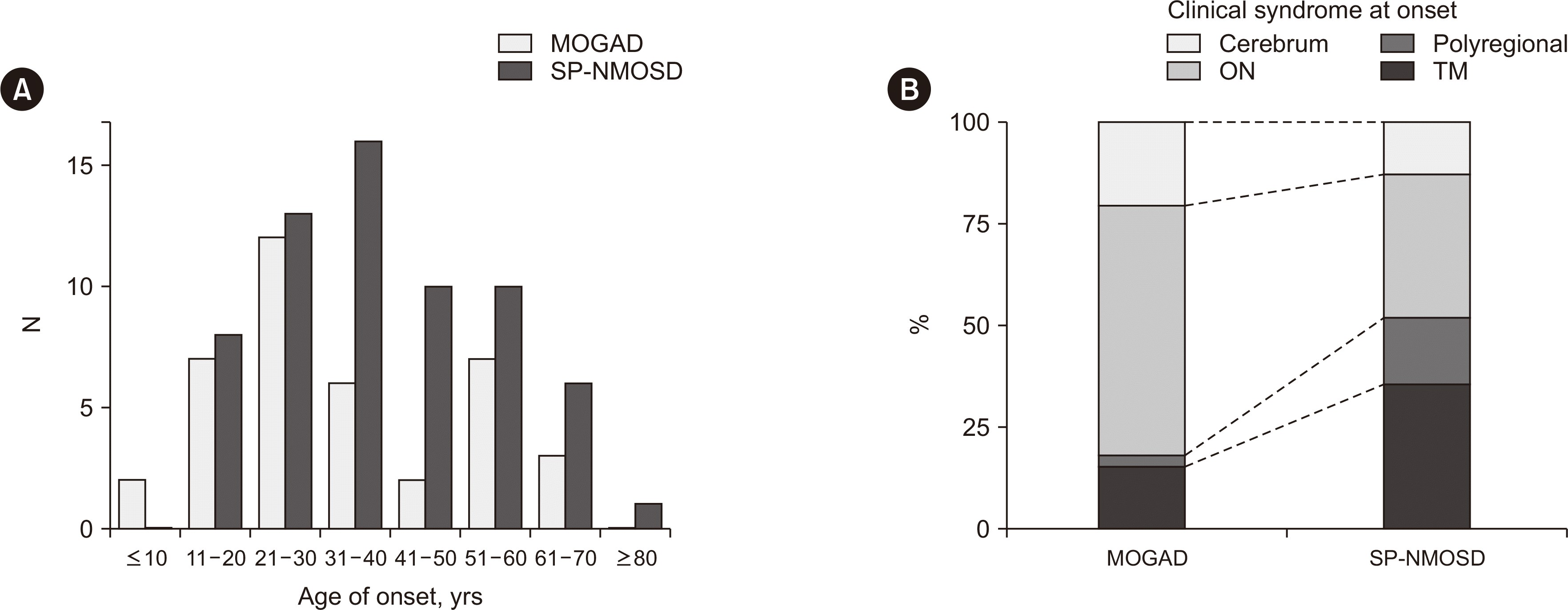
We recruited 29 additional patients with MOG-Abs and compared the clinical characteristics of 39 patients with MOG-Abs (22 female patients, 56.4%) with those of patients with seropositive NMOSD (N=66; 59 female patients, 89.4%). The disease duration was shorter in patients with MOG-Abs than in patients with AQP4-Abs (2.8±4.5 vs. 7.0±7.1 yrs, P<0.001), but the number of relapses was not different between the two groups. The NMOSD clinical phenotype was the most common in patients with MOG-Abs, whereas ON (61.5%) was the most frequent clinical syndrome at onset; TM (35.5%) was common in patients with AQP4-Abs (P<0.007) (Fig. 4). The clinical characteristics of the patients are shown in Table 2, according to the presence of different antibodies.
DISCUSSION
We established an in-house MOG-Ab CBA that showed reliable MOG-Ab test results with clinical implications. MOG-Ab has been studied in patients with MS for its potential as a disease-associated autoantibody. However, a study of MOG-Abs using ELISA and western blot analyses reported inconsistent results [16]. The use of a CBA to improve MOG-Ab testing enables detection of specific antibodies with clinical significance [13]. The clinical syndromes associated with MOG-Abs are diverse, including ON, seronegative NMOSD, myelitis, ADEM, and brainstem/cortical encephalitis [4-6, 17-20]. The frequency of these MOG-Ab phenotypes may differ according to the age of onset. In adults, ON is the most common manifestation, and ADEM/ADEM-like brain involvement is more common in children with MOG-Abs [21]. We also found that MOG-Abs were commonly present in ON. Seronegative NMOSD (17/39, 43.6%) was the most frequent clinical syndrome found in the additionally recruited patients with MOG-Abs, whereas ON was the most common clinical syndrome at onset. The enrolment in our multicenter registry (from which the samples were collected) was limited to adult patients. Furthermore, our registry consisted of only neurologists, which may have contributed to the observed high prevalence of NMOSD patients within the registry samples used in this study.
Our in-house CBA detected MOG-Abs in 10 patients (6.0%). The low detection rate of MOG-Abs can be explained by the high proportion of seropositive NMOSD (76/166, 45.8%) in our registry and the low seroprevalence of MOG-Abs in Korea. A previous report of MOG-Ab seroprevalences in Korea showed a low seroprevalence of MOG-Abs (6.1%) compared with that in Western countries [10, 22]. The results of the in-house MOG-Ab CBA were reliable when compared with those of the Oxford laboratory, and the CBA scores were well correlated between both laboratories; only two patients (1.2%) were positive for MOG-Abs by the in-house CBA but negative by the Oxford laboratory CBA (one patient with myelitis and the other with seronegative NMOSD). We found one patient with double seropositivity for MOG-Abs and AQP4-Abs, which is extremely rare [22, 23]. She showed recurrent ON with asymptomatic brain lesions and was receiving satralizumab treatment, which is an uncommon treatment option in Korea [24]. Two incongruent cases and one double-seropositive case are presented in Supplemental Data Table S3. The commercial MOG-Ab assay kit using fixed cells revealed one false-negative result and three false-positive results.
Previous comparative studies of MOG-Ab assays showed that live CBAs had superior positive predictive values compared with the commercial fixed CBA, which can generate false-positive and false-negative results [14, 25]. Moreover, an international multicenter investigation of the reproducibility of 11 MOG-Ab assays showed lower agreement within all CBAs, including a fixed-cell assay, than that of the live CBA [26]. The investigators tested 39 positive and 40 negative samples with seven live CBAs and one fixed CBA. The fixed CBA (a commercial MOG-Ab kit) was negative for five samples that were clearly positive with the other live CBAs, and one false-positive result was reported, suggesting utility in commercial fixed MOG-IgG testing in places where live MOG-IgG CBAs are unavailable, but this assay may miss 10%–15% of positive cases [26]. Consistent herewith, we found that false-positive cases were limited with the commercial CBA kit, and one false-negative result was obtained using the commercial CBA kit. Such false-positive results could be potentially harmful to patients by prompting the unnecessary use of immunosuppressive therapies based on misleading test results.
The clinical significance of MOG-Abs has increased as research has progressed. MOG-Abs are essential for diagnosing CNS inflammatory disorders [4]. The presence and titers of MOG-Abs can serve as predictors of recurrent or monophasic clinical courses [9, 27, 28]. Although diagnostic criteria or treatment guidelines for MOGAD are not yet established, accurate detection of MOG-Abs may be crucial in clinical practice and research. Our in-house MOG-Ab CBA showed comparable results with other laboratory tests and previous reports for MOG-Ab tests, and patients with MOG-Abs had different clinical features from those with AQP4-Ab, consistent with previous studies [29]. Therefore, the in-house MOG-Ab CBA may be useful in clinical practice for treating CNS inflammatory disorders, and further studies on the clinical implications of MOG-Abs are warranted.
Our study had several limitations. First, healthy controls were excluded. However, the risk of false-positive results has not yet been fully evaluated. Second, the demographic profiles of the enrolled registry samples were not controlled, and only adult patient samples were recruited, which limits generalization. Third, seronegative patients with NMOSD were not included in the comparison of clinical characteristics, which limits our understanding of the clinical differences between patients with NMOSD and MOG-IgG-positive patients. However, whether double-seronegative NMOSD is different from or the same as seropositive NMOSD remains controversial [30, 31]. Newly developed diagnostic criteria for MOGAD may aid in distinguishing it from other CNS-demyelinating diseases [32], and further research is required to resolve this issue. Finally, we evaluated the presence of MOG-Abs using only serum samples. However, recent studies have reported that some patients with MOGAD are only positive for MOG-Abs in the CSF, suggesting that intrathecal MOG-Ab production occurs frequently [7]. Further studies using serum and CSF samples are needed to understand the clinical characteristics of patients with MOG-Abs and the associated clinical implications.
In conclusion, we established an in-house MOG-Ab live CBA and validated it by comparing it with the Oxford MOG-Ab CBA, which showed distinctive clinical characteristics in patients with MOG-Abs. The clinical usefulness of MOG-Ab testing should be further evaluated in a prospective, long-term, large-scale study.
SUPPLEMENTARY MATERIALS
Supplementary materials can be found via https://doi.org/10.3343/alm.2024.44.1.56
 XML Download