Abstract
An 83-year-old male patient presented with a Marjolin’s ulcer on his lower leg, which developed 60 years after a bomb blast injury. The lesion was a 24×12-cm2 chronic ulcer located circumferentially around the mid-third lower leg. Considering the patient’s age, vessel status, and the extent of the defect after wide excision, which included a section of the tibia, reconstruction utilizing bilateral anterolateral thigh free flaps in a flow-through pattern under spinal-epidural anesthesia was planned. The operative time was 9 hours, and the patient fully recovered without any complications. The patient was able to walk without any orthosis, and no evidence of recurrence was found during a 3-year postoperative follow-up period.
Free flap reconstruction is inevitable in cases of vital structural exposure defects after cancer resection. Given that skin cancer is primarily diagnosed in elderly patients, reducing the operative time of resection and reconstruction is crucial to minimizing patient morbidity. In this case report, we present the successful bilateral anterolateral thigh (ALT) free flap reconstruction of an extensive Marjolin’s ulcer with tibial involvement in an 83-year-old male patient. To minimize the operative time, two teams performed simultaneous cancer resection and the first flap harvest, followed by a second flap harvest and microanastomosis. Both flaps were anastomosed using a flow-through chimeric pattern. All procedures were conducted under spinal-epidural anesthesia, considering the patient’s age and underlying diseases.
This study was approved by the Institutional Review Board of Soonchunhyang University Bucheon Hospital (No. 2023-03-011-001). Written informed consent was obtained from the patient for the publication of this report including all clinical images.
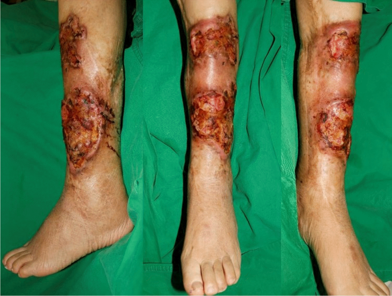
An 83-year-old male patient presented to our outpatient clinic with a chronic ulcer on his left lower leg. The ulcer was a result of a bomb blast injury that had occurred 60 years ago, and the patient underwent a skin graft procedure at that time. Despite the previous treatment, a recurrent ulcer had developed at the site of the original injury with severe pain and scarring, which raised a strong suspicion of Marjolin’s ulcer. A biopsy of the lesion confirmed the presence of squamous cell carcinoma (SCC). The dimensions of the entire lesion were 22×14 cm2, and preoperative magnetic resonance imaging revealed a mass extending to the anterolateral compartment muscle and tibial cortex with no regional lymph node invasion (Fig. 1).
Consequently, a total of 28×20-cm2 circumferential defects with additional tibial defects were expected after wide excision, and free flap reconstruction was planned. Preoperative computed tomography angiography revealed normal vascularity of the posterior tibial artery but long segmental occlusion of the anterior tibial artery, which could also be sacrificed during tumor resection. Therefore, bilateral ALT free flap reconstruction anastomosing the posterior tibial vessels was planned using a two-team approach to minimize operating time. Additionally, spinal-epidural anesthesia was administered during the entire surgery, as the patient was a high-risk older adult and had underlying diseases, including hypertension and depression.
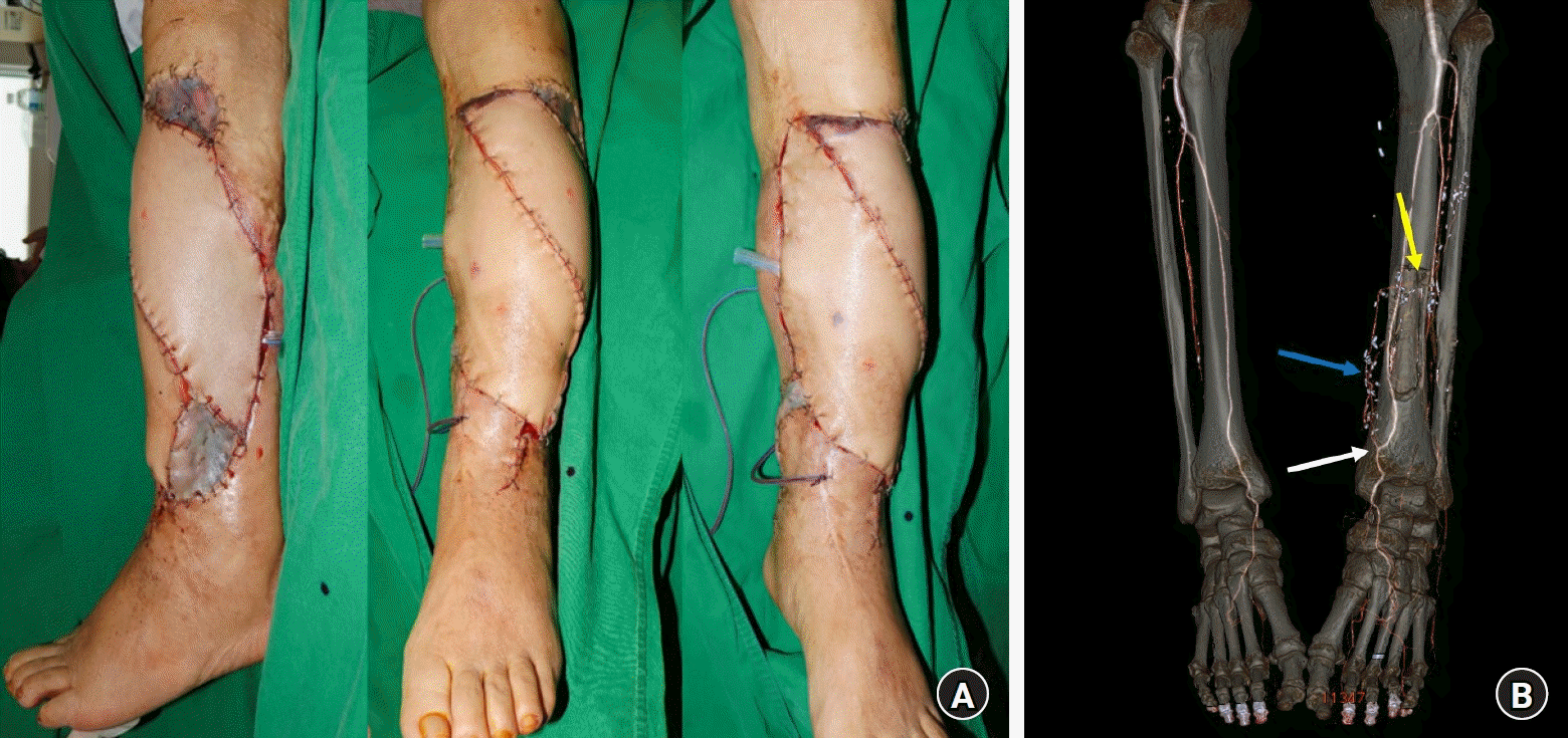
During the wide excision of the SCC, a 26×10-cm2 ALT free flap with a single perforator was simultaneously harvested from the right thigh, and a 28×10-cm2 ALT free flap with two perforators was harvested from the left thigh. While harvesting the first flap from the right side, the point where the distal perforator diverges from the descending branch of the lateral circumflex femoral artery was dissected and harvested as a “T” shape to serve as an additional anastomotic site with the second ALT flap in a flow-through technique. For the flap from the left thigh, turbocharging was performed between the two perforators that diverged from different branches. At the resection site, the descending branch of the lateral circumflex femoral artery of the left ALT flap was anastomosed to the left posterior tibial artery in an end-to-side fashion, and the accompanying vein of the flap was anastomosed to the vena comitans of the left posterior tibial artery in an end-to-end fashion. Then, the second ALT flap from the right thigh was anastomosed to the “T-shaped” distal branch of the lateral circumflex femoral artery from the first ALT flap in a chimeric pattern (Fig. 2). The entire operative time was 9 hours, and it ended after confirming the vascular patency of both flaps using indocyanine green angiography.
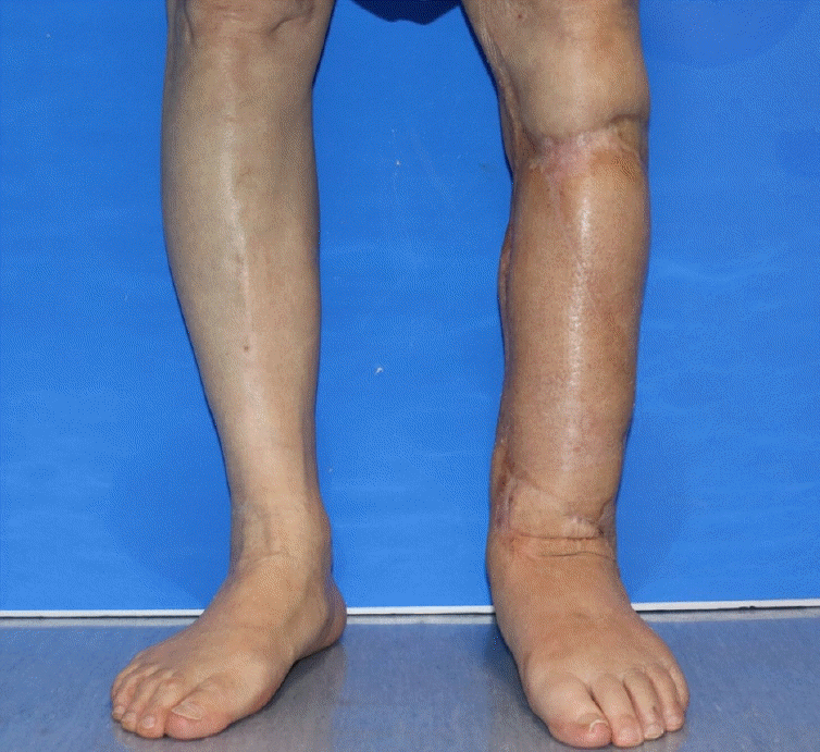
The flaps survived without complications, and the patient is currently able to walk without an orthosis. No evidence of recurrence was found during the 3-year follow-up period on magnetic resonance imaging and positron emission tomography (Fig. 3).
Marjolin’s ulcer is a rare and aggressive form of skin cancer that commonly occurs in scar tissues, chronic ulcers, and areas with chronic inflammation. Its incidence is estimated to be between 1% and 2% of all burn scars and usually presents as SCC. It is frequently identified during the examination of lesions that arise in scars or hard-to-heal chronic wounds, such as pressure sores and ulcers. Lesions can occur anywhere but are more common on the lower extremities, followed by the scalp, upper extremities, torso, and face [1].
Wide excision of the lesion, followed by resurfacing with a skin graft or locoregional flaps, is a common treatment for Marjolin’s ulcer. A particular concern in surgical treatment for Marjolin’s ulcer surgery is the resection-free margin, which is conventionally recommended to be at least 2 cm [2]. Due to the presence of concomitant deep-seated scars and three-dimensional tissue involvement, aggressive resection may expose vital structures and sometimes require amputation [3]. Furthermore, simultaneous contracture release and complete removal of the potential scar necessitate a greater number of defects to be resurfaced after resection. The residual wound usually requires flap transplantation to cover critical structures, such as the exposed joints, bones, and tendons.
For extensive defects in the extremities, free flap transplantation is inevitable for reconstruction in a single stage to allow the optimal restoration of the extremities both functionally and aesthetically. However, locating a suitable single donor site for extensive large-sized flap harvesting is challenging. Previous studies have reported using perforator flaps such as ALT flaps, thoracodorsal artery perforator flaps, and deep inferior epigastric artery perforator flaps for large defect reconstruction. These flaps are well-known large flaps with sufficient pedicle length and are available for donor-site primary closure. However, extensively large defects, as in our case, require multiple flaps for coverage, and the most efficient flaps in terms of factors such as patient morbidity, operative time, donor-site morbidity, and operative position should be considered. We selected a bilateral ALT flap with a long vascular pedicle that could be harvested in the supine position and offered a cosmetically acceptable donor site with minimal loss of function. Additionally, since preoperative computed tomography angiography revealed a single available recipient vessel (the posterior tibial artery), we selected a flap that could be easily connected in a flow-through-pattern chimeric flap.
The main advantage of conventional flow-through flaps is their ability to provide sufficient perfusion to the distal tissues, as they involve the anastomosis of a free flap with a vascular pedicle to both the proximal and distal ends. Therefore, flow-through flaps are mostly used when vascular reconstruction is needed along with soft tissue coverage. There are many reports regarding situations of severe trauma where flow-through flaps have prevented amputations both in the lower and upper extremities [4,5]. They can also be applied in cases requiring multiple flaps to connect sequentially to a second flap. The ALT flap is an ideal flow-through flap, as we can easily dissect and harvest the distal vascular stump for additional anastomosis during flap harvest. The descending branch of the lateral circumflex femoral artery generally has a large diameter; therefore, linking two ALT flaps in a flow-through pattern as a chimeric flap requires easier microanastomosis in shallow and wide microsurgical fields. Furthermore, additional surgical procedures, such as vascular grafts, which cause prolonged operation times and heightened complications, are not needed. As a result, the use of flow-through bilateral ALT flaps is deemed a highly effective option for extensive defect coverage.
We also considered the means of anesthesia for elderly patients who require longer operative times. Negative perceptions of elderly patients with reduced physiological reserves, multiple medical issues, and slower wound healing rates than younger individuals may pose challenges for reconstructive surgeons [6,7]. Additionally, elderly patients exhibit heightened sensitivity to anesthetic drugs, as well as an increased risk of respiratory and cardiac complications and cognitive dysfunction during the postoperative period. Therefore, we utilized combined spinal-epidural anesthesia, which has been shown to offer significant benefits over general anesthesia. These benefits include improved cardiac and respiratory stabilization, prolonged anesthesia duration, and expedited mobilization [8,9]. In free flap surgery, spinal-epidural anesthesia is known to increase the arterial maximal flow velocity of the free flap and decrease postoperative pain and opioid requirements [10].
In conclusion, a two-team approach under spinal-epidural anesthesia can minimize operative time, resulting in minimal patient morbidity. In cases of extensive defects where multiple flaps are required for coverage, utilizing flow-through-pattern chimeric free flaps can save operative time with minimal dissection of the recipient’s vessels and easier micro anastomoses.
References
1. Copcu E. Marjolin's ulcer: a preventable complication of burns? Plast Reconstr Surg. 2009; 124:156e–164e.

2. Copcu E, Aktas A, Sişman N, Oztan Y. Thirty-one cases of Marjolin's ulcer. Clin Exp Dermatol. 2003; 28:138–41.

3. Sharma S, Das N, Gupta V, Bera S, Bisht N. Lower extremity Marjolin's ulcer reconstruction with free anterolateral thigh flap: a case series of 11 patients. Cureus. 2020; 12:e11392.

4. Ozkan O, Ozgentaş HE, Dikici MB. Flow-through, functioning, free musculocutaneous flap transfer for restoration of a mangled extremity. J Reconstr Microsurg. 2005; 21:167–72.

5. Brandt K, Khouri RK, Upton J. Free flaps as flow-through vascular conduits for simultaneous coverage and revascularization of the hand or digit. Plast Reconstr Surg. 1996; 98:321–7.

6. Chick LR, Walton RL, Reus W, Colen L, Sasmor M. Free flaps in the elderly. Plast Reconstr Surg. 1992; 90:87–94.

7. Djokovic JL, Hedley-Whyte J. Prediction of outcome of surgery and anesthesia in patients over 80. JAMA. 1979; 242:2301–6.

8. Davis N, Lee M, Lin AY, et al. Postoperative cognitive function following general versus regional anesthesia: a systematic review. J Neurosurg Anesthesiol. 2014; 26:369–76.
Fig. 1.
Preoperative clinical photograph of a very large Marjolin’s ulcer on the left lower leg of the patient that developed due to a bomb blast injury 60 years ago.

Fig. 2.
(A) An immediate postoperative photograph of bilateral anterolateral thigh free flap reconstruction in a flow-through pattern. (B) Postoperative computed tomography angiography showing the anastomosis site on the posterior tibial artery (white arrow), the turbocharged site of the first anterolateral thigh (ALT) flap (blue arrow), and the anastomosis site of the first and second ALT flaps (yellow arrow).


 PDF
PDF Citation
 Print
Print


 XML Download