Journal List > Arch Hand Microsurg > v.28(3) > 1516083788

조갑 흑색종의 진단과 치료

Abstract

Subungual melanoma (SM) must be suspected in the initial workup of patients with pigmented nails. However, the differential diagnosis of SM from other nail disorders that involve nail pigmentation is challenging. Dermoscopic, clinical, and histopathological features are integrated for a definite diagnosis. The primary treatment of SM is surgery. Depending on the depth of invasion, amputation or functional surgery can be chosen. This article reviews the clinical, dermoscopic, and histopathological features of SM, as well as its diagnosis and treatment.

서론

조갑 흑색종은 조갑에 발생하는 암으로 피부 흑색종 중 약 0.7%–3.5%를 차지한다. 발생률은 인종에 따라 차이가 있는데, 유럽인은 0.18%–2.8%, 동양인은 10%–23%, 아프리카계 미국인은 25%로 보고된 바 있다[1,2]. 서양인과 비교하여 동양인에서는 발생률이 비교적 높다. 또한 주로 노년층에서 발생하며 정확한 진단의 지연으로 종종 진행된 상태로 발견된다[3].
조갑 흑색종의 병태생리는 아직 명확하지 않으나 외상, 자외선 노출, 가족력 등이 있다[4,5]. 외상은 흑색종의 위험인자로 알려져 있으나 인과관계에 대해 명확히 밝혀진 바는 없다[5,6]. 자외선의 영향은 일반적으로 없는 것으로 알려져 있으나 최근 조갑 흑색종의 유전적 연구에서는 자외선으로 발생하는 유전적 변이가 종양 돌연변이 부담에 가담하는 것으로 보고되었다[7,8]. 남녀 비율은 여러 연구에서 0.3–2.5:1로 다양하게 나타났다[9,10]. 다양한 조갑의 다른 병변들과 감별 진단을 요하며, 오진된 경우는 예후가 불량하다[11,12]. 조갑 흑색종의 조기 진단은 다른 다양한 감별 진단 때문에 쉽지 않다[13]. 조직 병리학적 분석을 위해 충분한 조직을 확보하기 위해서는 관련 의학 지식과 세밀한 기술이 필요하며[13], 조기 진단율을 높이기 위해 조갑 흑색종의 임상학적 소견에 대한 지식과 적절한 조직 검사의 조합이 필요하다. 진단이 되면 신속히 수술로 치료해야 하며, 진단 초기, 치료 과정 및 말기 환자에서 모두 다학제 치료가 필요하다.
조갑 흑색종은 불량한 예후를 지닌 악성 종양으로 여겨 과거에는 주로 절단술을 시도해 왔으나[14,15], 최근에는 기능과 미용적인 측면을 고려하여 절단을 최소화하면서 생존율을 유지하는 방향으로 치료가 바뀌고 있다[16,17].

진단

1. 임상적 소견

병변 부위의 색소가 선천적인지 후천적으로 발생했는지 반드시 확인해야 한다. 후천적으로 발생했을 경우 발생 시기, 인지 시점 및 인지 후 색소의 변화 양상 등의 정보가 진단에 도움이 될 수 있다. 많은 환자들이 색소의 변화 양상을 객관적으로 설명하기 어려워하는데, 이 경우 환자에게 직접 그림을 그려가면서 설명을 요청할 수 있다. 외상력, 아스피린 및 와파린과 같은 약 복용, 운동 경력 등을 통해 조갑하 혈종을 배제할 수 있다[18].
세로선 흑색 조갑(longitudinal melanonychia)은 조주름(nail fold)의 근위부에서 원위부에 이르는 짙은 검 갈색의 세로 방향의 띠 형태를 의미한다(Fig. 1). 양성 세로선 흑색 조갑과 비교하여 조갑 흑색종은 띠가 더 넓은 형태와 함께 사이사이 더 불규칙한 빈 공간의 다양한 색상의 띠가 나타난다[19,20]. 성인에서 빠른 시간 내 단측 손톱에서 세로선 흑색 조갑이 나타나면 악성을 강하게 의심해봐야 한다[20,21]. 상세한 병력 청취와 신체 검진을 통해 조갑하 혈종, 무좀균 감염, 단순 색소 침착, 모반 등 다양한 감별 질환들을 배제해야 한다[13,22].
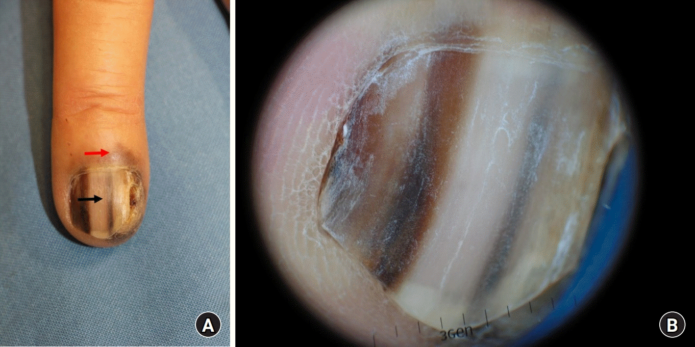
허친슨 사인(Hutchinson sign)은 조갑기질(nail matrix)과 손발톱바닥(nail bed)에 갈색 또는 흑색의 침착이 더 근위부와 가측 조주름 또는 하조피(hyponychium)로 번지는 소견이며 조갑 흑색종의 위험 신호이다[23,24] (Fig. 1). 비록 드물지라도 Laugier-Hunziker syndrome, Peutz-Jegher syndrome처럼 결합조직병과 같은 전신 질환에서도 손톱 침착 소견이 보일 수 있으며[1,25], 이러한 경우 구강과 생식기 점막의 병변으로 확인할 수 있다[26]. 약 6%–36%의 조갑 흑색종에서는 손톱 침착 소견이 없는데, 이러한 경우에 색소 침착 소견을 보이지 않는 조갑 진균증, 무사마귀와 화농성 육아종 등의 질환과 혼동되기도 한다[27]. 출혈, 손톱내 종괴, 조갑 이영양증, 손톱판 손실 등의 추가적인 임상 양상은 진행된 조갑 흑색종의 신호이다[28]. 이러한 임상 소견을 보이는 환자들은 조직 검사가 필요하다.
조갑 흑색종의 진단을 쉽게 하기 위해 ABCDEF 기준이 만들어졌다[29]. A는 연력(최고 발병률이 50–70대에 가장 많이 나타남)과 인종(흑인, 동양인, 북미 원주민)을, B는 흑갈색의 폭 3 mm 이상 병변을, C는 조갑의 띠의 변화를, D는 가장 흔히 침범되는 손가락을 말한다(엄지손가락>엄지발가락>집게 손가락, 단수지>다수지). E는 근위부 또는 가쪽 손발쪽 주름으로 색소 침착이 확장되는 것을 말하며, F는 흑색종 또는 이형성 모반(dyplastic nevi)등의 가족력을 나타낸다. ABCDEF 기준이 조갑 흑색종의 중요한 위험인자를 포함하긴 하나 실용성은 제한적이다[30]. 손톱판의 색소 침착된 대역폭(bandwidth) 비율이 양성 흑색조갑증과 조갑 흑색종을 구별하는 좋은 도구이며, 세로선 흑색 조갑에서 대역폭의 비율이 40% 이상이고 다른 조갑 흑색종의 의심 소견이 있을 경우 조직 검사가 추천된다[30]. 최근 Lee 등[31]이 조갑하 제자리 흑색종(subungual melanoma in situ) 진단을 위한 더 간단한 ABCD 기준을 만들었다. A는 18세 이상의 연령, B는 갈색 띠, C는 손톱 피부에 흑, 검, 청, 적, 흰색 및 다양한 색이 나타나는 경우, D는 one digit을 뜻한다. 이 기준에 의하면 세로선 흑색 조갑에서 A와 D 소견이 보이면서 B나 C 기준을 만족할 경우 조갑하 제자리 흑색종을 의심해야 한다.
피부 확대경(dermoscopy)은 육안으로 보이지 않는 피부의 세부 구조물들을 수십 배 확대해서 볼 수 있다. 조직 검사 없이 다양한 피부 질환 및 암을 쉽고 정확하게 진단할 수 있다. 이전에는 기름, 알코올, 젤 등의 침액(immersion liquid)와 피부를 접촉하여 관찰이 가능했다. 그러나 최신 피부 확대경은 편광을 이용하여 침액과 같은 접촉 매개가 불필요하다. 일반적으로 피부 확대경을 통해서 피부 질환의 개수, 혈관의 세부적인 모양, 및 그 외 질환마다 특이적인 병변 모양 등을 관찰하여 진단하게 된다[32].
피부 확대경은 비침습적으로 다양한 색소 병변과 초기 조갑 흑색종을 감별할 수 있는 유용한 도구이다[33,34]. 임상 소견만으로 초기 조갑 흑색종을 정확하게 진단하는 것은 매우 어려운 일이다. 임상 소견과 더불어 피부 확대경에서 특징적인 소견을 종합하면 진단의 정확성을 높일 수 있다[13,21]. 조갑 흑색종의 특징적인 피부 확대경 소견에는 허친슨 사인, 궤양 등이 있다[35]. 수술 중에 피부 확대경을 확인할 경우 단순 피부 확대경과 비교하여 직접적으로 손톱 침착의 위치를 확인할 수 있어 진단 정확성을 더 높일 수 있다[35,36]. 하지만 손톱 발조를 해야 하는 검사의 침습성 때문에 자주 시행되지 않는다.
Ohn 등[37]은 최근 조갑하 제자리 흑색종의 소견에 따라 예측 모델을 제시했다. 이 모델의 점수는 다음과 같다: 폭(세로선 흑색 조갑 ≥3 또는 6 mm), 색(다형색의 침착), 패턴(비대칭[asymmetry], 희미한 경계[border fading], 허친슨 사인). 세로선 흑색 조갑이 ≥3 mm일 경우 1점, 세로선 흑색 조갑이 ≥6 mm일 경우 2점, 다형색의 침착이 있을 경우 1점, 비대칭 패턴 2점, 희미한 경계 1점, 허친슨 사인 2점으로, 총 0에서 8점의 모델을 제시했다. 절사값(cutoff value)을 2점으로 할 경우 조갑하 제자리 흑색종의 진단의 민감도(sensitivity)는 100%였고, 3점으로 할 경우 89%의 민감도와 62%의 특이도(specificity)를 보였다.
세로선 흑색 조갑 소견을 보일 때 이러한 점수를 통해 악성 위험도를 계층화하는 것은 실용적이지만, 소아에서는 흑색종 발생률이 매우 낮고 세로선 흑색 조갑이 비전형적으로 나타날 수 있기 때문에 같은 모델을 적용하는 것은 바람직하지 않다[31,38]. 최근 연구에서는 궤양, 전 흑색조갑증(total melanonychia), 조갑 붕괴(nail plate destruction), 다색(polychromia) 등의 소견이 조갑 제자리 흑색종과 침습적 조갑 흑색종을 감별하는 수신자 조작 특성(receiver operating characteristic) 값을 0.902로 보고하였다[39].

2. 조직 검사 방법

병변의 위치와 세로선 흑색 조갑의 두께에 따라 다양한 손톱 조직 검사 방법이 존재한다[40]. 이론적으로 검사는 충분한 양의 조직을 확보하면서 흉터와 손가락 장애를 최소화해야 한다. 손톱 흑색종은 대부분 조갑기질의 멜라닌 세포(melanocyte)에서 기원하므로 조직 검사 검체는 조갑기질에서 채취해야 한다. 단, 진피 침범(dermal invasion)이 의심되는 경우 조갑기질보다는 손발톱바닥으로의 침투가 흔하여 세로 절개 생검(longitudinal incisional biopsy)이 추천된다[41].
펀치 생검은 세로선 흑색 조갑이 <3 mm일 때 유용하다. 이 검사 방법은 4–6 mm 크기의 펀치를 이용하여 손톱을 우선 제거한 후, 2 mm 펀치를 이용해 조갑기질 조직을 채취할 수 있다[13]. 검사 방법은 간단하지만 검사 중 조갑기질의 침착 여부를 정확하게 확인하기 어려워 위음성의 가능성이 있다[37].
색소 침착된 띠가 손톱의 가측 1/3 지점에 있을 경우 가쪽에서 세로 조갑 생검을 시행한다. 생검은 조갑기질의 가장 근위부에서 시작한다[42,43]. 침착된 띠가 3–6 mm 반경인 경우, 1 mm보다 얇은 굵기로 충분한 조직을 채취하면서 재상피화가 잘 되고 손톱 이영양증 발생률이 적은 표층 생검(shave biopsy)이 유용하다[42,44].
최근 연구에서는 세로선 흑색조갑증 의심 환자에서 CO2 레이저를 이용한 검사 방법도 있다[37]. 세로 절개 생검과 표층 생검을 혼용하는 경우 낮은 손톱 이영양증 발생률(25%)과 낮은 색소침착 재발률(13%)을 기대할 수 있다[45].

3. 조직학적 소견

조갑 흑색종에서 가장 중요한 조직학적 소견은 세포 형태(cell morphology), 구성 특징(architectural feature), 멜라닌 세포의 밀도, 피부의 염증성 변화 등이다. 세포 형태학적 평가 기준은 세포 크기, 모양, 멜라닌 세포의 염색성(chromasia) 등이 있다. 다른 피부 질환과 유사하게 핵 확장, 과염색증, 불규칙한 핵 모양, 현저히 보이는 핵소체(nucleolus) 등이 세포 모양의 비정형성으로 정의될 수 있다. 피부 기저층 또는 그 위에 존재하는 주변의 각질세포보다 크기가 큰 흩어진 비정형의 멜라닌 세포가 Fig. 2에서와 같이 보이고 임상적으로도 의심이 될 경우, 초기 병변을 시사한다[9,46]. 비정형 멜라닌 세포는 양성 질환보다 손톱 악성 흑색종에서 더 흔히 관찰된다. 구체적으로 비정형 멜라닌 세포의 비율이 수치로는 양성 조갑하 흑색 반점(subungual melanotic macule)에서 6.7%, 조갑하 제자리 흑색종에서 80%, 조갑 흑색종에서 100%이다[47]. 그 외에 단 세포 흑자성 성장 양식(single cell lentiginous growth pattern)과 파제트병(Paget disease) 모양의 퍼지는 형태도 악성을 시사하는 소견이다[46,47]. 반면 양성 조갑피질 모반에서는 증가된 멜라닌 세포가 둥지 형태로 피부 기저층 또는 그 위에 국한된 소견을 보인다[2]. 그 외에 조갑기질 활성화, 조갑하 흑색 반점, 흑색조갑증, 흑자 과색소 침착(lentigo hyperpigmentation) 등의 양성 질환은 조갑기질에서 멜라닌 세포 과형성을 동반하지 않은 증가된 멜라닌 침착 소견만 보인다[47,48].
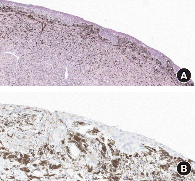
조직 검사 중 도움이 되는 지표는 상피 진피 접합(epidermal-dermal junction)에서 멜라닌 세포의 수(melanocyte count [MC]/millimeter)를 정량적으로 측정하는 것이다. 정상 손톱에서는 MC값이 4–9 (평균 7.7)이며 조갑하 흑색 반점에서는 5–31 (평균 15.3)이다[47]. 조갑 제자리 흑색종에서는 MC값이 39–136 (평균 58.9)이며 조갑 흑색종에서는 52–212 (평균 102)이다. MC값이 30보다 적을 경우 양성을, 40 이상일 경우 악성을 시사한다[47]. 그러나 약 22%의 조갑 제자리 흑색종에서 MC값이 40 미만으로, MC값의 신뢰성이 떨어진다[46]. 따라서 Park 등[46]은 조갑 제자리 흑색종이 임상적으로 강하게 의심될 때 산발적으로 과다 염색핵을 가진 비정형 멜라닌 세포의 수가 증가하여 있으면 조갑 제자리 흑색종 진단의 단서가 된다고 하였다. 조갑기질에서 멜라닌 세포를 확인하는 것이 어렵기 때문에 melan-A, HMB45, S-100, SOX-10, 또는 MiTF 면역화학 염색이 도움이 된다[46,49].
염증 세포의 침윤은 악성 멜라닌 세포에 대한 반응으로 나타날 수 있어 양성 질환보다는 조갑 흑색종을 시사하는 소견이다[50]. 하지만 초기의 조갑 흑색종에서는 염증 세포의 침윤이 나타나지 않을 수 있다[50,51].
초기의 조갑 흑색종은 정밀한 조직학적 분석을 해도 진단이 어려운 경우가 많다. 최근 한 연구는 양성 병변과 조갑 제자리 흑색종에서 p16, HMB45, Ki-67/melan-A 염색 등이 차이를 보이지 않았다고 보고하였다[49]. 따라서 조직학적으로 불분명한 상황에서는 완전 절제 또는 추가 조직 검사를 할 필요가 있다[46,52].
진단이 불분명한 경우에는 형광제자리부합법(fluorescence in situ hybridization, FISH)를 사용할 수 있다. 한 연구에서 7건의 조갑 흑색종에서 FISH를 시행하고 6p25 (RREB1) 증가(100%), 11q13 (CND1) 증가(86%), 6q23 (MYB) 상실과 8q24.1 (MYC) 증가(71%)를 관찰하였다[53]. 또 다른 연구에서는 23건의 조갑 흑색종 중 15건이 형광제자리부합법 검사 양성이었다[51].

치료

조갑 흑색종은 수술적 치료가 주를 이룬다. 하지만 병변이 뼈와 신전건과 인접해 있고 손톱의 해부학적 복잡성 때문에 수술적 치료가 쉽지 않다. 완전한 병변 제거를 위해 절단이 흔히 사용된다. 1960년대에는 중수지 관절(metacarpophalangeal joint) 또는 손목에서의 절단이 흔히 이루어졌다[54]. 하지만 조갑 흑색종 병변의 제거는 최대한 기능적 및 미용적으로 정상 구조물을 보존하는 방법으로 변화해왔다. 안전연(safety margin)이 확보되면 원위지 관절(distal interphalangeal joint)에서 절단을 해도 재발률과 생존율에 차이가 없다는 연구 결과가 쌓이면서 절제 범위가 더 줄기 시작했다[15]. 중수지관절에서 절단할 경우 엄지, 중지 또는 검지, 약지 또는 소지에서 각각 40%, 20%, 10%의 기능 장애를 보였으나, 원위지 관절에서 절단 시 중수지 관절에서 보이는 기능 장애가 절반이었다[55]. 조갑 제자리 흑색종 또는 깊지 않은 조갑 흑색종의 경우 생존율에 영향을 미치지 않으면서 절제를 최소화하는 기능적 수술(functional surgery) 치료가 표준이 되고 있다[56,57].
수술적 절제 범위를 정할 때는 절제연(resection margin)보다 병변의 깊이에 따라 정해야 한다. 한 연구에서는 조갑 제자리 흑색종과 브레슬로 두께(Breslow thickness) 0.5 mm 이하 조갑 흑색종에서 국소 재발률 차이가 없다고 보고하였다[58]. 조갑 제자리 흑색종의 치료는 조갑, 손발톱바닥과 조갑기질을 광범위하게 절제해야 한다[13,59]. 허친슨 사인이나 조갑에서 5 mm 안전연을 두고 병변 절제를 하면 충분하다[60,61]. 기적 수술을 시행할 때 조갑기질과 손가락 뼈가 매우 인접해 있어, 골막을 포함하여 조갑 전체 제거가 추천된다[61].
Kim 등[62]은 사체 연구에서 유용한 술기의 기능적 수술을 소개하였다. 기능적 수술 시 조갑기질의 가장 근위부 지점을 제거하면서 신전건 삽입 부위의 손상은 최소화하여 수술 후 원위부 관절의 근육 지연/긴장(extensor lag/stiffness)을 예방해야 한다. 이렇게 시행하기 위해서는 신전건의 주요 근육다발이 원위 수지골에 삽입하는 부위 가장 인접한 피부에서 절개를 해야 하고, 절개 후 신전건 삽입 부위부터 살짝 원위 부위에서 골막하 원위부 방향으로 제거해야 한다. 골막은 두 층으로 형성되어 있으며 바깥층은 관절낭으로 이어진다. 이 골막층은 100 μm까지 얇을 수 있으며 골 침범의 근원이 될 수 있다. 골 침범을 배제하기 위해서는 반드시 골막을 철저하게 제거해야 한다. 원위 수지골의 융기(tuberosity) 근처는 골막과 골의 단단한 부착성으로 박리가 어려울 수 있으며 피질골이 박리와 함께 부분적으로 부러질 수 있다. 이런 경우 발생하는 불규칙한 표면은 뼈갈기(burring)를 통해 교정할 수 있다. 갈고리 발톱 돌기(ungual process) 부위에서는 부분적 절단 또는 뼈갈기를 해야 침습이 적다고 나온 연구들이 있으나 논쟁의 여지가 있다.
또 다른 연구에서는 조갑 흑색종의 진피 침습 패턴에 따른 원격 전이와 예후를 보고했다[63]. 이 연구에서는 손톱을 해부학적으로 근위 손톱 주름의 등쪽 지붕(dorsal roof of proximal nail fold), 근위 손톱 주름의 복측 바닥(ventral floor of proximal nail fold), 조모 세포 조직(germinal matrix), 손발톱바닥, 그리고 하조피로 나누었다. 하조피 침습의 경우 통계적으로 유의미하게 브레슬로 두께(p=0.09), 림프절 전이(p=0.019), 원격 전이(p=0.036)가 높았으며, 무병 생존기간(p=0.001)은 낮았다. 기능과 미용적인 측면도 중요하지만, 조갑 흑색종에서 하조피 침습이 있을 경우 다른 패턴과 비교하여 진단, 치료, 및 감시를 더 신중하게 해야 한다.
또 다른 수술적 치료에는 모즈 수술(Mohs surgery)이 있으며 재발률은 문헌에 따라 0%–14%로 보고된다[64,65]. 하지만 이 방법은 가장자리에서 악성 멜라닌 세포를 확인해야 하는 까다로움이 있다. MART-1 염색을 통해서 민감도를 어느 정도 높일 수는 있다[65].
침습적인 조갑 흑색종의 경우 손발톱바닥과 손가락 뼈의 인접성 때문에 절단법이 선호된다. Dika 등[3]의 연구에서는 표본 수가 적어 일반화는 어렵지만 절단과 기능적 수술을 비교했는데, 조갑 흑색종 환자의 예후에 차이가 없었다. 침습적인 조갑 흑색종에서 기능적 수술과 절단 환자의 예후를 비교하는 연구가 아직은 부족하여 임상병리학적 소견, 수술자의 경험, 환자의 선호도 등 다양한 인자를 고려해야 한다. 진행된 조갑 흑색종에서 골 침범이 의심될 경우, 침습되지 않은 가장 원위부 관절면에서 절단을 시행해야 한다[15,66].
기능적 수술 후 발생하는 결손은 피부 이식, 국소 피판, 원거리 피판과 유리 피판 수술 등 다양한 방법으로 재건할 수 있다[67,68]. 피부 이식은 가장 간단한 방법으로 자주 이용되나 골 노출이 있는 경우 사용이 어렵다. 또한 부분적 손실로 인한 이차 치유 등이 필요할 수 있다. 진피 대체제(dermal substitute)를 이용하여 피부 이식 성공률을 높일 수 있으나 추가 수술이 필요하며 치유 기간이 길어진다. 국소 및 원거리 피판의 경우 조직이 부족하거나 회전 반경에 제한을 받는다. 하지만 재건 수술을 우선시하면 암 제거 수술을 보존적으로 하게 되어 종양성 결과(oncologic outcome)에 영향을 줄 수 있다. 유리 피판을 이용할 경우 이러한 문제를 해결할 수 있으며 Fig. 3와 같이 피부 이식을 동반한 전방거근근막(serratus anterior fascia) 피판, 피판 두께가 얇은 얕은엉덩휘돌이동맥 천공지(superficial circumflex iliac artery perforator) 피판, 그리고 Fig. 4, 5와 같이 유리 동맥화 정맥 피판(venous free flap) 등을 이용 할 수 있다[69].
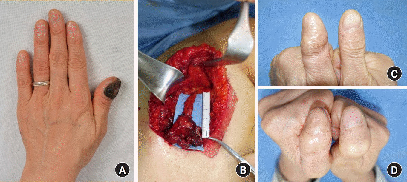
Lee 등[67]은 조갑 흑색종에서 기능적 수술 이후 발생한 결손에 대해 아주 얇은 얕은엉덩휘돌이동맥 천공지 피판을 이용해 재건했다. 피판은 얕은 피하지방층(superficial subcutaneous fat layer)으로 거상을 했으며 작은 가위를 이용해서 지방 소엽 하나 하나씩 탈지(defatting)함으로써 피판의 얇기를 최소화하여 피판 두께를 1.5–4 mm 정도로 얇게 만들 수 있었다. 이렇게 피판을 얇게 만들어도 2.5%에서만 피판 손실이 있었고 모든 피판이 생존하여 피판 관류에도 문제가 없었다.
Woo 등[70]은 조갑 흑색종에서 기능적 수술 이후 발생한 결손에 대해서 유리 동맥화 정맥 피판을 이용한 재건 환자들에 대해 후향적으로 연구했다. 전완 또는 발 등에서 최소 3개의 정맥이 포함되게 피판을 작도하여 근막상층(suprafascial layer)으로 피판 거상을 했다. 원위부 쪽의 수입 정맥(afferent vein)을 가락동맥(digital artery)과 연결하고 근위부 쪽의 원심 정맥(efferent vein)을 가락정맥(digital vein)과 연결했다. 공여부는 일차 봉합 또는 피부 이식을 이용하여 재건했다. 조갑 흑색종의 광범위 절제술 이후 발생하는 조직 결손은 크기가 작고 얇기 때문에 다른 유리 피판보다 유리 동맥화 정맥 피판이 결손 부위에 더 적합하다. 또한 유리 동맥화 정맥 피판은 공여 부위 합병증이 적고, 피판의 작도가 자유로우며, 수지부 피부와 비슷한 감촉의 피판을 사용할 수 있다.
감시 림프절 생검(sentinel lymph node biops)의 경우, 피부 흑색종에서는 무병 생존기간이 증가한다는 연구가 있으나 조갑 흑색종에서는 아직 근거가 명확하지 않다[71]. 피부 흑색종에서는 병변 깊이가 1 mm 이상인 경우, 궤양을 동반한 경우, 또는 유사 분열률이 >1/mm인 경우 감시 림프절 생검의 적응증이 된다[72,73]. 감시 림프절 생검은 침습적인 조갑 흑색종에서 병기 판정이나 예후를 알 수 있는 정보를 제공하나, 현재까지는 생존율을 높인다는 증거는 없다[6,10].
조갑 흑색종에서 항암요법을 사용하는 경우는 많지 않다. Dacarbazine 또는 cytoxan과 같은 항암제를 사용한 경우가 보고되어 있다[74,75]. 표적 또는 면역 치료는 3기 또는 4기 흑색종 환자에서 생존율을 증가시키는 것으로 보고되어 있다[57,76-79]. BRAF, KIT, MEK 억제제 등과 같은 표적 치료는 말단 또는 점막성 흑색종에서 좋은 효과를 보인다[80-83]. PD-1, CTLA-4 억제제와 같은 면역 치료제들은 BRAF, MEK 억제제 등이나 항암제보다 더 좋은 성적을 보인다[83]. 또 다른 연구에서는 anti–PD-1 억제 치료에서 효과가 미미했고 특히나 조갑 흑색종에서 낮은 반응률을 보였다[84]. 조갑 흑색종은 말단 흑색종의 한 아형이지만 생물분자학적으로 달라 같은 치료에 다른 반응을 보일 수 있다. 따라서 조갑 흑색종에서 면역 또는 표적 치료의 효과에 대해서는 추가적인 연구가 필요하다.

결론

조갑 흑색종의 정확한 진단을 위해서는 조갑 흑색종의 임상적, 병리학적, 피부 확대경 소견 등을 모두 이해해야 하고, 적절한 생검 방법을 통해 충분한 조직을 확보하면서 합병증은 줄여야 한다. 유전적으로 조갑 흑색종은 다른 흑색종과 차이점이 있으나 이에 대한 연구가 더 필요하다. 조갑 흑색종의 치료는 수술적 치료이고, 조갑 제자리 흑색종에서는 기능적 수술을 권장하며 골 침범 소견 시 절단을 해야 한다. 말기 조갑 흑색종에서 면역 및 표적 치료 약제의 효과는 아직 추가적인 연구가 필요한 상황이다.

Notes

The authors have nothing to disclose.

Funding

None.

References

1. De Giorgi V, Saggini A, Grazzini M, et al. Specific challenges in the management of subungual melanoma. Expert Rev Anticancer Ther. 2011; 11(5):749–61.
crossref
2. Nevares-Pomales OW, Sarriera-Lazaro CJ, Barrera-Llaurador J, et al. Pigmented lesions of the nail unit. Am J Dermatopathol. 2018; 40:793–804.
crossref
3. Dika E, Patrizi A, Fanti PA, et al. The prognosis of nail apparatus melanoma: 20 years of experience from a single institute. Dermatology. 2016; 232:177–84.
crossref
4. Banfield CC, Redburn JC, Dawber RP. The incidence and prognosis of nail apparatus melanoma: a retrospective study of 105 patients in four English regions. Br J Dermatol. 1998; 139:276–9.
crossref
5. Möhrle M, Häfner HM. Is subungual melanoma related to trauma? Dermatology. 2002; 204:259–61.
crossref
6. Chakera AH, Thompson JF. ASO author reflections: subungual melanomas of the hand present diagnostic and therapeutic challenges. Ann Surg Oncol. 2019; 26:1044–5.
crossref
7. Newell F, Wilmott JS, Johansson PA, et al. Whole-genome sequencing of acral melanoma reveals genomic complexity and diversity. Nat Commun. 2020; 11:5259.
crossref
8. Hayward NK, Wilmott JS, Waddell N, et al. Whole-genome landscapes of major melanoma subtypes. Nature. 2017; 545:175–80.
crossref
9. Tan KB, Moncrieff M, Thompson JF, et al. Subungual melanoma: a study of 124 cases highlighting features of early lesions, potential pitfalls in diagnosis, and guidelines for histologic reporting. Am J Surg Pathol. 2007; 31:1902–12.
10. Reilly DJ, Aksakal G, Gilmour RF, et al. Subungual melanoma: management in the modern era. J Plast Reconstr Aesthet Surg. 2017; 70:1746–52.
crossref
11. Ishihara Y, Matsumoto K, Kawachi S, Saida T. Detection of early lesions of “ungual” malignant melanoma. Int J Dermatol. 1993; 32:44–7.
crossref
12. Shukla VK, Hughes LE. Differential diagnosis of subungual melanoma from a surgical point of view. Br J Surg. 1989; 76:1156–60.
crossref
13. Braun RP, Baran R, Le Gal FA, et al. Diagnosis and management of nail pigmentations. J Am Acad Dermatol. 2007; 56:835–47.
crossref
14. Gregorcyk S, Shelton RM, Ladaga LE, Perry RR. Pathologic fracture secondary to subungual melanoma. J Surg Oncol. 1996; 61:230–3.
crossref
15. Nguyen JT, Bakri K, Nguyen EC, Johnson CH, Moran SL. Surgical management of subungual melanoma: mayo clinic experience of 124 cases. Ann Plast Surg. 2013; 71:346–54.
16. Moehrle M, Metzger S, Schippert W, Garbe C, Rassner G, Breuninger H. “Functional” surgery in subungual melanoma. Dermatol Surg. 2003; 29:366–74.
crossref
17. Lazar A, Abimelec P, Dumontier C. Full thickness skin graft for nail unit reconstruction. J Hand Surg Br. 2005; 30:194–8.
crossref
18. Pham F, Boespflug A, Duru G, et al. Dermatoscopic and clinical features of congenital or congenital-type nail matrix nevi: a multicenter prospective cohort study by the International Dermoscopy Society. J Am Acad Dermatol. 2022; 87:551–8.
crossref
19. Duarte AF, Correia O, Barros AM, Ventura F, Haneke E. Nail melanoma in situ: clinical, dermoscopic, pathologic clues, and steps for minimally invasive treatment. Dermatol Surg. 2015; 41:59–68.
20. Goettmann S, Moulonguet I, Zaraa I. In situ nail unit melanoma: epidemiological and clinic-pathologic features with conservative treatment and long-term follow-up. J Eur Acad Dermatol Venereol. 2018; 32:2300–6.
crossref
21. Ronger S, Touzet S, Ligeron C, et al. Dermoscopic examination of nail pigmentation. Arch Dermatol. 2002; 138:1327–33.
crossref
22. Mun JH, Kim GW, Jwa SW, et al. Dermoscopy of subungual haemorrhage: its usefulness in differential diagnosis from nail-unit melanoma. Br J Dermatol. 2013; 168:1224–9.
crossref
23. Saida T, Ohshima Y. Clinical and histopathologic characteristics of early lesions of subungual malignant melanoma. Cancer. 1989; 63:556–60.
crossref
24. Yun SJ, Kim SJ. Images in clinical medicine: Hutchinson’s nail sign. N Engl J Med. 2011; 364:e38.
25. Baran LR, Ruben BS, Kechijian P, Thomas L. Non-melanoma Hutchinson’s sign: a reappraisal of this important, remarkable melanoma simulant. J Eur Acad Dermatol Venereol. 2018; 32:495–501.
crossref
26. Cooper C, Arva NC, Lee C, et al. A clinical, histopathologic, and outcome study of melanonychia striata in childhood. J Am Acad Dermatol. 2015; 72:773–9.
crossref
27. Güneş P, Göktay F. Melanocytic lesions of the nail unit. Dermatopathology (Basel). 2018; 5:98–107.
crossref
28. Lee WJ, Lee JH, Won CH, et al. Nail apparatus melanoma: a comparative, clinicoprognostic study of the initial clinical and morphological characteristics of 49 patients. J Am Acad Dermatol. 2015; 73:213–20.
crossref
29. Levit EK, Kagen MH, Scher RK, Grossman M, Altman E. The ABC rule for clinical detection of subungual melanoma. J Am Acad Dermatol. 2000; 42(2 Pt 1):269–74.
30. Ko D, Oromendia C, Scher R, Lipner SR. Retrospective single-center study evaluating clinical and dermoscopic features of longitudinal melanonychia, ABCDEF criteria, and risk of malignancy. J Am Acad Dermatol. 2019; 80:1272–83.
crossref
31. Lee JH, Park JH, Lee JH, Lee DY. Early detection of subungual melanoma in situ: proposal of ABCD strategy in clinical practice based on case series. Ann Dermatol. 2018; 30:36–40.
32. Zalaudek I, Argenziano G, Di Stefani A, et al. Dermoscopy in general dermatology. Dermatology. 2006; 212:7–18.
33. Kittler H, Marghoob AA, Argenziano G, et al. Standardization of terminology in dermoscopy/dermatoscopy: results of the third consensus conference of the International Society of Dermoscopy. J Am Acad Dermatol. 2016; 74:1093–106.
crossref
34. Light RJ. Measures of response agreement for qualitative data: some generalizations and alternatives. Psychol Bull. 1971; 76:365–77.
crossref
35. Di Chiacchio N, Hirata SH, Enokihara MY, Michalany NS, Fabbrocini G, Tosti A. Dermatologists’ accuracy in early diagnosis of melanoma of the nail matrix. Arch Dermatol. 2010; 146:382–7.
crossref
36. Hirata SH, Yamada S, Enokihara MY, et al. Patterns of nail matrix and bed of longitudinal melanonychia by intraoperative dermatoscopy. J Am Acad Dermatol. 2011; 65:297–303.
crossref
37. Ohn J, Jo G, Cho Y, Sheu SL, Cho KH, Mun JH. Assessment of a predictive scoring model for dermoscopy of subungual melanoma in situ. JAMA Dermatol. 2018; 154:890–6.
crossref
38. Ohn J, Choe YS, Mun JH. Dermoscopic features of nail matrix nevus (NMN) in adults and children: a comparative analysis. J Am Acad Dermatol. 2016; 75:535–40.
crossref
39. Ohn J, Hur K, Cho Y, et al. Developing a predictive model for distinguishing invasive nail unit melanoma from nail unit melanoma in situ. J Eur Acad Dermatol Venereol. 2021; 35:906–11.
40. Grover C, Bansal S. Nail biopsy: a user’s manual. Indian Dermatol Online J. 2018; 9:3–15.
crossref
41. Shin HT, Jang KT, Mun GH, Lee DY, Lee JB. Histopathological analysis of the progression pattern of subungual melanoma: late tendency of dermal invasion in the nail matrix area. Mod Pathol. 2014; 27:1461–7.
crossref
42. Haneke E, Baran R. Longitudinal melanonychia. Dermatol Surg. 2001; 27:580–4.
crossref
43. de Berker DA. Lateral longitudinal nail biopsy. Australas J Dermatol. 2001; 42:142–4.
44. Richert B, Theunis A, Norrenberg S, André J. Tangential excision of pigmented nail matrix lesions responsible for longitudinal melanonychia: evaluation of the technique on a series of 30 patients. J Am Acad Dermatol. 2013; 69:96–104.
crossref
45. Zhou Y, Chen W, Liu ZR, Liu J, Huang FR, Wang DG. Modified shave surgery combined with nail window technique for the treatment of longitudinal melanonychia: evaluation of the method on a series of 67 cases. J Am Acad Dermatol. 2019; 81:717–22.
crossref
46. Park SW, Jang KT, Lee JH, et al. Scattered atypical melanocytes with hyperchromatic nuclei in the nail matrix: diagnostic clue for early subungual melanoma in situ. J Cutan Pathol. 2016; 43:41–52.
crossref
47. Amin B, Nehal KS, Jungbluth AA, et al. Histologic distinction between subungual lentigo and melanoma. Am J Surg Pathol. 2008; 32:835–43.
crossref
48. Husain S, Scher RK, Silvers DN, Ackerman AB. Melanotic macule of nail unit and its clinicopathologic spectrum. J Am Acad Dermatol. 2006; 54:664–7.
crossref
49. Chu A, André J, Rich P, Leachman S, Thompson CT. Immunohistochemical characterization of benign activation of junctional melanocytes and melanoma in situ of the nail unit. J Cutan Pathol. 2019; 46:479–83.
crossref
50. Darmawan CC, Jo G, Montenegro SE, et al. Early detection of acral melanoma: a review of clinical, dermoscopic, histopathologic, and molecular characteristics. J Am Acad Dermatol. 2019; 81:805–12.
51. Ren M, Ren J, Cai X, et al. Clinicopathological, immunohistochemical and fluorescence in-situ hybridisation features of early subungual melanoma: an analysis of 65 cases. Histopathology. 2021; 78:717–26.
crossref
52. Ohn J, Bae JM, Lim JS, et al. Acral lentiginous melanoma, indolent subtype diagnosed by en bloc excision: a case report. Ann Dermatol. 2017; 29:327–30.
crossref
53. Romano RC, Shon W, Sukov WR. Malignant melanoma of the nail apparatus: a fluorescence in situ hybridization analysis of 7 cases. Int J Surg Pathol. 2016; 24:512–8.
54. Dasgupta T, Brasfield R. Subungual melanoma: 25-year review of cases. Ann Surg. 1965; 161:545–52.
crossref
55. Rondinelli RD. Changes for the new AMA Guides to impairment ratings, 6th edition: implications and applications for physician disability evaluations. PM R. 2009; 1:643–56.
56. Chow WT, Bhat W, Magdub S, Orlando A. In situ subungual melanoma: digit salvaging clearance. J Plast Reconstr Aesthet Surg. 2013; 66:274–6.
crossref
57. Cochran AM, Buchanan PJ, Bueno RA Jr, Neumeister MW. Subungual melanoma: a review of current treatment. Plast Reconstr Surg. 2014; 134:259–73.
58. Jo G, Cho SI, Choi S, Mun JH. Functional surgery versus amputation for in situ or minimally invasive nail melanoma: a meta-analysis. J Am Acad Dermatol. 2019; 81:917–22.
crossref
59. Flores-Terry M, Romero-Aguilera G, Mendoza C, et al. Functional surgery for malignant subungual tumors: a case series and literature review. Actas Dermosifiliogr (Engl Ed). 2018; 109:712–21.
crossref
60. Sureda N, Phan A, Poulalhon N, Balme B, Dalle S, Thomas L. Conservative surgical management of subungual (matrix derived) melanoma: report of seven cases and literature review. Br J Dermatol. 2011; 165:852–8.
crossref
61. Jo G, Hur K, Cho SI, Mun JH. Secondary intention healing after functional surgery for in situ or minimally invasive nail melanoma. Acta Derm Venereol. 2020; 100:adv00179.
crossref
62. Kim BJ, Kim J, Hu J, Kwak Y, Kwon ST. Functional surgery for subungual melanoma: surgical tips based on histological analysis of 21 cadavers. Dermatol Surg. 2022; 48:7–11.
crossref
63. Yoo H, Kim H, Kwon ST, et al. Tumor invasion in the hyponychium is associated with distant metastasis and poor prognosis in subungual melanoma: a histologic landscape of 44 cases. J Am Acad Dermatol. 2022; 86:1027–34.
crossref
64. Brodland DG. The treatment of nail apparatus melanoma with Mohs micrographic surgery. Dermatol Surg. 2001; 27:269–73.
crossref
65. Matsumoto A, Strickland N, Nijhawan RI, Srivastava D. Nail unit melanoma in situ treated with mohs micrographic surgery. Dermatol Surg. 2021; 47:98–103.
crossref
66. Finley RK 3rd, Driscoll DL, Blumenson LE, Karakousis CP. Subungual melanoma: an eighteen-year review. Surgery. 1994; 116:96–100.
67. Lee KT, Park BY, Kim EJ, et al. Superthin SCIP flap for reconstruction of subungual melanoma: aesthetic functional surgery. Plast Reconstr Surg. 2017; 140:1278–89.
crossref
68. Park JU, Kim K, Kwon ST. Venous free flaps for the treatment of skin cancers of the digits. Ann Plast Surg. 2015; 74:536–42.
crossref
69. Yun MJ, Park JU, Kwon ST. Surgical options for malignant skin tumors of the hand. Arch Plast Surg. 2013; 40:238–43.
crossref
70. Woo SJ, Ma IZ, Kwon ST, Park SO, Kim BJ, Hong KY. Long-term results of wide local excision with concurrent venous free flap reconstruction in subungual melanoma. Arch Hand Microsurg. 2022; 27:240–6.
crossref
71. Morton DL, Thompson JF, Cochran AJ, et al. Final trial report of sentinel-node biopsy versus nodal observation in melanoma. N Engl J Med. 2014; 370:599–609.
crossref
72. Martin DE, English JC, Goitz RJ. Subungual malignant melanoma. J Hand Surg Am. 2011; 36:704–7.
crossref
73. Hinchcliff KM, Pereira C. Subungual tumors: an algorithmic approach. J Hand Surg Am. 2019; 44:588–98.
crossref
74. Bormann G, Marsch WC, Haerting J, Helmbold P. Concomitant traumas influence prognosis in melanomas of the nail apparatus. Br J Dermatol. 2006; 155:76–80.
crossref
75. Sinno S, Wilson S, Billig J, Shapiro R, Choi M. Primary melanoma of the hand: an algorithmic approach to surgical management. J Plast Surg Hand Surg. 2015; 49:339–45.
crossref
76. Pasquali S, Hadjinicolaou AV, Chiarion Sileni V, Rossi CR, Mocellin S. Systemic treatments for metastatic cutaneous melanoma. Cochrane Database Syst Rev. 2018; 2:CD011123.
crossref
77. Chen YA, Teer JK, Eroglu Z, et al. Translational pathology, genomics and the development of systemic therapies for acral melanoma. Semin Cancer Biol. 2020; 61:149–57.
crossref
78. Swetter SM, Tsao H, Bichakjian CK, et al. Guidelines of care for the management of primary cutaneous melanoma. J Am Acad Dermatol. 2019; 80:208–50.
crossref
79. Curti BD, Faries MB. Recent advances in the treatment of melanoma. N Engl J Med. 2021; 384:2229–40.
crossref
80. Pavri SN, Clune J, Ariyan S, Narayan D. Malignant melanoma: beyond the basics. Plast Reconstr Surg. 2016; 138:330e–340e.
crossref
81. Guo J, Carvajal RD, Dummer R, et al. Efficacy and safety of nilotinib in patients with KIT-mutated metastatic or inoperable melanoma: final results from the global, single-arm, phase II TEAM trial. Ann Oncol. 2017; 28:1380–7.
crossref
82. Woodman SE, Davies MA. Targeting KIT in melanoma: a paradigm of molecular medicine and targeted therapeutics. Biochem Pharmacol. 2010; 80:568–74.
crossref
83. Zaremba A, Murali R, Jansen P, et al. Clinical and genetic analysis of melanomas arising in acral sites. Eur J Cancer. 2019; 119:66–76.
crossref
84. Nakamura Y, Namikawa K, Yoshino K, et al. Anti-PD1 checkpoint inhibitor therapy in acral melanoma: a multicenter study of 193 Japanese patients. Ann Oncol. 2020; 31:1198–206.
crossref

Fig. 1.
Nail malignant melanoma in a 74-year-old female patient. (A) Patient presenting longitudinal melanonychia (black arrow) and Hutchinson sign (red arrow) involving the left second digit. (B) Dermoscopy showing irregular, multicolor pattern.Written informed consent was obtained for publication of these images.
ahm-22-0072f1.tif
Fig. 2.
Histopathology of melanoma in situ of a 51-year-old female patient showing atypical melanocytes. (A) H&E stain, ×400. (B) HMB45 stain, ×400.
ahm-22-0072f2.tif
Fig. 3.
(A) A 56-year-old female patient presented with left thumb subungual melanoma with a thickness of 6 mm. (B) Elevation of serratus anterior fascial free flap from the left side through a lazy S-shaped incision near the anterior border of latissimus dorsi muscle. Micro-anastomosis of serratus collateral artery to the ulnar digital proper artery was done. Flap was placed with the deeper side turning up and covered with split-thickness skin graft. (C) Appearance of the foot 2 years postoperatively with no recurrence. The length, width, and thickness of the thumb tip were preserved. Two-point discrimination was 6.5 mm at the middle of flap in the left distal phalanx. (D) Clinical photograph of reconstructed nail apparatus and acral skin in interphalangeal joint (IPJ) flexion. Range of motion at the IPJ was 85° which was similar to the contralateral side thumb. The patient was very satisfied with the functional and esthetic results. Written informed consent was obtained for publication of these images.
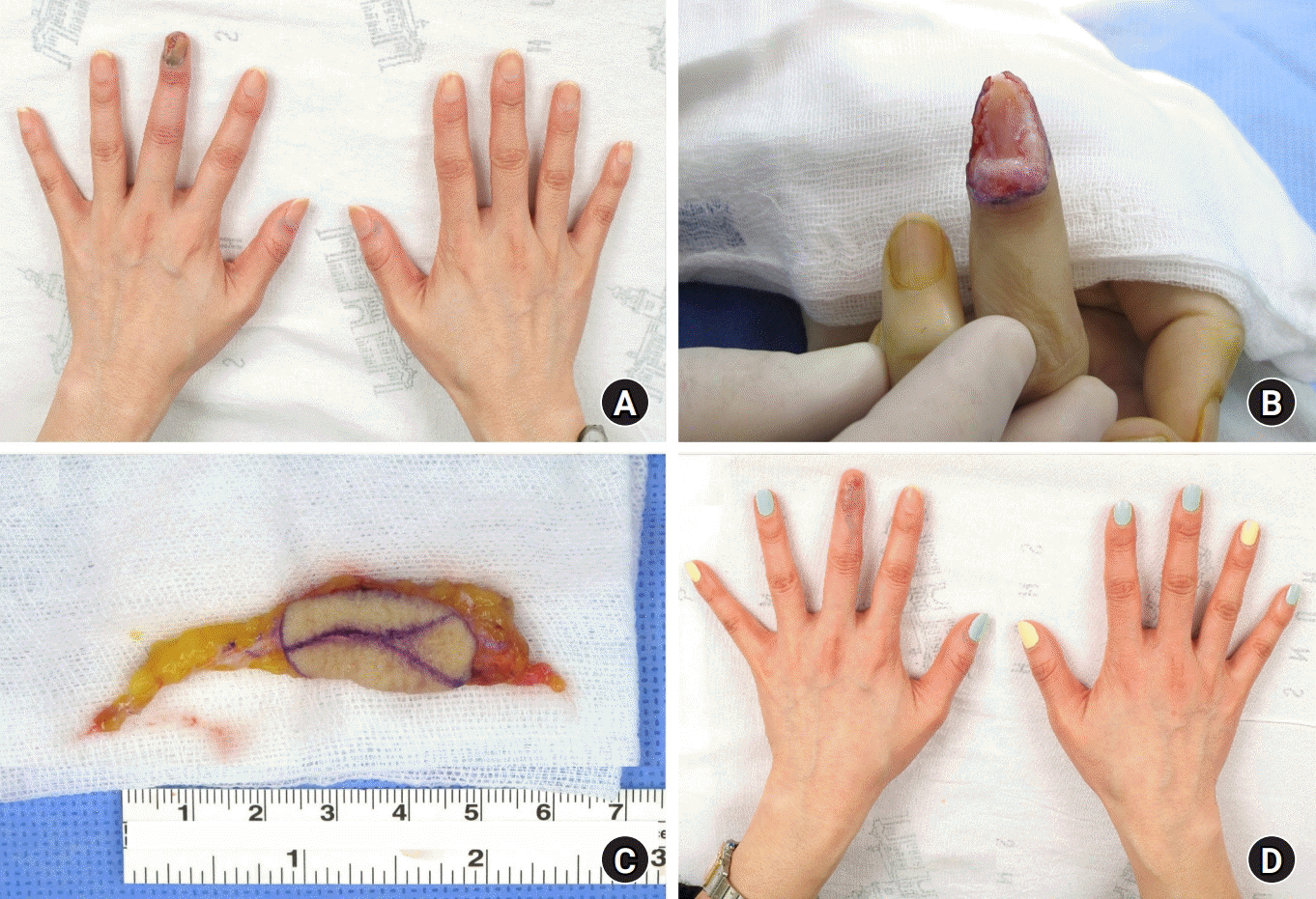
ahm-22-0072f3.tif
Fig. 4.
A 36-year-old female patient with subungual melanoma on the left third finger. (A) Preoperative clinical photograph. (B) En bloc excision of the tumor and nail complex with a 5-mm margin. (C) A venous flap harvested from the volar side of the left forearm was anastomosed with one digital artery and two digital veins. (D) Natural contour of the reconstructed finger without any complications at the 8-month follow-up. Reprinted from Woo et al. [70], according to the Creative Commons License.
ahm-22-0072f4.tif
Fig. 5.
A 64-year-old female patient with subungual melanoma on the right first toe. (A) Preoperative clinical photograph. (B) After inset of a venous flap harvested from the dorsal side of the right foot to allow reversed blood flow with the against-valve type. (C) Clinical photograph of a skin defect that occurred at the center of the flap due to trauma 3 months after surgery. (D) Completely healed flap without further treatment at a 5-year follow-up after debridement and skin grafting for the skin defect. Reprinted from Woo et al. [70], according to the Creative Commons License.
ahm-22-0072f5.tif
TOOLS
Similar articles