Journal List > Ann Rehabil Med > v.47(Suppl 1) > 1516083708

Lee, Son, Kim, and Park: Association of Diaphragm Thickness and Respiratory Muscle Strength With Indices of Sarcopenia

Abstract

Objective

To evaluate the relationship between respiratory muscle strength, diaphragm thickness (DT), and indices of sarcopenia.

Methods

This study included 45 healthy elderly volunteers (21 male and 24 female) aged 65 years or older. Sarcopenia indices, including hand grip strength (HGS) and appendicular skeletal muscle mass/body mass index (ASM/BMI), were measured using a hand grip dynamometer and bioimpedance analysis, respectively. Calf circumference (CC) and gait speed were also measured. Maximal inspiratory pressure (MIP) and maximal expiratory pressure (MEP) were obtained using a spirometer, as a measure of respiratory muscle strength. DT was evaluated through ultrasonography. The association between indices of sarcopenia, respiratory muscle strength, and DT was evaluated using Spearman’s rank correlation test, and univariate and multiple regression analysis.

Results

ASM/BMI (r=0.609, p<0.01), CC (r=0.499, p<0.01), HGS (r=0.759, p<0.01), and gait speed (r=0.319, p<0.05) were significantly correlated with DT. In the univariate linear regression analysis, MIP was significantly associated with age (p=0.003), DT (p<0.001), HGS (p=0.002), CC (p=0.013), and gait speed (p=0.026). MEP was significantly associated with sex (p=0.001), BMI (p=0.033), ASM/BMI (p=0.003), DT (p<0.001), HGS (p<0.001), CC (p=0.001) and gait speed (p=0.004). In the multiple linear regression analysis, age (p=0.001), DT (p<0.001), and ASM/BMI (p=0.008) showed significant association with MIP. DT (p<0.001) and gait speed (p=0.050) were associated with MEP.

Conclusion

Our findings suggest that respiratory muscle strength is associated with DT and indices of sarcopenia. Further prospective studies with larger sample sizes are needed to confirm these findings.

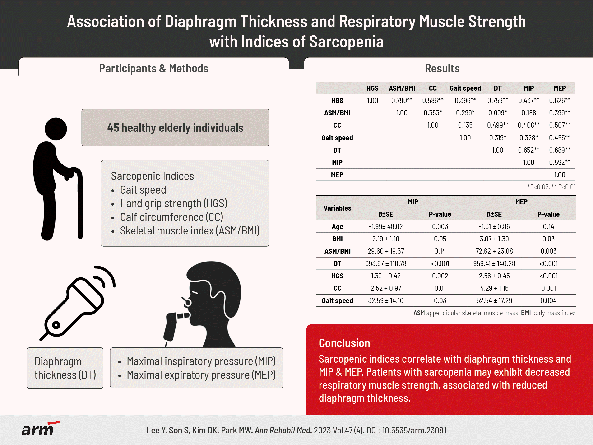
GRAPHICAL ABSTRACT

INTRODUCTION

Sarcopenia is a geriatric syndrome associated with loss of skeletal muscle mass and muscle strength [1]. Sarcopenia commonly occurs as an age-related process and is also influenced by malnutrition, inactivity, disease, and other iatrogenic factors [2]. Sarcopenia is associated with low quality of life, increased risk of falls and fractures, disability, and loss of independence [3].
The association between sarcopenia and respiratory diseases has been previously documented. One study reported a high prevalence of about 60% of sarcopenia in patients with respiratory failure [4]. On the other hand, a cross-sectional study based on the 2008–2011 Korean National Health and Nutritional Examination Survey showed that lower skeletal muscle mass is associated with reduced respiratory function in the elderly [5]. Reduced respiratory muscle strength can also impact respiratory health. A study conducted in Japan reported that respiratory muscle weakness and lower body trunk muscle mass increased the risk for pneumonia in older people [6]. A systematic review of adults with respiratory muscle weakness after stroke showed that respiratory muscle strength training decreased the risk of respiratory complications [7].
Diaphragm thickness (DT) can be decreased in a range of disease states, including sarcopenia. Ultrasonographic evaluation of the diaphragm showed that DT is reduced in patients on prolonged mechanical ventilation [8]. A study by Deniz et al. [9] revealed a significant reduction in DT among individuals with sarcopenia compared to non-sarcopenic elderly individuals. This reduction in DT is concerning, as it can contribute to diaphragm dysfunction and respiratory complications [10]. Recently, the concept of respiratory sarcopenia has emerged and ultrasonographic evaluation of DT has been proposed as a measure of respiratory muscle mass [11]. However, evaluation of the diaphragm by ultrasound is usually performed in the intensive care unit setting, and is not routinely measured in sarcopenia patients [12].
While previous studies have examined the correlation between respiratory muscle strength and sarcopenia [13], as well as DT and sarcopenia [9], there is a dearth of research investigating the association among all three factors simultaneously. Hence, our study aimed to assess the relationship between respiratory muscle strength, diaphragm muscle, and indices of sarcopenia within a single investigation.

METHODS

Study population

Healthy adult volunteers (25 male and 25 female) aged 65 years or older were consecutively recruited for this cross-sectional study through advertisements. Participants with functional problems due to lung disease (such as lung cancer, history of lung surgery, chronic obstructive pulmonary disease, asthma, or tuberculosis), diseases which can affect sarcopenia (such as stroke, spinal cord injury, or peripheral neuropathy), or a history of major joint surgery were excluded. They were informed of the purpose and nature of the study and signed the written consent form. The study was approved by the Institutional Review Board of Chung-Ang University Hospital (No. 1751-003-281).

Skeletal muscle mass assessment

Bioelectrical impedance analysis (BIA), InBody S10 (Biospace) was used to measure skeletal muscle mass. BIA is a non-invasive, easy to administer tool for measuring body composition [14]. The participants were instructed to avoid eating or doing exercises at least 8 hours before the study. After measuring the height and weight, electrodes were attached to the four extremities of the participants in the supine position. Appendicular skeletal muscle mass (ASM) was obtained through the body composition analysis. Appendicular skeletal muscle mass/body mass index (ASM/BMI) was calculated as follows [15]:
ASM/BMI=appendicular skeletal muscle (kg)/body mass index
Body mass index=weight (kg)/height (m)2

Thigh and calf circumference measurement

Thigh and calf circumference (CC) was measured with the patient in the supine position. The left knee was raised to form a 90° between the calf and the thigh [16]. The tape measure was placed around the left calf and thigh, and the maximal circumference was measured without compressing the subcutaneous tissue.

Muscle strength and physical performance measurements

Handgrip strength (HGS) was measured using a hand-grip dynamometer, T.K.K.5401 (Takei Scientific Instruments). Participants were asked to assume the following position: adduct and neutrally rotate the shoulder, flex the elbow to 90°, and place the forearm in a neutral position, with the wrist between 0° and 30° extension and between 0° and 15° ulnar deviation while sitting in a straight-backed chair [17]. Instruction was given to squeeze the grip handle as hard as possible for 3 seconds, and the maximum contraction force (kg) was recorded. The tests were performed three times in each hand with a 60-second rest between each trial. The average of the three values was used for the analysis.
Gait speed was measured to evaluate physical performance, and function of the lower extremities. Gait speed was evaluated on a hard surface by measuring the time taken to walk 4 m at one’s usual walking pace [18]. The participant walked a total 9-m distance, with 2.5 m at the start and end used for acceleration and deceleration. The measurements of three trials were averaged and used for the analysis.

Respiratory muscle strength

Maximal expiratory pressure (MEP) and maximal inspiratory pressure (MIP) were used as a measure of expiratory and inspiratory muscle strength [19]. MEP and MIP were measured in the sitting position using the portable spirometer (Pony FX; COSMED) [20]. To minimize errors, an experienced operator coached the subjects to completely seal their lips around the mouthpiece to prevent air leakage. Participants were encouraged to maximally expire for MEP measurements and to maximally inspire for MIP measurements. At least five trials were performed under supervision and the maximum value between trials which varied by less than 20% were recorded [21]. Each test was performed with a 1-minute break.

DT measurement

DT was measured by B-mode ultrasound using a 7.5 MHz linear transducer (SONOACE R7, Samsung Medison Inc.). The measurement of the diaphragm was conducted at the right side, at the zone of apposition in the 8th or 9th intercostal space as described by De bruin et al. [22]. The probe was placed between the anterior and mix-axillary lines. The participant was in the sitting position and measurements of the diaphragm were taken at the end of expiration (DTe) and inspiration (DTi) during quiet breathing by a single experienced physician. The mean value of DTe and DTi was used as a measure of DT. Thickening fraction (TF) of the diaphragm during quiet breathing was also calculated as follows [23].
TF=DTi-DTeDTe×100

Statistical analysis

The baseline characteristics and measurement of participants are presented as the mean±standard deviation. The Mann–Whitney test was used to compare differences between the sexes. The Spearman’s rank correlation test was used to evaluate correlation between the DT and other indices of sarcopenia. Linear regression analysis was used to evaluate association between respiratory muscle strength, DT and indices of sarcopenia. Multiple linear regression analysis with backward elimination was performed to identify factors predictive of respiratory muscle strength. Statistical significance was defined as a p-value of less than 0.05. Statistical analysis was performed using the IBM SPSS Statistics ver. 19.0 (IBM Corp.).

RESULTS

Baseline characteristics

A total of 25 male and 25 female participants were recruited. Five participants with a history of lung disease (chronic obstructive pulmonary disease and asthma) were excluded. The baseline characteristics of the participants are shown in Table 1. The mean age was 76.76±1.13 years for male (n=21), and 76.42±1.03 years for female (n=24). Height, weight, HGS, CC, MEP, DT, and ASM/BMI were significantly different according to sex. Measures of gait speed, HGS, and CC were comparable to previously published normal range for age and sex [24].

Respiratory muscle strength, DT, and indices of sarcopenia

HGS (r=0.759, p<0.01), ASM/BMI (r=0.609, p<0.01), CC (r=0.499, p<0.01), and gait speed (r=0.319, p<0.05) were significantly correlated with DT. Additionally, HGS (r=0.437, p<0.01), CC (r=0.408, p<0.01), gait speed (r=0.328, p<0.05), and DT (r=0.652, p<0.01) showed significant correlations with MIP. Lastly, significant correlations were found between HGS (r=0.626, p<0.01), ASM/BMI (r=0.399, p<0.01), CC (r=0.507, p<0.01), gait speed (r=0.592, p<0.01), and DT (r=0.689, p<0.01) with MEP (Table 2).
In the univariate linear regression analysis, MIP was significantly associated with age (p=0.003), DTi (p<0.001), DTe (p<0.001), DT (p<0.001), HGS (p=0.002), CC (p=0.013), and gait speed (p=0.026). MEP was significantly associated with sex (p=0.001), BMI (p=0.033), ASM/BMI (p=0.003), DTi (p<0.001), DTe (p<0.001), DT (p<0.001), HGS (p<0.001), CC (p=0.001), and gait speed (p=0.004; Table 3).
In the multiple linear regression analysis with backward elimination, age (p=0.001), DT (p<0.001), and ASM/BMI (p=0.008) showed significant association with MIP. DT (p<0.001) and gait speed (p=0.050) were found to be associated with MEP (Table 4).

DISCUSSION

In this study we have demonstrated that indices of sarcopenia, DT, MIP, and MEP were intercorrelated with each other. In the univariate analysis, DTi, DTe, DT, HGS, CC, and gait speed were significantly associated with both MIP and MEP. Age was significantly associated with MIP and sex and ASM/BMI with MEP only. In the multivariate linear regression analysis DT showed significant association with both MIP and MEP. Age and ASM/BMI were significantly associated with MIP.
The diaphragm is a skeletal muscle like limb muscles. It is composed of roughly equal proportions of slow and fast fibers [25]. The main difference lies in structure and not composition. Compared to limb muscles, diaphragm fibers have smaller cross sectional area, allowing efficient oxygen supply and increased resistance to fatigue [25]. Due to similarity in fiber composition, the diaphragm muscle may be affected in situations where skeletal muscle wasting occurs, such as sarcopenia. An animal study found that sarcopenic rats induced by genetic modification have thinner diaphragm and weaker respiratory muscle strength than normal rats [26]. In their study of 30 sarcopenic and 30 non-sarcopenic elderly patients aged over 65, Deniz et al. [9] reported that DT was significantly reduced in the sarcopenic compared to the non-sarcopenic elderly individuals. Similarly, in our study of health elderly people, indices of sarcopenia were significantly associated with DT. Our findings suggest that the diaphragm may be affected in sarcopenia.
Respiratory muscle strength is closely related to diaphragmatic, abdominal, and intercostal muscle strength [21] and can be reduced in a range of diseases such as stroke [27], spinal cord injury [28], and neuromuscular disease [29]. Findings from previous studies suggest that respiratory muscle strength may also be reduced in sarcopenia. In a study on healthy elderly by Shin et al. [30], skeletal muscle mass index was significantly associated with MIP. In a cross-sectional study by Ohara et al. [31], MEP and MIP were associated with sarcopenia indicators such as muscle mass, hand grip strength, and gait speed. Our study findings were similar to prior research, illustrating a correlation between respiratory muscle strength and indicators of sarcopenia.
The diaphragm is a primary inspiratory muscle which contracts during inhalation and relaxes during exhalation. Its correlation with MIP is apparent, given its fundamental role, and has been reported previously [32]. However, the reason DT was associated with MEP is not readily discernible. One possible explanation is that expiration was enhanced by greater elastic recoil of the rib cage and lung after stronger inspiration. Another possibility is that the DT was increased in subjects with already strong expiratory muscles. In a study by Souza et al. [33], elderly female who underwent inspiratory muscle strengthening training showed significant increases in MIP, MEP, and DT compared to the control group, indicating a positive correlation between respiratory muscle strength and DT. Our study demonstrated that DT is significantly associated with both inspiratory and expiratory muscle strength.
In the multiple regression analysis, MIP was negatively correlated with age and ASM/BMI, and positively correlated with DT. Age related decrease in MIP has been described before [34]. Possible explanations include age related muscle atrophy and loss of fast twitch fibers [34]. The association between MIP and skeletal muscle mass have also been reported in previous studies. In a study by Ro et al. [35], skeletal muscle mass was significantly associated with MIP in both young male and female. Similarly, Shin et al. [30] also reported that skeletal muscle mass showed significant correlation with MIP in the healthy elderly. Contrary to our study results, in both studies, the correlation was positive. However, they did not evaluate DT, a significant determinant of inspiratory strength. The reason skeletal muscle mass, which is a measure of limb muscle, was significantly negatively associated with MIP is unclear. Obesity may have been a contributing factor. In general, people who are obese tend to have larger muscle mass [36]. Obesity can affect respiratory function through mechanical factors and metabolic effects associated with proinflammatory state [37]. However, measures of obesity and central obesity such as waist circumference, and lipid profile were not evaluated. Skeletal muscle mass was measured using BIA, which may not be as accurate as dual-energy X-ray absorptiometry (DXA) [38]. If surrogate measures such as DXA, limb muscle ultrasound, magnetic resonance imaging were used results could have been different. Further studies are needed to validate this.
MEP was also significantly correlated with DT but not ASM/BMI in the multiple regression analysis. The significant positive correlation between ASM/BMI and MEP in the univariate linear regression analysis was not observed in the multiple regression analysis. This may be due to the fact that ASM/BMI primarily reflects limb muscle mass rather than trunk muscles, which are more closely associated with expiratory muscle strength [21]. The results seem to indicate that the ASM/BMI has a lesser role in predicting expiratory strength. Therefore, it may be necessary to measure DT separately as an additional indicator of respiratory sarcopenia [39].
There are some limitations to this study. First, this was a cross-sectional study of healthy elderly volunteers with small sample size. Causal relationships cannot be confirmed and caution is needed in generalizing the findings. Second, skeletal muscle mass was measured by BIA. BIA may overestimate skeletal muscle mass compared to DXA [38]. However, studies have shown that BIA was reliable in measuring muscle mass, and strongly correlated with skeletal muscle measurement by DXA [40]. Third, TF of the diaphragm was measured only during quiet breathing and not maximal breathing. TF during maximal breathing may be better correlated with maximal respiratory pressure. Lastly, other possible confounding factors such as abdominal muscles involved in respiration were not evaluated.
This was the first study to demonstrate correlation between respiratory muscle strength, DT, and skeletal muscle mass in healthy Korean elderly. Sarcopenia patients may have decreased respiratory muscle strength associated with reduced DT. Therefore, assessment of respiratory muscle strength and DT may be needed in sarcopenia patients to prevent respiratory functional decline. Further studies are necessary to evaluate changes in DT in patients with sarcopenia and whether early interventions may help prevent pulmonary complications.

Notes

No potential conflict of interest relevant to this article was reported.

None.

Conceptualization: Son S, Kim DK. Methodology: Son S, Park MW, Kim DK. Formal analysis: Lee Y, Son S, Park MW, Kim DK. Visualization: Lee Y, Son S. Writing – original draft: Lee Y, Son S. Writing – review and editing: Lee Y, Park MW, Kim DK. Approval of final manuscript: all authors.

REFERENCES

1. Cruz-Jentoft AJ, Landi F, Topinková E, Michel JP. Understanding sarcopenia as a geriatric syndrome. Curr Opin Clin Nutr Metab Care. 2010; 13:1–7.
crossref
2. Cruz-Jentoft AJ, Sayer AA. Sarcopenia. Lancet 2019;393:2636-46. Erratum in: Lancet. 2019; 393:2590.
3. Cruz-Jentoft AJ, Bahat G, Bauer J, Boirie Y, Bruyère O, Cederholm T, et al. Sarcopenia: revised European consensus on definition and diagnosis. Age Ageing 2019;48:16-31. Erratum in: Age Ageing. 2019; 48:601.
4. Sheean PM, Peterson SJ, Gomez Perez S, Troy KL, Patel A, Sclamberg JS, et al. The prevalence of sarcopenia in patients with respiratory failure classified as normally nourished using computed tomography and subjective global assessment. JPEN J Parenter Enteral Nutr. 2014; 38:873–9.
crossref
5. Jeon YK, Shin MJ, Kim MH, Mok JH, Kim SS, Kim BH, et al. Low pulmonary function is related with a high risk of sarcopenia in community-dwelling older adults: the Korea National Health and Nutrition Examination Survey (KNHANES) 2008-2011. Osteoporos Int. 2015; 26:2423–9.
crossref
6. Okazaki T, Suzukamo Y, Miyatake M, Komatsu R, Yaekashiwa M, Nihei M, et al. Respiratory muscle weakness as a risk factor for pneumonia in older people. Gerontology. 2021; 67:581–90.
crossref
7. Menezes KK, Nascimento LR, Ada L, Polese JC, Avelino PR, Teixeira-Salmela LF. Respiratory muscle training increases respiratory muscle strength and reduces respiratory complications after stroke: a systematic review. J Physiother 2016;62:138-44. Erratum in: J Physiother. 2018; 64:73.
crossref
8. Goligher EC, Dres M, Fan E, Rubenfeld GD, Scales DC, Herridge MS, et al. Mechanical ventilation-induced diaphragm atrophy strongly impacts clinical outcomes. Am J Respir Crit Care Med. 2018; 197:204–13.
crossref
9. Deniz O, Coteli S, Karatoprak NB, Pence MC, Varan HD, Kizilarslanoglu MC, et al. Diaphragmatic muscle thickness in older people with and without sarcopenia. Aging Clin Exp Res. 2021; 33:573–80.
crossref
10. Cavayas YA, Eljaiek R, Rodrigue É, Lamarche Y, Girard M, Wang HT, et al. Preoperative diaphragm function is associated with postoperative pulmonary complications after cardiac surgery. Crit Care Med. 2019; 47:e966–74.
crossref
11. Sato S, Miyazaki S, Tamaki A, Yoshimura Y, Arai H, Fujiwara D, et al. Respiratory sarcopenia: a position paper by four professional organizations. Geriatr Gerontol Int. 2023; 23:5–15.
crossref
12. Le Neindre A, Philippart F, Luperto M, Wormser J, Morel-Sapene J, Aho SL, et al. Diagnostic accuracy of diaphragm ultrasound to predict weaning outcome: a systematic review and meta-analysis. Int J Nurs Stud. 2021; 117:103890.
crossref
13. Martínez-Arnau FM, Buigues C, Fonfría-Vivas R, Cauli O. Respiratory muscle strengths and their association with lean mass and handgrip strengths in older institutionalized individuals. J Clin Med. 2020; 9:2727.
crossref
14. Walter-Kroker A, Kroker A, Mattiucci-Guehlke M, Glaab T. A practical guide to bioelectrical impedance analysis using the example of chronic obstructive pulmonary disease. Nutr J. 2011; 10:35.
crossref
15. Cawthon PM, Peters KW, Shardell MD, McLean RR, Dam TT, Kenny AM, et al. Cutpoints for low appendicular lean mass that identify older adults with clinically significant weakness. J Gerontol A Biol Sci Med Sci. 2014; 69:567–75.
crossref
16. Bonnefoy M, Jauffret M, Kostka T, Jusot JF. Usefulness of calf circumference measurement in assessing the nutritional state of hospitalized elderly people. Gerontology. 2002; 48:162–9.
crossref
17. Innes E. Handgrip strength testing: a review of the literature. Aust Occup Ther J. 1999; 46:120–40.
crossref
18. Bianchi L, Ferrucci L, Cherubini A, Maggio M, Bandinelli S, Savino E, et al. The predictive value of the EWGSOP definition of sarcopenia: results from the InCHIANTI study. J Gerontol A Biol Sci Med Sci. 2016; 71:259–64.
crossref
19. Gomes-Neto M, Saquetto MB, Silva CM, Carvalho VO, Ribeiro N, Conceição CS. Effects of respiratory muscle training on respiratory function, respiratory muscle strength, and exercise tolerance in patients poststroke: a systematic review with meta-analysis. Arch Phys Med Rehabil. 2016; 97:1994–2001.
crossref
20. Bambra G, Jalota L, Kapoor C, Mills PK, Vempilly JJ, Jain VV. Office spirometry correlates with laboratory spirometry in patients with symptomatic asthma and COPD. Clin Respir J. 2017; 11:805–11.
crossref
21. American Thoracic Society (ATS)/European Respiratory Society (ERS). ATS/ERS statement on respiratory muscle testing. Am J Respir Crit Care Med. 2002; 166:518–624.
22. De Bruin PF, Ueki J, Bush A, Khan Y, Watson A, Pride NB. Diaphragm thickness and inspiratory strength in patients with Duchenne muscular dystrophy. Thorax. 1997; 52:472–5.
crossref
23. Santana PV, Cardenas LZ, Albuquerque ALP, Carvalho CRR, Caruso P. Diaphragmatic ultrasound: a review of its methodological aspects and clinical uses. J Bras Pneumol. 2020; 46:e20200064.
24. Kim M, Won CW. Sarcopenia in Korean community-dwelling adults aged 70 years and older: application of screening and diagnostic tools from the Asian Working Group for Sarcopenia 2019 update. J Am Med Dir Assoc. 2020; 21:752–8.
25. Polla B, D'Antona G, Bottinelli R, Reggiani C. Respiratory muscle fibres: specialisation and plasticity. Thorax. 2004; 59:808–17.
crossref
26. Greising SM, Mantilla CB, Gorman BA, Ermilov LG, Sieck GC. Diaphragm muscle sarcopenia in aging mice. Exp Gerontol. 2013; 48:881–7.
crossref
27. Pollock RD, Rafferty GF, Moxham J, Kalra L. Respiratory muscle strength and training in stroke and neurology: a systematic review. Int J Stroke. 2013; 8:124–30.
crossref
28. Berlowitz DJ, Tamplin J. Respiratory muscle training for cervical spinal cord injury. Cochrane Database Syst Rev. 2013; (7):CD008507.
crossref
29. Silva IS, Pedrosa R, Azevedo IG, Forbes AM, Fregonezi GA, Dourado Junior ME, et al. Respiratory muscle training in children and adults with neuromuscular disease. Cochrane Database Syst Rev. 2019; 9:CD011711.
crossref
30. Shin HI, Kim DK, Seo KM, Kang SH, Lee SY, Son S. Relation between respiratory muscle strength and skeletal muscle mass and hand grip strength in the healthy elderly. Ann Rehabil Med. 2017; 41:686–92.
crossref
31. Ohara DG, Pegorari MS, Oliveira Dos Santos NL, de Fátima Ribeiro Silva C, Monteiro RL, Matos AP, et al. Respiratory muscle strength as a discriminator of sarcopenia in community-dwelling elderly: a cross-sectional study. J Nutr Health Aging. 2018; 22:952–8.
crossref
32. Summerhill EM, Angov N, Garber C, McCool FD. Respiratory muscle strength in the physically active elderly. Lung. 2007; 185:315–20.
crossref
33. Souza H, Rocha T, Pessoa M, Rattes C, Brandão D, Fregonezi G, et al. Effects of inspiratory muscle training in elderly women on respiratory muscle strength, diaphragm thickness and mobility. J Gerontol A Biol Sci Med Sci. 2014; 69:1545–53.
crossref
34. Sharma G, Goodwin J. Effect of aging on respiratory system physiology and immunology. Clin Interv Aging. 2006; 1:253–60.
35. Ro HJ, Kim DK, Lee SY, Seo KM, Kang SH, Suh HC. Relationship between respiratory muscle strength and conventional sarcopenic indices in young adults: a preliminary study. Ann Rehabil Med. 2015; 39:880–7.
36. Lear SA, Kohli S, Bondy GP, Tchernof A, Sniderman AD. Ethnic variation in fat and lean body mass and the association with insulin resistance. J Clin Endocrinol Metab. 2009; 94:4696–702.
37. Hegewald MJ. Impact of obesity on pulmonary function: current understanding and knowledge gaps. Curr Opin Pulm Med. 2021; 27:132–40.
38. Buckinx F, Reginster JY, Dardenne N, Croisiser JL, Kaux JF, Beaudart C, et al. Concordance between muscle mass assessed by bioelectrical impedance analysis and by dual energy X-ray absorptiometry: a cross-sectional study. BMC Musculoskelet Disord. 2015; 16:60.
39. Nagano A, Wakabayashi H, Maeda K, Kokura Y, Miyazaki S, Mori T, et al. Respiratory sarcopenia and sarcopenic respiratory disability: concepts, diagnosis, and treatment. J Nutr Health Aging. 2021; 25:507–15.
40. Yamada Y, Nishizawa M, Uchiyama T, Kasahara Y, Shindo M, Miyachi M, et al. Developing and validating an age-independent equation using multi-frequency bioelectrical impedance analysis for estimation of appendicular skeletal muscle mass and establishing a cutoff for sarcopenia. Int J Environ Res Public Health. 2017; 14:809.

arm-23081f1.tif
Table 1.
Baseline characteristics
Characteristics Total (n=45) Male (n=21) Female (n=24) p-value
Age 76.58±0.76 76.76±1.13 76.42±1.03 0.864
Height (cm) 155.02±1.32 162.86±0.81 148.17±1.17 <0.001
Weight (kg) 58.22±1.49 64.14±1.75 53.04±1.76 <0.001
BMI (km/m2) 24.14±3.06 24.15±2.58 24.13±3.49 0.891
Gait speed (m/s) 1.02±0.04 1.06±0.05 0.98±0.05 0.270
HGS (kg) 21.61±1.13 28.03±1.17 15.99±0.76 <0.001
CC (cm) 32.97±0.50 34.41±0.70 31.71±0.63 0.011
TC (cm) 38.31±0.55 38.33±0.74 38.29±0.82 0.973
MIP (cmH2O) 66.49±3.45 71.81±5.63 61.83±4.05 0.255
MEP (cmH2O) 106.36±4.39 121.76±6.16 92.88±4.83 0.001
DTi (cm) 0.20±0.02 0.22±0.02 0.19±0.01 <0.001
DTe (cm) 0.18±0.02 0.19±0.02 0.16±0.02 <0.001
DT (cm) 0.19±0.03 0.20±0.00 0.17±0.00 <0.001
TF 15.04±5.56 14.66±4.86 15.38±6.20 0.458
ASM/BMI 0.76±0.03 0.91±0.02 0.62±0.02 <0.001

BMI, body mass index; HGS, hand grip strength; CC, calf circumference; TC, thigh circumference; MIP, maximal inspiratory pressure; MEP, maximal expiratory pressure; DTi, diaphragm thickness at the end of inspiration; DTe, diaphragm thickness at the end of expiration; TF, thickening fraction; ASM/BMI, appendicular skeletal muscle mass/body mass index.

Table 2.
Spearman’s correlation analysis of indices of sarcopenia, respiratory muscle strength, and DT
HGS ASM/BMI CC Gait speed DT MIP MEP
HGS 1.00 0.790** 0.586** 0.396** 0.759** 0.437** 0.626**
ASM/BMI 1.00 0.353* 0.299* 0.609** 0.188 0.399**
CC 1.00 0.135 0.499** 0.408** 0.507**
Gait speed 1.00 0.319* 0.328* 0.455**
DT 1.00 0.652** 0.689**
MIP 1.00 0.592**
MEP 1.00

DT, diaphragm thickness; HGS, hand grip strength; ASM/BMI, appendicular skeletal muscle mass/body mass index; CC, calf circumference; MIP, maximal inspiratory pressure; MEP, maximal expiratory pressure.

* p<0.05 and

** p<0.01.

Table 3.
Associations of age, sex, indices of sarcopenia, and DT with MIP and MEP by univariate linear regression analysis
Variables MIP
MEP
β±SE p-value β±SE p-value
Age -1.99±48.02 0.003 -1.31±0.86 0.136
Sex -9.98±6.82 0.151 -28.89±7.74 0.001
BMI 2.19±1.10 0.053 3.07±1.39 0.033
ASM/BMI 29.60±19.57 0.138 72.62±23.08 0.003
DTi 670.17±116.99 <0.001 912.46±140.81 <0.001
DTe 763.33±127.56 <0.001 904.16±171.51 <0.001
DT 693.67±118.78 <0.001 959.41±140.28 <0.001
TF -0.50±0.63 0.427 0.48±0.80 0.553
HGS 1.39±0.42 0.002 2.56±0.45 <0.001
CC 2.52±0.97 0.013 4.29±1.16 0.001
Gait speed 32.59±14.10 0.026 52.54±17.29 0.004

DT, diaphragm thickness; MIP, maximal inspiratory pressure; MEP, maximal expiratory pressure; SE, standard error; BMI, body mass index; ASM/BMI, appendicular skeletal muscle mass/body mass index; DTi, diaphragm thickness at the end of inspiration; DTe, diaphragm thickness at the end of expiration; DT, diaphragm thickness; TF, thickening fraction; HGS, hand grip strength; CC, calf circumference.

Table 4.
Associations of age, sex, indices of sarcopenia, and DT with MIP and MEP by multiple linear regression analysis
Variables MIP
MEP
β±SE p-value β±SE p-value
Age -1.66±0.44 0.001 - -
ASM/BMI -45.01±16.06 0.008 - -
DT 852.80±126.98 <0.001 869.70±142.67 <0.001
Gait speed - - 27.01±13.41 0.050

DT, diaphragm thickness; MIP, maximal inspiratory pressure; MEP, maximal expiratory pressure; SE, standard error; ASM/BMI, appendicular skeletal muscle mass/body mass index.

TOOLS
Similar articles