

 PDF
PDF Citation
 Print
Print


INTRODUCTION
Situs inversus totalis (SIT) is a rare phenomenon that presents as an inversion of cardiac and abdominalbody organs, unlike their left-right orientation. A congenital anomaly with a flip in the organ orientation leads to difficulties in diagnoses and surgical procedures. Mirizzi syndrome, a condition where the common hepatic duct or common bile duct (CBD) is obstructed by gallstone, is a rare condition. While the global incidence of Mirrizzi syndrome is approximately 0.7% and the incidence of SIT is 1 in 6,500, what’s more, the co-occurrence of Mirizzi syndrome in patients with SIT is extremely rare [1,2]. Gallbladder in sinistroposition, or the presence of the gallbladder on the left side, is highly unusual in people with SIT [3].
Ventricular septal defect (VSD) is distinguished by an improper communication between the right and left ventricles and the creation of a shunt, which is the primary mechanism of hemodynamic compromise. The majority of VSDs close on their own, however, if they do not, big defects can lead to complications such as pulmonary arterial hypertension and ventricular dysfunction and an increased risk of arrhythmias. VSD develops during the intricate embryologic heart morphogenesis, when there is a developmental defect or an interruption of the interventricular septum formation [4].
Transposition of the great arteries (TGA), sometimes known as full transposition, is a congenital heart abnormality characterized by atrioventricular concordance as well as ventriculoarterial (VA) discordance. In VA, the morphological left atrium is connected to the morphological left ventricle, which is where the pulmonary trunk arises, and the morphological right atrium is connected to the morphological right ventricle, which gives rise to all or most of the aorta [5].
This is a case report of an SIT patient with Mirizzi syndrome type III who successfully underwent laparoscopic cholecystectomy and choledocholithotomy.
Go to : 
CASE
A 32-year-old female patient arrived at the hospital with obstructive jaundice and cholangitis that had lasted for 10 days, accompanied by mild abdominal discomfort, and fever-associated chills. There was no history of vomiting, abdominal pain, or distention of the abdomen. In the past, no similar complaints or symptoms had been observed. Total bilirubin levels were 2.2 mg/dL, while direct bilirubin levels were 0.7 mg/dL. The white blood cell count was 12,870 cells/cumm. The patient was diabetic, with an HbA1C level of 8.5%.
On abdominal ultrasonography (USG) imaging, the gallbladder was left-sided, dilated and had multiple stones with dilatation of the CBD.
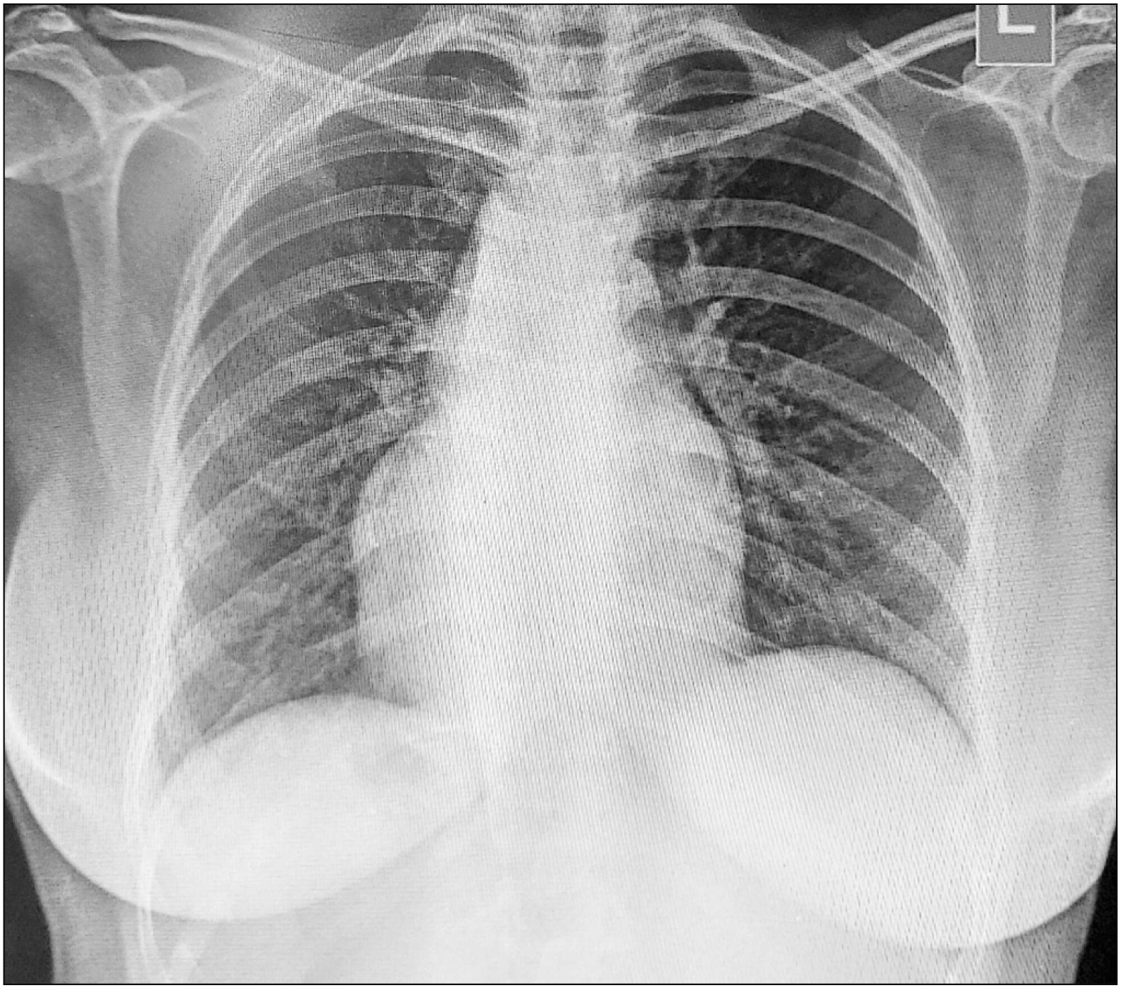
The abdominal USG revealed the manifestation of situs inversus. A chest X-ray revealed dextrocardia, and SIT (Fig. 1). She was also diagnosed with dextrocardia, atrioventricular discordance, VA discordance, significant atrioventricular valve regurgitation (moderate mitral regurgitation and mild aortic regurgitation), L-transposition of the great arteries, and a large ventricle septal defect with an ejection fraction of 40%.
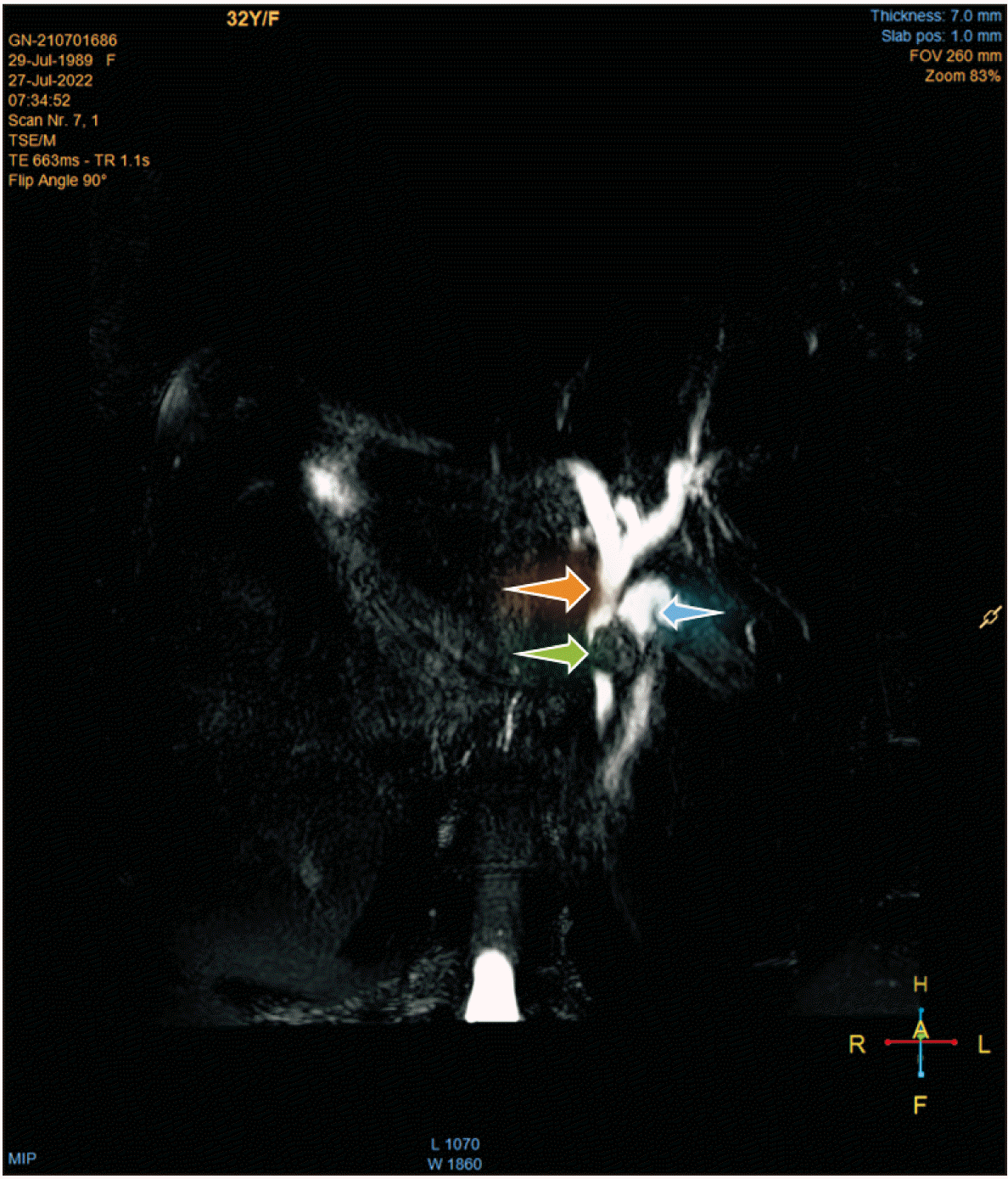
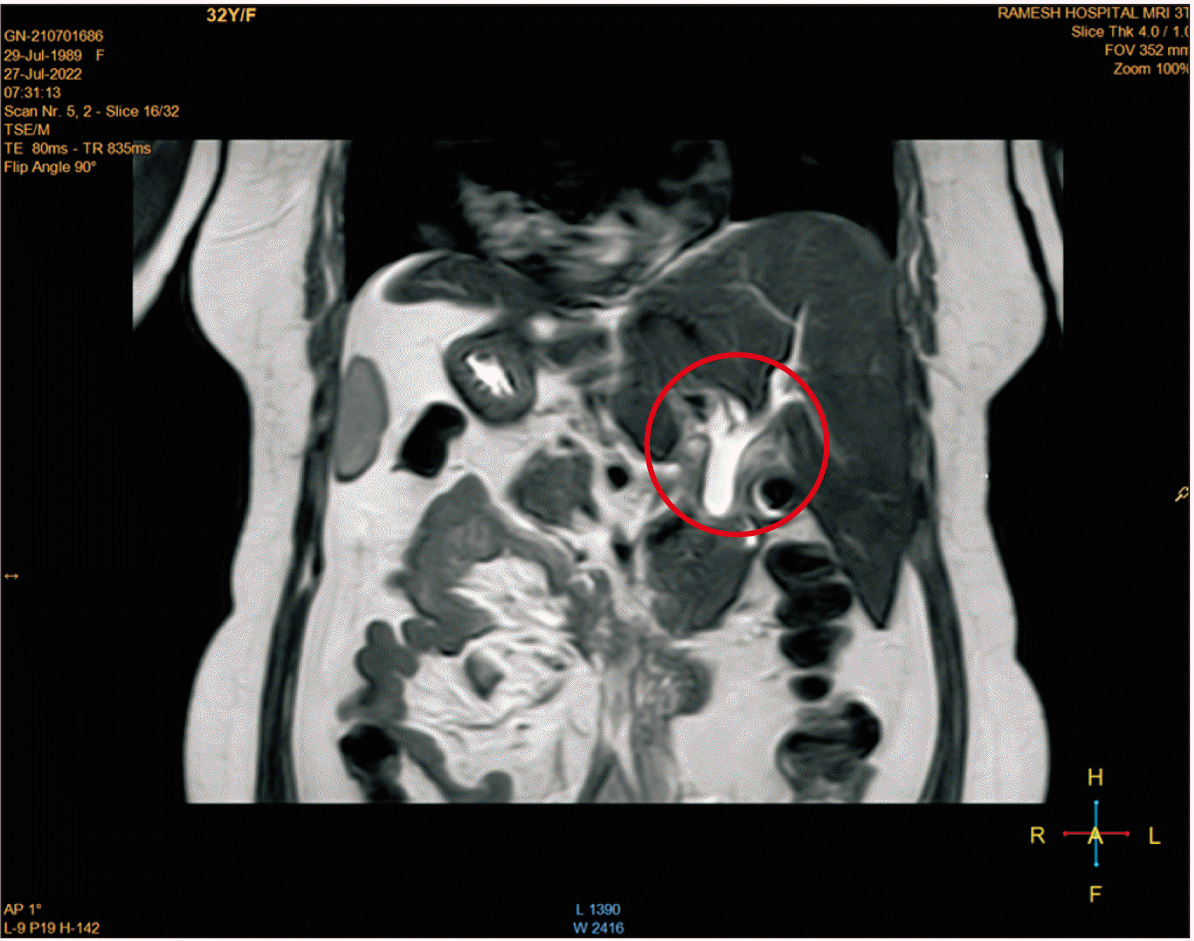
A magnetic resonance cholangiopancreatography (MRCP) revealed a moderately dilated gallbladder and a normal gallbladder wall thickness (Fig. 2). There was an 18 mm calculus along with gallbladder sludge, in the middle segment of the cystic duct, which was compressing the middle CBD with moderate dilatation of the upper CBD and bilobar intrahepatic biliary radical dilatation (IHBRD) (Fig. 3). The pancreas was of normal size and form.
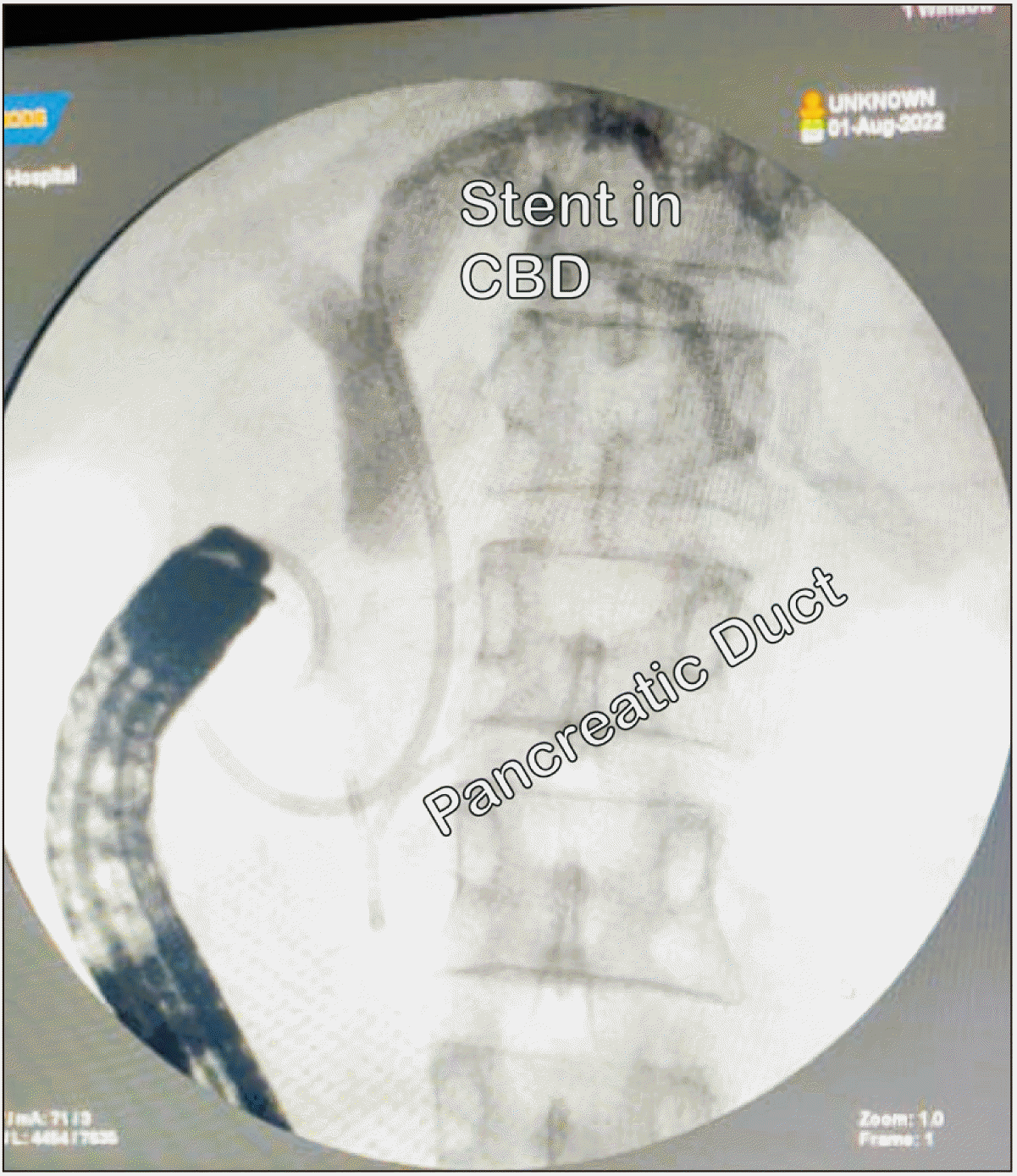
Therapeutic endoscopic retrograde cholangiopancreatography (ERCP) along with CBD and pancreatic duct stenting were done to reduce cholangitis (Fig. 4). During ERCP, attempts to remove the CBD stone were unsuccessful because it was within the gallbladder, which would not budge. As a result, the patient was monitored, and a plan, which included an elective laparoscopic cholecystectomy, CBD choledochotomy, and stone removal, after 6 weeks was made. The timeframe of 6 weeks was chosen in view of complete recovery from cholangitis, to prevent any hindrance to the surgery.
The patient was hospitalized with significant stomach discomfort, fever, and chills during the fourth week. Blood tests revealed a rise in C-reactive protein to 76.9 mg/L, as well as a white blood cell count of 14,900 cells/cumm. The values of the total bilirubin and direct bilirubin were within the normal range, 0.9 mg/dL and 0.5 mg/dL, respectively. The patient was diagnosed with acute febrile illness based on the test results. The patient underwent conservative treatment for fever and pain, and a cholecystectomy was scheduled four weeks after the fever episode.
After 8 weeks, the total bilirubin level was 0.5 mg/dL, while the direct bilirubin level was 0.2 mg/dL. The white blood cell count was 10,100 cells/cumm. A preoperative USG indicated a dilated gallbladder with an edematous, irregular wall and 16 mm of calculus in the neck region. In addition, there was an in situ CBD stent and a 13 mm calculus on the proximal CBD. There was evidence of bilobar IHBRD.
In consideration of the congenital defect and the potential necessity to convert the laparoscopic treatment to an open one, the patient was appropriately briefed, and high-risk consents were obtained. The surgical procedure was carried out under general anesthesia with endotracheal intubation.
The patient was put in supine Anti-Trendelenburg posture with a left tilt of approximately 25 degrees. The surgeon stood on the right side of the patient, as opposed to the left side which is the standard position during surgery. On the left side of the abdomen, exactly opposite the typical right-sided ports, four standard ports were created, including two 10 mm ports in the epigastric and umbilical regions and two 5 mm working ports in the left hypochondrium.
The omentum was adhered to the liver and duodenum, which were themselves glued to the gallbladder. All these numerous adhesions were initially separated. The gallbladder was held at its fundus with a toothed grasper and then raised for traction. The Calot’s triangle was dissected, but the cystic duct could not be distinguished.
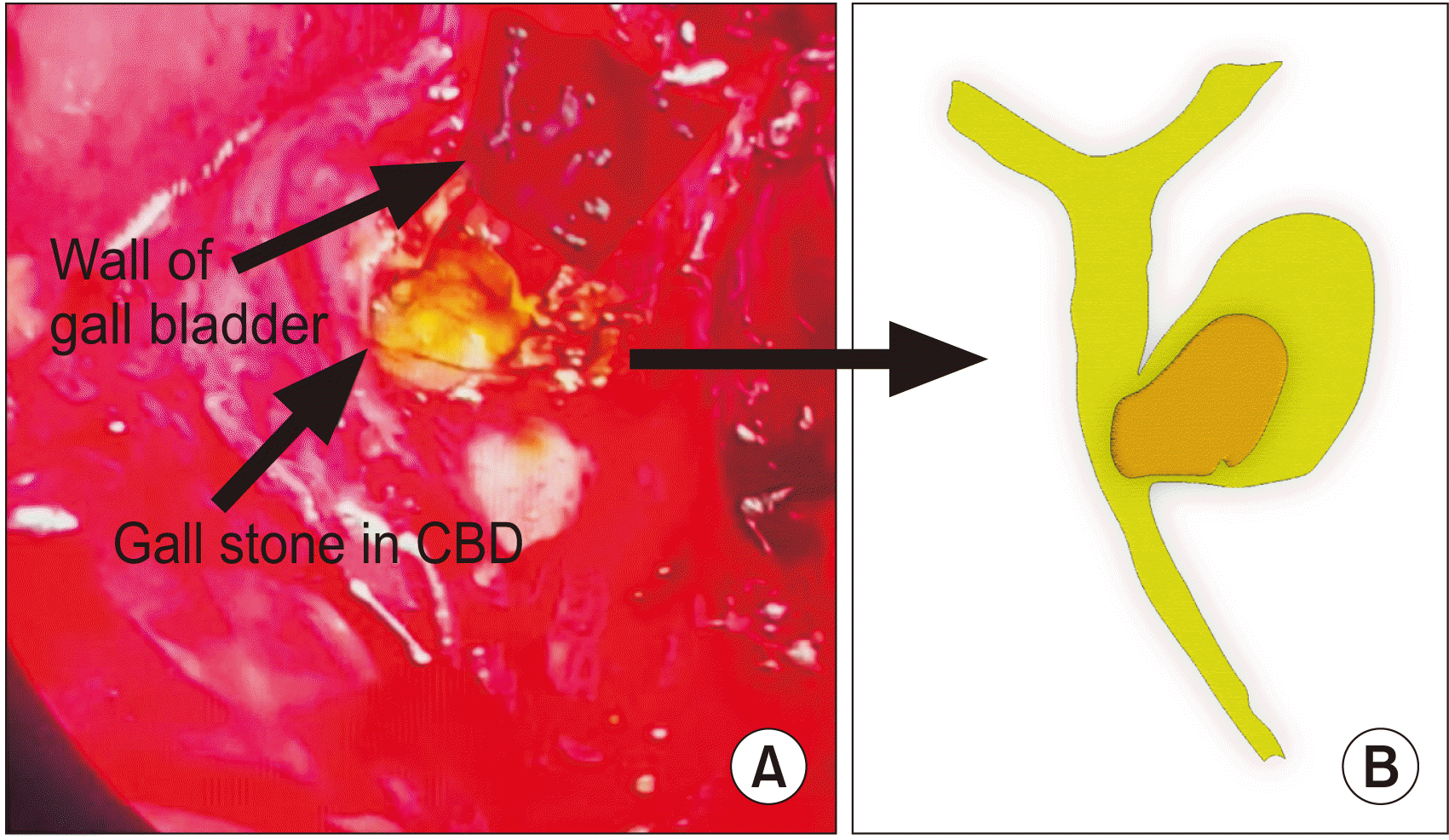
It was discovered that the gallbladder was directly connected to the CBD. In addition, the duodenum was adhered to the Calot’s triangle region. The duodenum was carefully separated from the CBD, the gallbladder was opened close to the CBD, and a gallstone was removed from the neck of the gallbladder. It was observed that the neck of the gallbladder opened straight into the CBD. In addition, it was discovered that the second gallstone had corroded the CBD and occupied two-thirds of its circumference, although the CBD stent remained in place (Fig. 5). The presence of this gallstone deep within the CBD confirmed the Mirizzi syndrome as type III.
Therefore, to remove the stone from the CBD, the CBD was opened up and examined. After the stone was removed, 2.0 vicryl was used for intermittent endo-sutures to suture the CBD. Cholecystectomy was performed after coagulating the cystic artery with bipolar cautery. A tube drain was inserted, and all the ports were closed in layers.
The surgery lasted for 2 hours and 50 minutes from the time of incision to the point of skin closure. The blood loss was negligible, (a total of 20 mL). The postoperative total bilirubin level was 0.6 mg/dL, while the direct bilirubin level was 0.2 mg/dL. The postoperative period was uneventful, and the patient was discharged two days after the successful procedure.
Following surgery, the patient was observed for one month and the CBD stent was removed. Upon the removal of the stent, the patient's total bilirubin and direct bilirubin levels were 0.8 mg/dL and 0.3 mg/dL, respectively. After a 3-month follow-up, USG abdomen showed that the patient was found to have a slight thickness of the CBD wall with lumen measuring 5.2 mm but no abnormalities in the gallbladder region. Total bilirubin was 0.7 mg/dL, while direct bilirubin was 0.2 mg/dL. With no episodes of stomach pain, cholangitis, or jaundice after the cholecystectomy, and the patient was healthy and active.
Go to :
DISCUSSION
SIT, the total inversion of organs, can be described simply as the mirror image of a normal human organ’s anatomy. The organs that are ordinarily located on the left side of the body tend to be inverted. After a detailed physical examination, SIT may be suspected; however, the widespread availability of medical imaging and routine screening programs makes it easy to validate the findings and find more information and pathologies. Traditional imaging modalities, such as ultrasound (US) and plain film X-ray, are typically the first-choice diagnostic imaging modality [6].
Pablo Luis Mirizzi (1893–1964), one of the foremost biliary surgeons, was the first to describe Mirizzi syndrome [7]. A biliary surgeon who is aware of the patient's danger faces a unique challenge in identifying and treating Mirizzi syndrome. In addition, surgery for Mirizzi syndrome is very risky due to the considerable rise in the risk of intraoperative biliary damage during cholecystectomy. Due to the varied clinical presentation, there is currently no standard treatment for Mirizzi syndrome. After a thorough analysis of the local circumstances and anatomy, surgical treatment should be planned [8]. There are 5 types of Mirizzi syndrome based on the position of the gallstone in the gallbladder in relation to CBD (Fig. 6, Table 1).
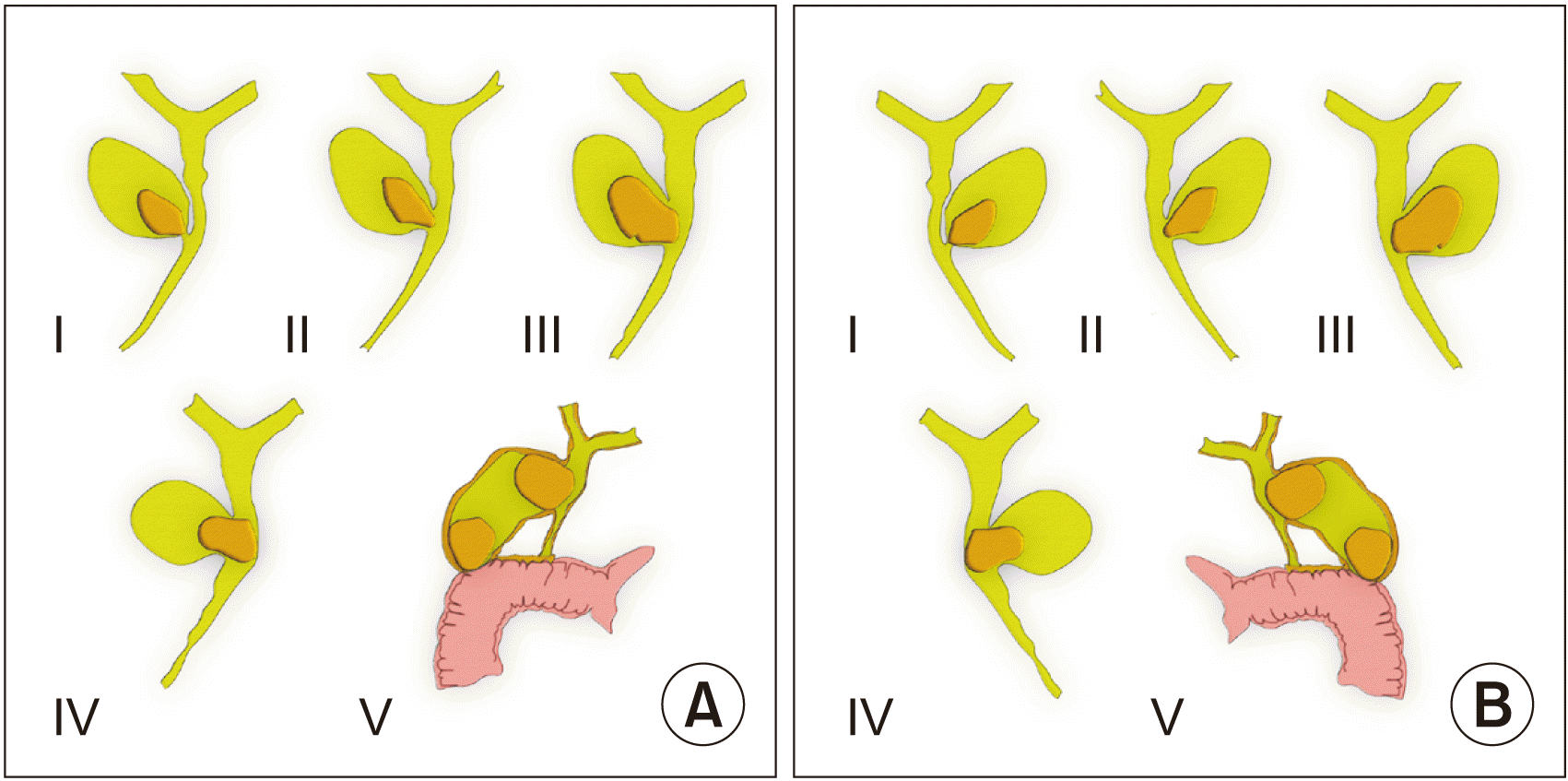 | Fig. 6(A) Types of Mirizzi syndrome (I–V represent Mirizzi syndrome types I–V). (B) The same types of Mirizzi syndromes present in a patient of situs inversus (seen in inverted position).
|
Table 1
Types of Mirizzi syndrome
![]()
VSD is one of the most common congenital defects of the heart, accounting for up to 40% of all cardiac anomalies [9]. Not only is VSD a common isolated cardiac malformation, but it is also an integral component of several complicated malformations, such as tetralogy of Fallot and univentricular atrioventricular connection. Multiple factors are likely to contribute to the development of ventricular septal abnormalities [10].
In hearts with TGA, the atriums and ventricles retain their normal structure, and conduction tissue takes a similar distribution. Typically, a right-sided subaortic infundibulum separates the tricuspid from the aortic valve, and there is fibrous continuity between the mitral and pulmonary valves. This fact is constant and regardless of the geographical relationship between the two large vessels. The exact etiology of TGA is still unknown. The definitive diagnosis, however, is determined by echocardiography which gives a precise morphological and functional examination of the heart, allowing for the identification of the unique characteristics of TGA [5].
Laparoscopic cholecystectomy, an improved and less risky surgical technique, is currently used for simple and quick surgical treatments. Laparoscopic surgery necessitates more caution, especially with SIT patients. Although not contraindicated, laparoscopic cholecystectomy in patients with situs inversus is technically challenging. The surgeon’s position, their assistance, and the dissection ports must be adjusted. Given the surgeons’ lack of familiarity with patients whose organs are in the contralateral position, it may be difficult for a right-handed surgeon to do a lopsided operation due to the possibility of arm crossing [11-13].
Due to the uniqueness of the presentation of this case, we recommend the following treatment steps for future similar cases and prospective studies on quicker and safer therapeutic approaches.
Cholecystectomy in situs inversus totalis patient
• The left-hand ports will be mirror images of the right-hand ports. To minimize intraoperative complications, one must precisely outline the anatomy with MRCP prior to surgery so that it can be performed in the least amount of time with the safest outcome.
• The CO2 flow rate should be very low and gradual while producing pneumoperitoneum, and the pressure should be maintained between 10 and 12 mm of Hg, as opposed to the normal 15 mm of Hg, in the presence of cardiac issues, to reduce the stress on the heart.
Management of type III Mirizzi syndrome
• When cholangitis is present, it is advisable to do ERCP despite the presence of sludge in the CBD.
• Following ERCP, cholangitis and acute cholecystitis should be managed pharmacologically, and it is always safe to manage conservatively for a minimum of six weeks to avoid intra- and postoperative complications during laparoscopic cholecystectomy.
• Cholangitis can be managed pharmacologically during that 6-week period, even if the patient experiences recurrent bouts of pain.
• As a CBD stent will already be in place after ERCP, laparoscopic CBD exploration, stone removal, and suturing can be accomplished with relative simplicity. The patient can be discharged from the hospital the next day because T-tube implantation and drainage follow-up is no longer required over the course of six weeks.
Go to :
 XML Download