Journal List > Ann Hepatobiliary Pancreat Surg > v.27(3) > 1516083699

Ghosh, R, Singh, Malage, Sharma, Kumar, Singh, Behari, Kumar, and Saxena: Surgery for symptomatic hepatic hemangioma: Resection vs. enucleation, an experience over two decades

Abstract

Backgrounds/Aims

Hemangiomas are the most common benign liver lesions; however, they are usually asymptomatic and seldom require surgery. Enucleation and resection are the most commonly performed surgical procedures for symptomatic lesions. This study aims to compare the outcomes of these two surgical techniques.

Methods

A retrospective analysis of symptomatic hepatic hemangiomas (HH) operated upon between 2000 and 2021. Patients were categorized into the enucleation and resection groups. Demographic profile, intraoperative bleeding, and morbidity (Clavien-Dindo Grade) were compared. Independent t-test and chi-square tests were used for continuous and categorical variables respectively. p-value of < 0.05 was considered significant.

Results

Sixteen symptomatic HH patients aged 30 to 66 years underwent surgery (enucleation = 8, resection = 8) and majority were females (n = 10 [62.5%]). Fifteen patients presented with abdominal pain, and one patient had an interval increase in the size of the lesion from 9 to 12 cm. The size of hemangiomas varied from 6 to 23 cm. The median blood loss (enucleation: 350 vs. resection: 600 mL), operative time (enucleation: 5.8 vs. resection: 7.5 hours), and postoperative hospital stay (enucleation: 6.5 vs. resection: 11 days) were greater in the resection group (statistically insignificant). In the resection group, morbidity was significantly higher (62.6% vs. 12.5%, p = 0.05), including one mortality. All patients remained asymptomatic during the follow-up.

Conclusions

Enucleation was simpler with less morbidity as compared to resection in our series. However, considering the small number of patients, further studies are needed with comparable groups to confirm the superiority of enucleation over resection.

INTRODUCTION

Hemangiomas are the most common benign lesions in the liver, but very few require treatment. The incidence ranges from 0.4% to 20% in the overall population [1,2]. Most of the lesions are asymptomatic, small, and detected incidentally. In general, such benign lesions do not increase in size and do not cause any symptoms. Large hepatic hemangiomas (HH) may cause symptoms as a result of capsular stretching or compression of the surrounding structures. Rarely, they can cause bleeding, obstructive jaundice, or consumption coagulopathy that requires intervention [3]. Asymptomatic HH are followed-up unless there is a diagnostic uncertainty or an interval increase in their size. Symptomatic HH require treatment; however, it is necessary to rule out other causes before considering surgical treatment. Surgical management of hemangiomas includes liver resection, enucleation, hepatic artery ligation, and transplantation. Enucleation and resection are the most commonly performed surgical interventions [4-7]. The standard surgical technique is still controversial. This study aims to compare the outcomes of these two surgical procedures in terms of intraoperative difficulty and postoperative outcomes.

MATERIALS AND METHODS

Study design and population

A retrospective analysis of a prospectively maintained database of HH patients who underwent surgery between 2000 and 2021 was performed. Patients who were asymptomatic, had no interval increase in size and no atypical imaging features, and were managed non-operatively were excluded. A written informed consent was obtained from all participants.

Evaluation and management

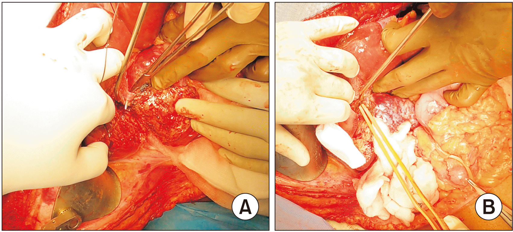
Initial suspicion was based on abdominal ultrasound (USG), which was performed to evaluate abdominal pain. Contrast-enhanced computed tomography (CECT) or Magnetic resonance imaging (MRI) of the abdomen was performed to assess the lesion and its relationship with the surrounding structures (Fig. 1). Fig. 1 shows the location of hemangiomas in different hepatic segments and their management. Tissue diagnosis was not necessary as CECT or MRI findings were characteristic of hemangioma, and it carries a risk of bleeding. Following the operational fitness assessment, all patients were counseled about the benign nature of the disease and surgery was conducted for symptom relief. Resection was preferred when the lesion was abutting major vascular structures, hemangioma replaced most of the parenchyma confined to the anatomical boundary, or the lesion was surrounded by normal liver parenchyma all around (Fig. 1). Enucleation was performed when the lesion was subcapsular and located away from intra-hepatic major vascular structures. All patients were subjected to open surgery. In all patients, generous hepatic mobilization was carried out on the side of the lesion. The hepatoduodenal ligament was looped in all cases for the Pringle maneuver. For enucleation, after incising the capsule of the liver using monopolar energy, bipolar energy was used to shelve out the hemangioma from the compressed surrounding liver parenchyma. Feeding vessels were secured with suture ligation or Liga clip, depending on the size of the vessel (Fig. 2). At the end of the procedure, hemostasis and biliostasis was ensured and a drain was placed near the cut surface. Fig. 3 demonstrates preserved intra-hepatic vasculature following enucleation. In case of resection, standard anatomical liver resection was performed. Cases with multiple lesions, in which both resection and enucleation were used in combination based on the anatomy, were classified into the resection group. Morbidity was reported according to the Clavien-Dindo grading (CDG) system [8]. Postoperatively, patients were regularly followed up with physical assessment and ultrasonography if needed.

Data extraction and statistical analysis

Patients’ demographic profile, surgical procedure, and postoperative outcomes were retrieved from a prospectively maintained electronic database and follow-up cards. Continuous variables were reported as mean ± standard deviation in case of normally distributed data or median and interquartile range (IQR) in case of skewed data. Categorical variables were reported as percentages. All statistical analyses were performed using the Statistical Package for Social Sciences (SPSS) v.20 software (IBM Corp.). Continuous variables were compared with an independent t-test or Mann–Whitney U test, and categorical variables were compared with a chi-square test. p-value of < 0.05 was considered significant.

RESULTS

Patient information and clinical presentation

A total of 16 patients underwent surgery for symptomatic HH. The age of the patients varied from 30 years to 66 years, and the majority of patients were female (n = 10, 62.5%). Patients in the enucleation group were younger compared to those in the resection group (38.6 vs. 50.2 years; p = 0.01). Sex distribution was equal in the resection group; however, there were more females in the enucleation group. Co-morbid medical conditions (37.5% vs. 25.0%; p = 0.23) and size of the HH (13 vs. 9.9 cm; p = 0.51) were higher in the resection group. Fifteen patients presented with abdominal pain, and one patient had an interval increase in the size of the lesion from 9 to 12 cm. There were no signs of Kasabach–Merritt syndrome in any of our patients. Table 1 provides a detailed clinical profile of patients, surgery, and results.

Laboratory and radiological intervention

All patients were evaluated by abdominal USG and CECT. MRI was performed in two patients (one patient had multiple hemangiomas and another patient had an interval increase in the size of the lesion). Fine needle aspiration cytology (FNAC) was carried out in three patients before being referred to our institute, without any yield. Multiple hemangiomas were found in four patients. One patient had multiple small hemangiomas scattered all over the liver; hence, only the largest lesion was enucleated for symptom relief. Blood parameters, including complete blood count, liver function test, renal function test, and coagulation profile, were normal in all cases. The maximum diameter of hemangiomas ranged from 6 to 23 cm; the diameter of hemangiomas was larger in the resection group, as mentioned previously.

Treatment and outcome

Enucleation and resection were conducted in eight patients each. One patient who underwent right hepatectomy for right lobe lesion and enucleation for left lobe hemangioma was included in the resection group. Median blood loss (enucleation: 350 vs resection: 600 mL) and operative time (enucleation: 5.8 vs. resection: 7.5 hours) were greater in the resection group, although the differences were not statistically significant. Overall morbidity was significantly higher in the resection group (62.6% vs. 12.5%; p = 0.05), including one case of mortality in the resection group (Table 2). Pelvic fluid collection was noted in a patient after hepatectomy, which was managed through percutaneous drainage (CDG-IIIa). Postoperative bleeding was observed in one patient who had undergone enucleation; bleeding from the right inferior phrenic vein and posterolateral aspect of the inferior vena cava was noted and both bleeding sites were securely sutured on re-exploration (CDG-IIIb). Postoperative ascites (n = 1) and pleural effusion (n = 2) were managed conservatively with diuretics. Postoperative hospital stay was longer following resection (11 vs. 6.5 days; p = 0.15), although statistically insignificant. One patient in the resection group suffered massive myocardial infarction, and could not be revived (CDG-V). During follow-up, the remaining patients remained asymptomatic.

DISCUSSION

Hepatic hemangioma is the most common type of benign liver tumor, which is common in young females and has an incidence of 0.4% to 20% [1,2]. Majority (n = 10, 62.5%) of patients in the present series were female. Patients in the resection group were significantly older than those in the enucleation group. Previous studies have also documented similar findings; however, the difference was not significant [9-11]. The size of hemangioma may vary from 1 to 50 cm [12]. In the present series, the size ranged from 6 to 23 cm. In accordance with the other studies, hemangiomas in the resection group were larger than those in the enucleation group. With progression of age, there may be an increase in the size of lesion and more chance of undergoing hepatic resection. Patients with asymptomatic HH were being followed up and offered intervention in case of an interval increase in size. Symptomatic hemangiomas are usually of a larger size (> 5 cm), and symptoms vary from abdominal pain, discomfort, and distension secondary to capsular stretching, to symptoms due to pressure effect on the surrounding structures (early satiety, jaundice, and postprandial bloating). Rarely, they can cause high output cardiac failure [13-16]. Cases of Kasabach–Merritt syndrome (thrombocytopenia, disseminated intravascular coagulation, and systemic hemorrhage) [17,18] and spontaneous rupture [19] have been rarely reported. In our series, none of the patients had symptoms due to pressure effect, Kasabach–Merritt syndrome, or spontaneous rupture. Imaging evaluation includes USG abdomen, CECT abdomen, and MRI. USG has a sensitivity and specificity of 96.9% and 60.3%, respectively, and additional contrast-enhanced USG can demonstrate peripheral nodular enhancement with centripetal filling in the late phases to improve the diagnostic yield [20-22]. CECT demonstrates typical peripheral nodular enhancement and centripetal filling with 98.3% sensitivity and 55% specificity [20]. In our series, all patients underwent CECT following USG abdomen.On MRI, hemangiomas are hypointense on T1-weighted and hyperintense on T2-weighted images with characteristics similar to those on CECT after contrast injection [20-23]. Two patients in our series had undergone MRI; one patient had undergone MRI for better characterization of the lesion and the other patient had undergone MRI to avoid multiple radiation exposure where the patient had an interval increase in the size (follow-up of the lesion). Biopsy is not recommended due to the risk of bleeding, and it has poor diagnostic output [24-26]. However, FNAC was performed in three patients before being referred to our institute, without any effectiveness.Symptomatic patients should be evaluated for other etiologies, as Farges et al. (1995) [26] have demonstrated the associated causes of pain in 54% of patients with hemangiomas.
Surgery is the most commonly performed treatment for symptomatic HH, which includes enucleation, resection, hepatic artery ligation, and liver transplantation. Enucleation or resection is most frequently performed, and the choice of treatment depends upon the location, size, relationship with major vascular pedicles, and surgeons’ preference. Several studies have demonstrated the advantage of enucleation over resection [9,10,27]. A meta-analysis has also shown that normal hepatic parenchyma may be preserved by enucleation and is associated with fewer postoperative complications [27]. Similar results were also documented in our series. There was one patient with morbidity in the enucleation group, but it was a serious life-threatening morbidity, which required re-exploration for secondary hemorrhage (CDG-IIIb). Compressed major vessels in close relation to a large HH may result in a major bleed during enucleation, and utmost importance should be given to correct identification of feeder vessels and major vascular pedicles in close relation to the tumor. Overall enucleation provides a less complex surgery with less blood loss, less operative time, lesser complications, and lesser hospital stay along with preservation of maximum functional liver parenchyma as compared to resection, which seems logical in case of a benign tumor being operated upon only for symptom relief.
Resection is a more complex surgery with higher morbidity, but it has less chance of bile leak and bleeding due to the anatomical resection plane, and it is logical to perform resection in specific situations i.e., a hemangioma abutting major vascular structures or replacing most of the parenchyma confined to an anatomical plane or surrounded by hepatic parenchyma all around where enucleation is not feasible. Although enucleation is a simpler procedure, it may result in a larger raw surface area and higher risk of bleeding or bile leak. In our study, enucleation fared better compared to resection surgery. However, there was a selection bias and it was a small cohort to draw any definite conclusion. Further studies are needed with a large sample size among comparable patient population to demonstrate the superiority of either enucleation or resection in case of HH. With the present status, both procedures yield comparable results after executing a sound surgical technique in specific situations, and they should be used judiciously according to the anatomy of the lesion.
Minimal access surgery for hemangiomas has also been reported (laparoscopic and robotic) to provide good results [19,20]. Rarely, liver transplantation has been performed for hemangiomas or unresectable lesions causing severe symptoms or failure of other non-surgical treatment modalities [28,29].
Both resection and enucleation procedures have a role in the management of hepatic hemangioma, depending on the anatomy of the lesion. But enucleation should be considered as the procedure of choice whenever the anatomy permits, as it is a simpler procedure with less morbidity and it preserves maximum future liver remnant considering the benign nature of the disease.

Notes

FUNDING

None.

CONFLICT OF INTEREST

No potential conflict of interest relevant to this article was reported.

AUTHOR CONTRIBUTIONS

Conceptualization: AS. Data curation: NKG. Methodology: All authors. Visualization: NKG, AS. Writing - original draft: NKG, AS. Writing - review & editing: All authors.

REFERENCES

1. Zhou JX, Huang JW, Wu H, Zeng Y. 2013; Successful liver resection in a giant hemangioma with intestinal obstruction after embolization. World J Gastroenterol. 19:2974–2978. DOI: 10.3748/wjg.v19.i19.2974. PMID: 23704832. PMCID: PMC3660824.
2. Caseiro-Alves F, Brito J, Araujo AE, Belo-Soares P, Rodrigues H, Cipriano A, et al. 2007; Liver haemangioma: common and uncommon findings and how to improve the differential diagnosis. Eur Radiol. 17:1544–1554. DOI: 10.1007/s00330-006-0503-z. PMID: 17260159.
3. Choi BY, Nguyen MH. 2005; The diagnosis and management of benign hepatic tumors. J Clin Gastroenterol. 39:401–412. DOI: 10.1097/01.mcg.0000159226.63037.a2. PMID: 15815209.
4. Starzl TE, Koep LJ, Weil R 3rd, Fennell RH, Iwatsuki S, Kano T, et al. 1980; Excisional treatment of cavernous hemangioma of the liver. Ann Surg. 192:25–27. DOI: 10.1097/00000658-198007000-00004. PMID: 7406559. PMCID: PMC1344800.
5. Alper A, Ariogul O, Emre A, Uras A, Okten A. 1988; Treatment of liver hemangiomas by enucleation. Arch Surg. 123:660–661. DOI: 10.1001/archsurg.1988.01400290146027. PMID: 3358693.
6. Nishida O, Satoh N, Alam AS, Uchino J. 1988; The effect of hepatic artery ligation for irresectable cavernous hemangioma of the liver. Am Surg. 54:483–486. PMID: 3395024.
7. Tepetes K, Selby R, Webb M, Madariaga JR, Iwatsuki S, Starzl TE. 1995; Orthotopic liver transplantation for benign hepatic neoplasms. Arch Surg. 130:153–156. DOI: 10.1001/archsurg.1995.01430020043005. PMID: 7848084.
8. Clavien PA, Barkun J, de Oliveira ML, Vauthey JN, Dindo D, Schulick RD, et al. 2009; The Clavien-Dindo classification of surgical complications: five-year experience. Ann Surg. 250:187–196. DOI: 10.1097/SLA.0b013e3181b13ca2. PMID: 19638912.
9. Hamaloglu E, Altun H, Ozdemir A, Ozenc A. 2005; Giant liver hemangioma: therapy by enucleation or liver resection. World J Surg. 29:890–893. DOI: 10.1007/s00268-005-7661-z. PMID: 15951941.
10. Singh RK, Kapoor S, Sahni P, Chattopadhyay TK. 2007; Giant haemangioma of the liver: is enucleation better than resection? Ann R Coll Surg Engl. 89:490–493. DOI: 10.1308/003588407X202038. PMID: 17688721. PMCID: PMC2048596.
11. Yang Z, Tan H, Liu X, Sun Y. 2017; Extremely giant liver hemangioma (50 cm) with Kasabach-Merritt syndrome. J Gastrointest Surg. 21:1748–1749. DOI: 10.1007/s11605-017-3429-7. PMID: 28424986.
12. Liu X, Yang Z, Tan H, Zhou W, Su Y. 2018; Fever of unknown origin caused by giant hepatic hemangioma. J Gastrointest Surg. 22:366–367. DOI: 10.1007/s11605-017-3522-y. PMID: 28785931.
13. Smith AA, Nelson M. 2016; High-output heart failure from a hepatic hemangioma with exertion-induced hypoxia. Am J Cardiol. 117:157–158. DOI: 10.1016/j.amjcard.2015.10.019. PMID: 26525213.
14. Mikami T, Hirata K, Oikawa I, Kimura M, Kimura H. 1998; Hemobilia caused by a giant benign hemangioma of the liver: report of a case. Surg Today. 28:948–952. DOI: 10.1007/s005950050259. PMID: 9744407.
15. Huang SA, Tu HM, Harney JW, Venihaki M, Butte AJ, Kozakewich HP, et al. 2000; Severe hypothyroidism caused by type 3 iodothyronine deiodinase in infantile hemangiomas. N Engl J Med. 343:185–189. DOI: 10.1056/NEJM200007203430305. PMID: 10900278.
16. Sakamoto Y, Kokudo N, Watadani T, Shibahara J, Yamamoto M, Yamaue H. 2017; Proposal of size-based surgical indication criteria for liver hemangioma based on a nationwide survey in Japan. J Hepatobiliary Pancreat Sci. 24:417–425. DOI: 10.1002/jhbp.464. PMID: 28516570.
17. Miura JT, Amini A, Schmocker R, Nichols S, Sukato D, Winslow ER, et al. 2014; Surgical management of hepatic hemangiomas: a multi-institutional experience. HPB (Oxford). 16:924–928. DOI: 10.1111/hpb.12291. PMID: 24946109. PMCID: PMC4238859.
18. Jain V, Ramachandran V, Garg R, Pal S, Gamanagatti SR, Srivastava DN. 2010; Spontaneous rupture of a giant hepatic hemangioma - sequential management with transcatheter arterial embolization and resection. Saudi J Gastroenterol. 16:116–119. DOI: 10.4103/1319-3767.61240. PMID: 20339183. PMCID: PMC3016500.
19. Toro A, Mahfouz AE, Ardiri A, Malaguarnera M, Malaguarnera G, Loria F, et al. 2014; What is changing in indications and treatment of hepatic hemangiomas. A review. Ann Hepatol. 13:327–339. DOI: 10.1016/S1665-2681(19)30839-7. PMID: 24927603.
20. Dietrich CF, Mertens JC, Braden B, Schuessler G, Ott M, Ignee A. 2007; Contrast-enhanced ultrasound of histologically proven liver hemangiomas. Hepatology. 45:1139–1145. DOI: 10.1002/hep.21615. PMID: 17464990.
21. Quaia E, Calliada F, Bertolotto M, Rossi S, Garioni L, Rosa L, et al. 2004; Characterization of focal liver lesions with contrast-specific US modes and a sulfur hexafluoride-filled microbubble contrast agent: diagnostic performance and confidence. Radiology. 232:420–430. DOI: 10.1148/radiol.2322031401. PMID: 15286314.
22. McFarland EG, Mayo-Smith WW, Saini S, Hahn PF, Goldberg MA, Lee MJ. 1994; Hepatic hemangiomas and malignant tumors: improved differentiation with heavily T2-weighted conventional spin-echo MR imaging. Radiology. 193:43–47. DOI: 10.1148/radiology.193.1.8090920. PMID: 8090920.
23. Terriff BA, Gibney RG, Scudamore CH. 1990; Fatality from fine-needle aspiration biopsy of a hepatic hemangioma. AJR Am J Roentgenol. 154:203–204. DOI: 10.2214/ajr.154.1.2104717. PMID: 2104717.
24. Davies R. 1993; Haemorrhage after fine-needle aspiration biopsy of an hepatic haemangioma. Med J Aust. 158:364. DOI: 10.5694/j.1326-5377.1993.tb121823.x. PMID: 8474396.
25. Taavitsainen M, Airaksinen T, Kreula J, Päivänsalo M. 1990; Fine-needle aspiration biopsy of liver hemangioma. Acta Radiol. 31:69–71. DOI: 10.1177/028418519003100113. PMID: 2187514.
26. Farges O, Daradkeh S, Bismuth H. 1995; Cavernous hemangiomas of the liver: are there any indications for resection? World J Surg. 19:19–24. DOI: 10.1007/BF00316974. PMID: 7740805.
27. Liu Y, Wei X, Wang K, Shan Q, Dai H, Xie H, et al. 2017; Enucleation versus anatomic resection for giant hepatic hemangioma: a meta-analysis. Gastrointest Tumors. 3:153–162. DOI: 10.1159/000455846. PMID: 28611982. PMCID: PMC5465724.
28. Prodromidou A, Machairas N, Garoufalia Z, Kostakis ID, Tsaparas P, Paspala A, et al. 2019; Liver transplantation for giant hepatic hemangioma: a systematic review. Transplant Proc. 51:440–442. DOI: 10.1016/j.transproceed.2019.01.018. PMID: 30879561.
29. Longeville JH, de la Hall P, Dolan P, Holt AW, Lillie PE, Williams JA, et al. 1997; Treatment of a giant haemangioma of the liver with Kasabach-Merritt syndrome by orthotopic liver transplant a case report. HPB Surg. 10:159–162. DOI: 10.1155/1997/10136. PMID: 9174860. PMCID: PMC2423854.
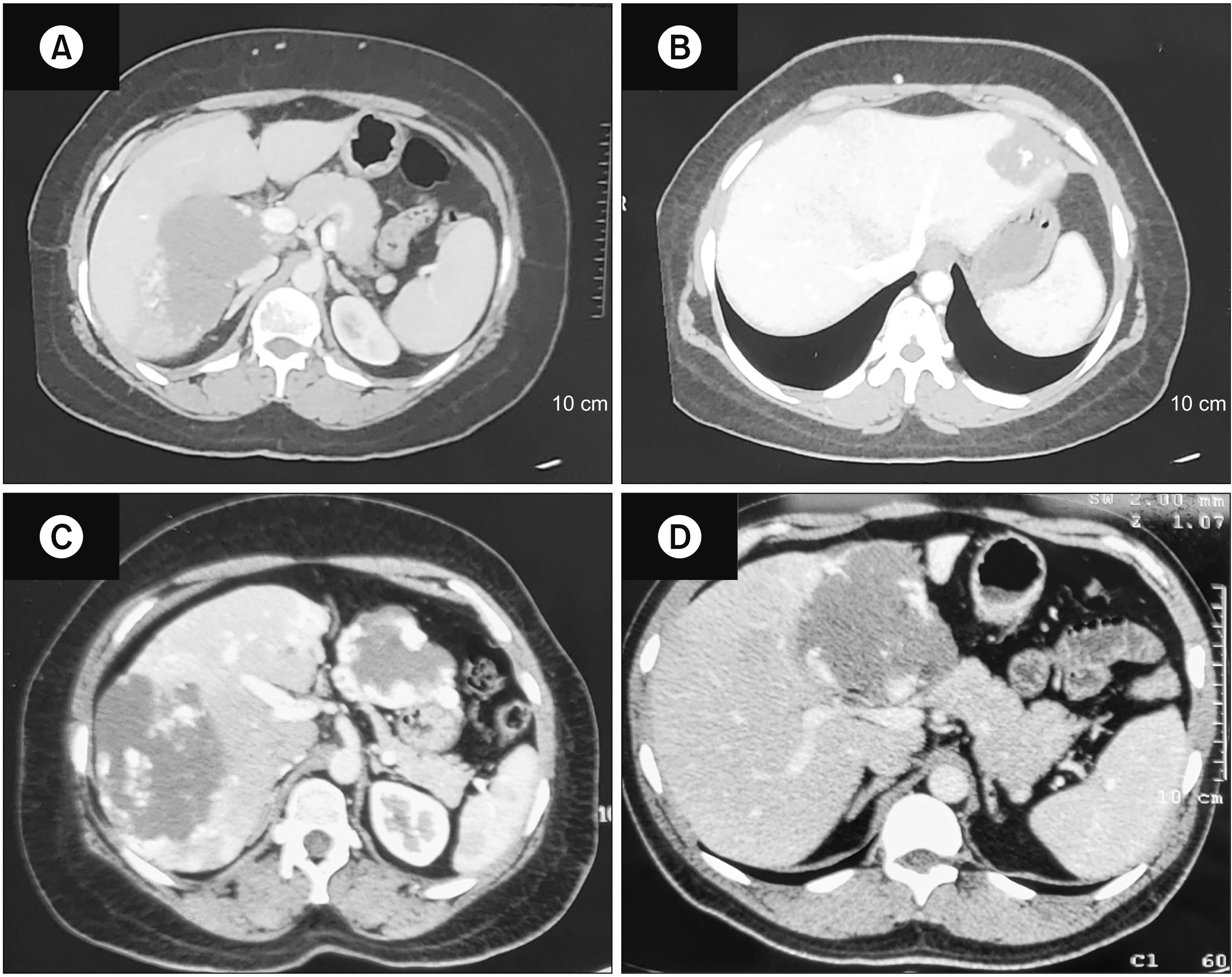
Fig. 1
Contrast-enhanced computed tomography of the liver showing hemangiomas in different segments of the liver. (A) Segment VI, (B) segment II, (C) segments VI and VII, and (D) segments IVb and V.
ahbps-27-3-258-f1.tif
Fig. 2
Intraoperative pictures. (A) Ligation of the feeding vessel, (B) creation of a plane between the lesion and liver parenchyma.
ahbps-27-3-258-f2.tif
Fig. 3
(A) A figure post-enucleation showing a preserved hepatic vein branch; (B) enucleated specimens from multiple segments.
ahbps-27-3-258-f3.tif
Table 1
Summaries of all patients
S. no. Age (yr)/Sex Location (segments) Clinical features (symptoms & signs) Associated condition Maximum diameter (cm) Surgery Morbidity (Clavien-Dindo grade) Hospital stays (day) Blood loss (mL)
1 33/F VI, VII Pain abdomen, constipation - 10 Enucleation Bleeding (IIIb) 14 800
2 40/M V, VI, VII, VIII Pain abdomen, palpable liver - 14 Right hepatectomy Ascites (I) 11 500
3 31/F IVb Pain abdomen Cholelithiasis 8.5 Enucleation - 6 100
4 43/F VI Pain abdomen, palpable liver - 6 Enucleation - 5 400
5 37/F III, V, VI Pain abdomen Hypothyroidism, cholelithiasis 8.5 Enucleation - 3 300
6 30/M V, VIII Pain abdomen - 12 Enucleation (robotic→open) - 7 300
7 48/M III, IV Abdominal fullness Cholelithiasis 13.1 Enucleation - 6 50
8 44/F IV Pain abdomen and palpable liver - 17.5 Enucleation - 7 400
9 56/M II, III, IV Pain abdomen and epigastric lump Cholelithiasis 15.4 Left hepatectomy Pleural effusion (I) 15 300
10 59/F III, V-VIII Pain abdomen Hypothyroidism 10 Right hepatectomy, Enucleation of segment III lesion (3 × 3 cm) - 8 600
11 66/M V, VI Pain abdomen and epigastric lump Hypothyroidism 7 Right hepatectomy Pelvic collection (IIIa) 35 500
12 36/F V-VIII Pain abdomen - 23 Right hepatectomy - 8 600
13 40/F V-VIII Pain abdomen, hepatomegaly - 13 Right hepatectomy - 5 1,500
14 63/M V-VIII Enlarging hemangioma Hypertension 12 Right hepatectomy Pleural effusion (IIIa) 14 1,000
15 43/F VII, II-III Pain abdomen Hypothyroidism, hypertension 7 Enucleation - 11 800
16 42/F V-VIII Pain abdomen - 10 Right hepatectomy Mortality (V) 1 600

S. no, serial no; M, male; F, female.

Table 2
Comparison of enucleation vs. hepatectomy
S. no. Parameters Enucleation Resection p-value
1 Age (yr) 38.6 ± 6.7 50.2 ± 11.9 0.01
2 Sex (M : F) 2:6 4:4 0.33
3 Comorbidity 2 (25.0) 3 (37.5) 0.23
4 Size (cm) 9.9 ± 3.8 13 ± 4.8 0.51
5 Blood loss (mL) 350 (150–700) 600 (500–800) 0.64
6 Operating time duration (h) 5.8 ± 1.9 7.5 ± 1.6 0.91
7 Morbidity 1 (12.5) 5 (62.6) 0.05
8 Hospital stay (day) 6.5 (5.2–10) 11 (8–15) 0.15

Values are presented as mean ± standard deviation, number (%), or median (interquartile range).

S. no, serial no; M, male; F, female.

TOOLS
Similar articles