

 PDF
PDF Citation
 Print
Print


INTRODUCTION
The N-methyl-D-aspartate (NMDA) receptor is a receptor for glutamate and ion channel proteins and comprises GluN1, GluN2, and GluN3 subunits [1]. It is involved in cell signal transmission via calcium, synaptic plasticity regulation, and learning and memory functions [2]. Excessive glutamate overstimulates the nerves in the brain, causing damage and death. Further, NMDA antagonists inhibit the action of NMDA receptors, affect glutamate neurotransmission, and prevent the toxic effects of increased glutamate levels [3]. Therefore, their role in neurodegenerative diseases has been extensively studied [4]. Drugs such as ifenprodil, ketamine, memantine, and amantadine have been clinically approved to prevent glutamate-induced excitatory toxicity. Memantine prevents neuronal cell death without affecting normal glutamate signal transmission [5].
Although no cure for Alzheimer's disease is currently available, memantine, a non-competitive (channel-blocking) NMDA receptor antagonist, may efficiently slow Alzheimer's disease [6]. Memantine inhibits glutamine hyperactivity in Alzheimer's disease by modulating NMDA receptors via autophagy [7,8]. Autophagy protects against numerous diseases, including neurodegenerative and cardiovascular diseases and aging. The accumulation of toxic proteins (such as tau) in nerve cells can be caused by reduced autophagy [9]. In neurological diseases, autophagy dysfunction is implicated in the accumulation of misfolded proteins and formation of protein aggregates, leading to cytotoxicity and, ultimately, neuronal cell death. Autophagy is a destructive mechanism by which lysosomes naturally degrade unnecessary or non-functional cellular components, such as modified proteins or damaged organelles, to maintain cell homeostasis and function [10]. The amino acids of these proteins, which are degraded via autophagy, are converted and reused as raw materials for new proteins.
Ifenprodil, a GluN2B selective NMDA receptor antagonist, was tested in clinical trials for treating drug addiction, idiopathic pulmonary fibrosis, and COVID-19 [11]. It improves long-term neurological deficits by blocking glutamate-induced excitotoxicity, thereby reducing apoptosis in primary cortical neurons and high-glutamate-elevated endothelial permeability in human brain microvascular endothelial cells [12].
Autophagy protects against numerous diseases and plays an important role in the retina. However, the relationship between NMDA receptor antagonists and age-related macular degeneration (AMD) remains unclear. Notably, we previously found that the NMDA receptor antagonist Ro 25-6981 degrades lipofuscin via autophagy in human retinal pigment epithelial cells (ARPE-19). N-retinylidene-N-retinylethanolamine (A2E) is a major autofluorescent component of the retinal pigment epithelial lipofuscin, which is a granular partridge and autofluorescent pigment in lysosomes [13]. It is rarely detected in individuals younger than 10 years of age and linearly accumulates with age. Lipofuscin accumulation commonly increases in elderly individuals, and intracellular lipofuscin is a widely accepted biomarker for cellular aging [14]. Lipofuscin is engulfed and degraded in lysosomes; however, when lysosomes become dysfunctional due to aging, engulfed lipofuscin can accumulate and reduce lysosomal degradation. Similarly, normal retinal pigment epithelial cells remove lipofuscin from the lysosomes via autophagy [15]. In contrast, when autophagy of retinal pigment epithelial cells is damaged due to aging, unremoved lipofuscin continues to accumulate in the lysosomes of retinal pigment epithelial cells [16]. Eventually, this chain reaction causes AMD, a retina-related disease. Therefore, A2E ablation is important in retinal diseases [17,18]. In this study, we aimed to test whether the NMDA receptor antagonists, memantine and ifenprodil, induced autophagy in ARPE-19 cells to eliminate A2E. Our study findings indicated that lipofuscin is removed through autophagy in ARPE-19 cells, thereby providing a new perspective on the relationship between A2E ablation and NMDA antagonist-mediated autophagy in ARPE-19 cells.
METHODS
Cell culture and materials
The human RPE cell line (ARPE-19) was acquired from the American Type Culture Collection and cultured in Dulbecco’s modified Eagle’s medium F-12 (WELGENE) supplemented with 10% fetal bovine serum at 37°C in a humidified incubator containing 5% CO2. Fluorescence-labeled A2E (A2E-BDP) was synthesized as previously described [19]. A2E was synthesized by AptaBio. Memantine and ifenprodil were purchased from Sigma-Aldrich.
Cell viability assay
ARPE-19 cells were cultured in 24-well plates at a density of 2 × 10⁴ cells/well. Cells were treated with specific concentrations of memantine and ifenprodil at 37°C for 24 h. Cell viability was determined using an EZ-Cytox assay kit (DoGenBio). Absorbance was measured at 450 nm using a BioTek microplate reader.
A2E-BDP assay
A2E-BDP assay was used to confirm A2E degradation in ARPE-19 cells plated at 5 × 10³ cells/well in a 96-well plate. The cells were initially treated with 10 μM A2E-BDP for 24 h and then with the compound of interest for 48 h. Subsequently, A2E-BDP fluorescence intensity was measured using a microplate reader (VICTOR X3; PerkinElmer Inc.).
A2E treatment and confocal microscopy
The ARPE-19 cells were cultured in six-well plates at 2 × 10⁴ cells/well and treated with 5 μM A2E three times at 48 h intervals. They were then treated with memantine or ifenprodil twice at 24 h intervals, fixed in 4% paraformaldehyde for 10 min, and washed three times with phosphate-buffered saline (PBS) for 5 min. Nuclei were stained with 1 μg/ml Hoechst 33342 (Thermo Scientific) for 5 min, washed three times with PBS, and imaged using a confocal fluorescence microscope (Nikon ECLIPSE Ti; Nikon).
Western blot analysis
The ARPE-19 cells were harvested on ice in a radioimmunoprecipitation assay buffer containing protease inhibitors. Proteins were separated using sodium dodecyl sulfate-polyacrylamide gel electrophoresis and transferred onto Immun-Blot polyvinylidene fluoride membranes (Bio-Rad). Subsequently, the membranes were blocked with 10% skim milk in Tris-buffered saline + Tween 20 (TBS-T) for 2 h, probed with the specified primary antibodies (anti-phospho-p62 [S349], anti-p62, anti-LC3, and anti-ATG5 [Cell Signaling Technology] and β-actin [Santa Cruz Biotechnology, Inc.]) overnight at 4°C, washed three times for 10 min with TBS-T, and incubated with secondary antibodies in TBS-T for 2 h. They were then washed three times for 10 min with TBS-T and developed using Immobilon Western Chemiluminescent HRP Substrate (Sigma-Aldrich). Protein expression levels were measured using a ChemiDoc XRS+ imaging system (Bio-Rad).
RNA interference and transfection
The human RPE cells were seeded in six-well plates at 2 × 10⁴ cells/well and transfected with small interfering RNAs targeting ATG5 mRNA (siATG5) or non-specific siRNA (siNS). Next, RNA interference was performed using Oligofectamine (Invitrogen) according to the manufacturer’s protocol. The target mRNA sequences were: siNS, sense 5′-UUCUCCGAACGUGUCACGUTT-3′ and antisense 5′-ACGUGACACGUUCGGAGAATT-3′; and siATG5, sense 5′-AUUCAUGGUGAGCCAdTdT-3′ and antisense 5′-UGGCUCAAUUCCAUGAAUCdTdT-3′.
RESULTS
NMDA antagonist screening for A2E degradation
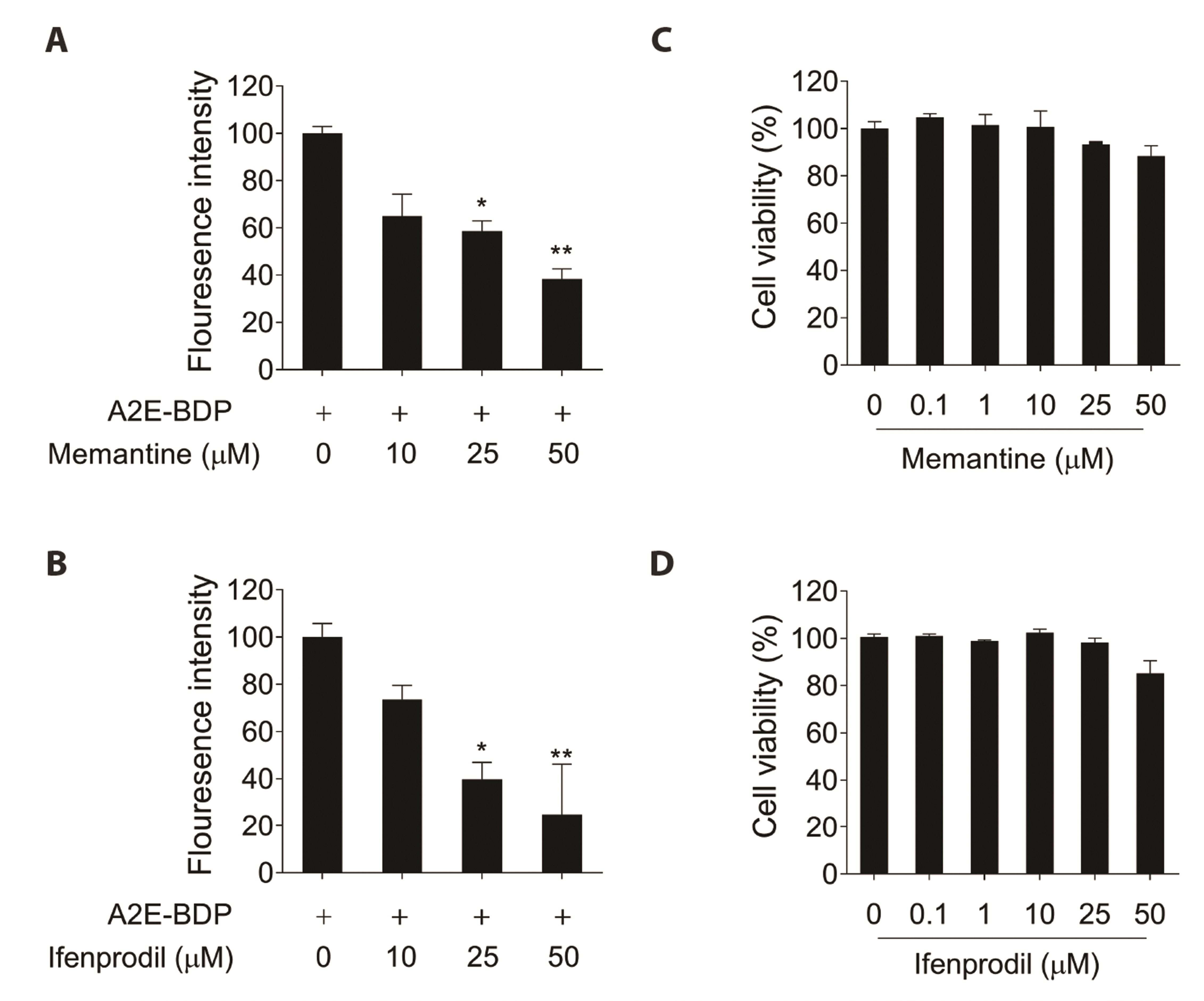
We have previously confirmed that the NMDA receptor antagonist Ro 25-6981 significantly degrades A2E [20]. Accordingly, we screened clinically approved NMDA receptor antagonists to determine whether they could effectively degrade A2E. Among the clinically approved NMDA receptor antagonists, memantine and ifenprodil, in a concentration-dependent manner, decreased the fluorescence intensity of ARPE-19 cells that was increased by A2E-BDP treatment (Fig. 1A, B). The concentrations used in the experiments were not cytotoxic, indicating that A2E clearance by ARPE-19 cells was not due to cytotoxicity (Fig. 1C, D). These results indicated that the NMDA receptor antagonists effectively removed A2E-BDP accumulated in ARPE-19 cells.
Memantine and ifenprodil degrade A2E in ARPE-19 cells
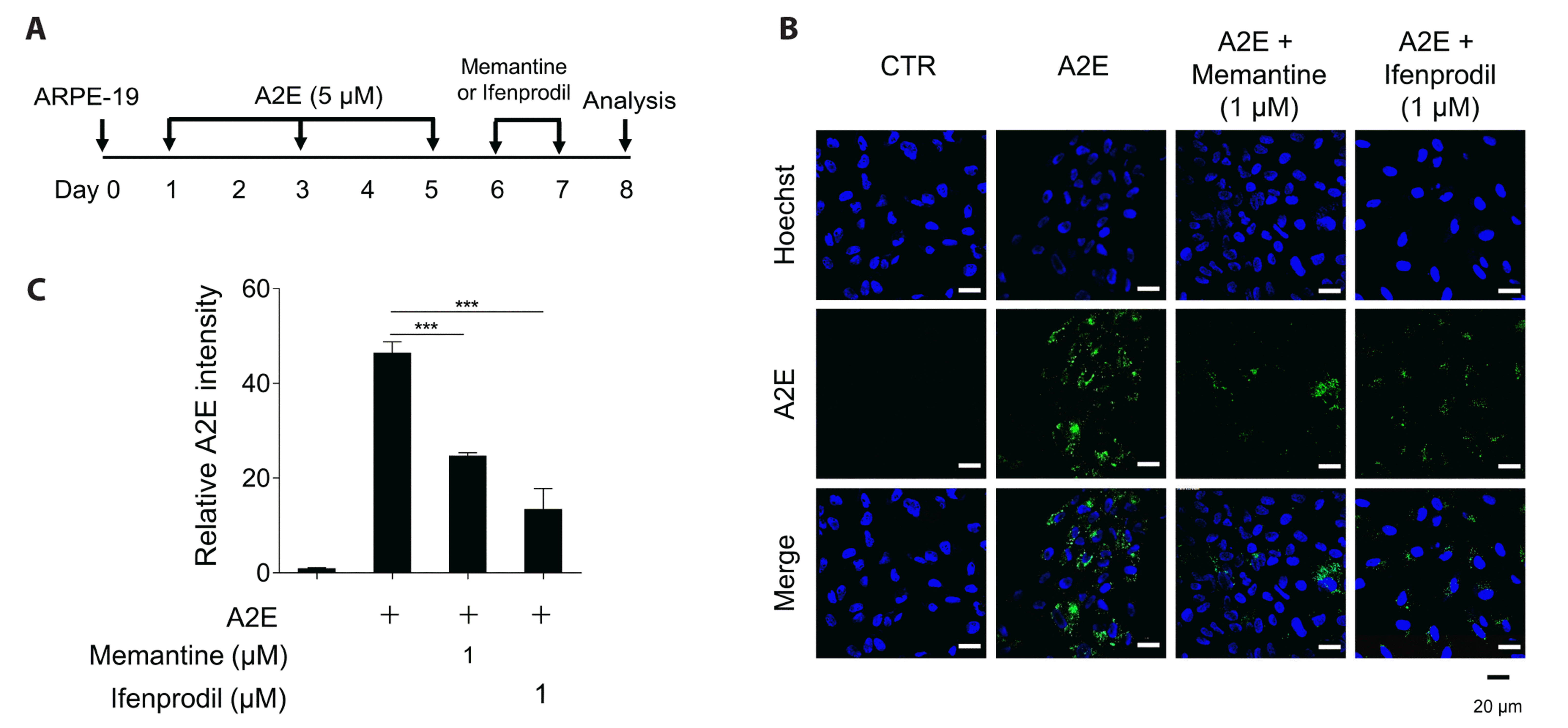
In the above A2E-BDP assay, although memantine and ifenprodil degraded A2E, the remaining fluorophore, BDP, emitted fluorescence. Therefore, higher concentrations of the compounds were required to reduce the fluorescence intensity. Thus, we further investigated whether they could sufficiently degrade A2E at concentrations lower than those used in the previous experiments. To construct an A2E accumulation cell model, ARPE-19 cells were treated with A2E three times at two-day intervals. The cells were then treated with memantine and ifenprodil (Fig. 2A), and A2E degradation was observed using confocal microscopy. Green fluorescence was observed in the lysosomes of ARPE-19 cells owing to A2E accumulation, which was significantly reduced after treatment with memantine and ifenprodil (Fig. 2B). In addition, the quantification of A2E fluorescence intensity indicated statistically significant results, and ifenprodil degraded A2E at a concentration lower than that of memantine (Fig. 2C). Confocal microscopy results confirmed that memantine and ifenprodil effectively degraded A2E in ARPE-19 cells, even at low concentrations that were not cytotoxic.
Memantine- and ifenprodil-mediated autophagy activation in ARPE-19 cells
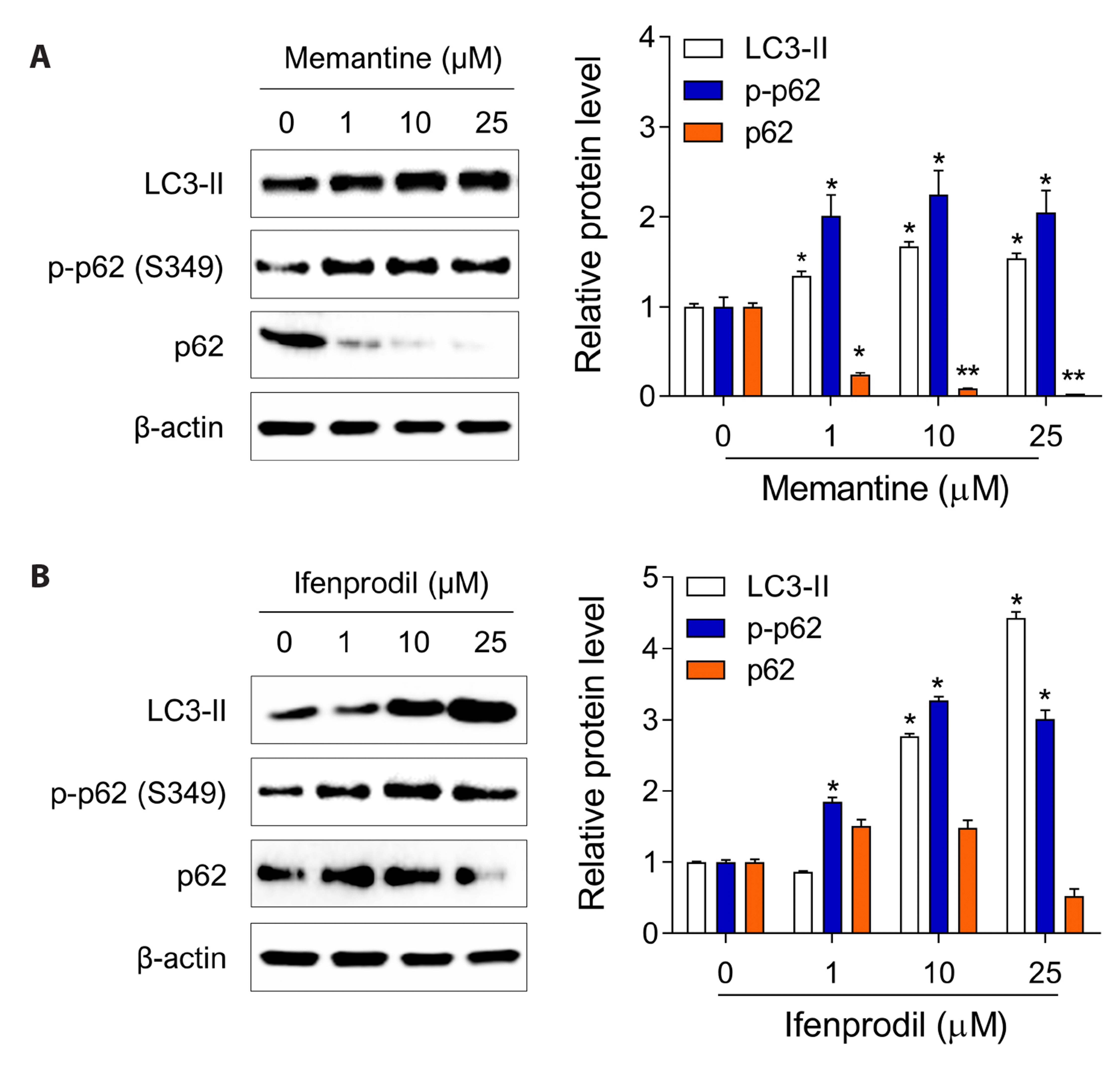
Previous studies have reported that memantine is a novel potential therapeutic agent for neurodegenerative diseases that enhances autophagic flux. When autophagy is activated, the outer membrane of the autophagosome fuses with the lysosome, and the inner substance is degraded in the autolysosome. Autophagy activation was assessed by the level of microtubule-associated protein 1A/1B-light chain3-II (LC3-II) proteins, a well-known autophagy marker [21]. Further, LC3 forms autophagosomes and is associated with autolysosomes. During autophagy, LC3 is converted to LC3-І by protease autophagy-related 4 (ATG4), which is further converted to LC3-II by phosphatidylethanolamine (PE) [22]. In ARPE-19 cells, memantine and ifenprodil significantly increased the LC3-II protein level in a concentration-dependent manner (Fig. 3). The phosphorylation level of serine 349 residue of p62 was measured as another autophagy marker. Previous studies have reported that p62 S349 phosphorylation promotes p62 ubiquitination and induces autophagy [23,24]. Phosphorylated p62 interacts with LC3 through the LC3 interacting region to form autophagosomes [25,26]. Treatment with memantine and ifenprodil significantly increased the p-p62 levels in ARPE-19 cells in a concentration-dependent manner (Fig. 3). These results suggested that memantine and ifenprodil activated autophagy in the ARPE-19 cells.
Involvement of autophagy in memantine- and ifenprodil-mediated A2E degradation
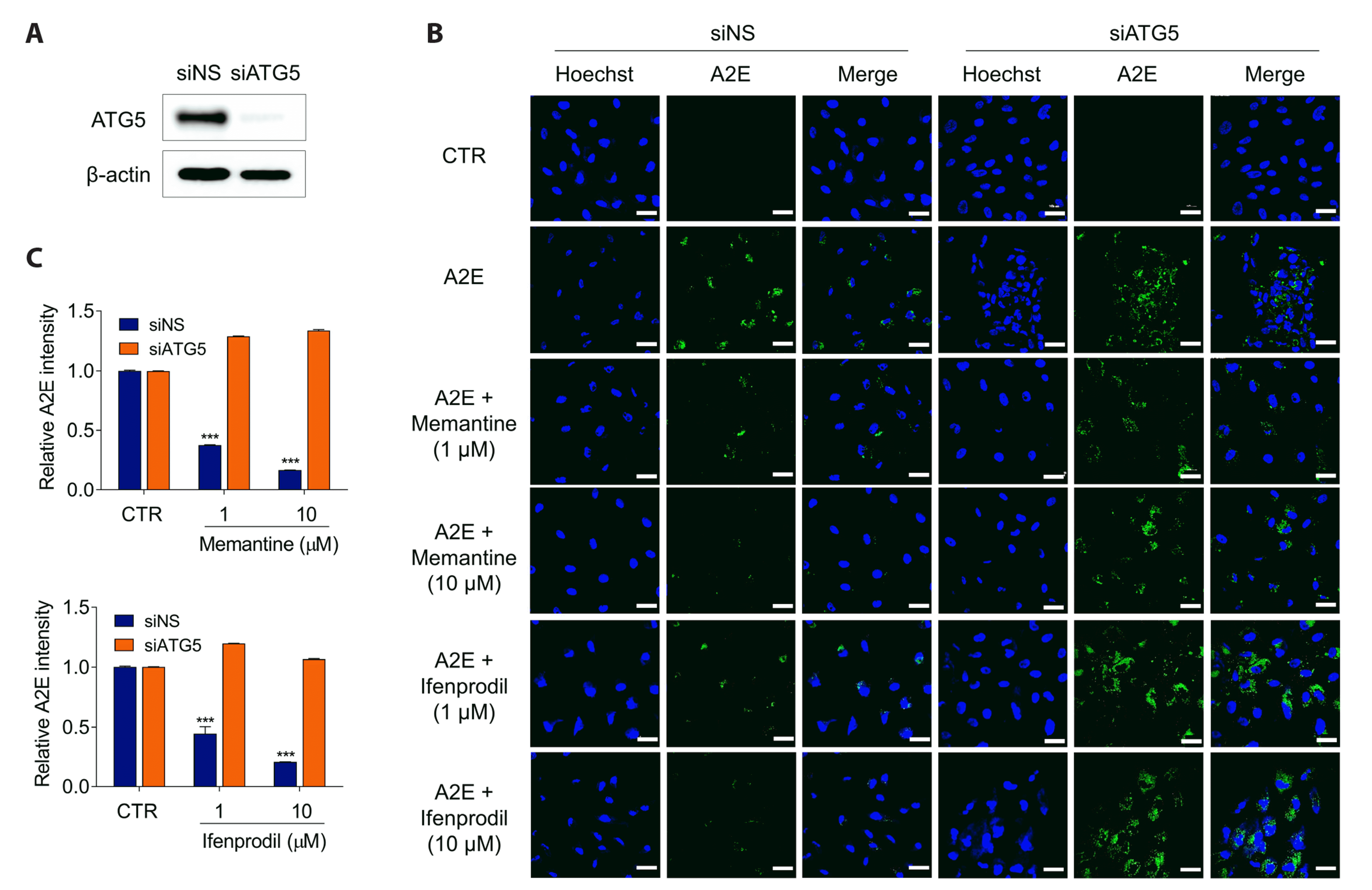
To confirm whether memantine- and ifenprodil-mediated A2E degradation is due to autophagy, autophagy-related 5 (ATG5), a key element that forms the phagophore membrane in autophagy vesicles, was silenced via RNA interference (Fig. 4A). Notably, ATG5 is a key protein involved in the extension of phagophore membranes in autophagic vesicles [27]. It is subsequently activated by ATG7 to form a complex with ATG12 and ATG16L1 [28]. The ATG12–ATG5:ATG16L complex thus formed is responsible for the elongation of phagophores in the autophagy pathway and binds the C-terminus of LC3-І to PE in the phospholipid bilayer to form LC3-II (LC3–PE conjugate) [29]. In siNS-transfected control cells, memantine and ifenprodil treatment effectively reduced A2E accumulation in ARPE-19 cells. In contrast, siATG5-transfected ARPE-19 cells did not form phagocytic membranes; therefore, memantine and ifenprodil treatment did not degrade A2E (Fig. 4B). Quantification of A2E intensity indicated that both memantine and ifenprodil effectively ablated A2E via an autophagic mechanism (Fig. 4C). These results indicated that autophagy is involved in A2E degradation in ARPE-19 cells.
DISCUSSION
The theory of excitotoxicity induced by glutamate has led to the discovery of new molecules that can be used to treat neurodegenerative diseases by blocking NMDA receptors. Among the NMDA receptor antagonists, memantine has been approved for the treatment of dementia and Alzheimer's disease, but no other NMDA receptor antagonists have successfully passed clinical trials since [30,31]. Therefore, we conducted a drug repositioning experiment using NMDA receptor antagonists approved as therapeutics. In this study, the NMDA receptor antagonists memantine and ifenprodil were investigated as novel drug repositioning targets capable of diffusing across the blood–retinal barrier (BRB). Our study was conducted to determine whether NMDA antagonists can sufficiently clear A2E by passing through the BRB.
Notably, AMD is a common retinal disease that causes vision loss in individuals aged > 50 years [32]. The retina is a tissue located at the back of the eyeball that detects light and transmits visual information to the brain. Healthy retinal cells fail to survive AMD. Further, AMD affects the macula, located at the center of the retina, thereby reducing visual acuity. Aging, smoking, genetic predisposition, and oxidative stress, which are well-known factors associated with the occurrence of dry AMD, induce the accumulation of lipofuscin, the most important risk factor for dry macular degeneration [33]. Notably, lipofuscin is a complex mixture of oxidized proteins and degradation residues, including A2E. Further, A2E exposure to blue light (450 nm) induces RPE cell death via A2E oxidation or phototoxicity [34,35], suggesting that A2E degradation in RPE cells can be important in preventing AMD. Additionally, autophagy is a clearance mechanism for lipofuscin [36]. Autophagy-related proteins are expressed at high levels in the retina, and impaired autophagy and mitochondrial damage are hallmarks of an aging retina in AMD. Retinal autophagy markedly reduces as aging progresses. Decreased autophagic activity is associated with several retinal diseases, including AMD, diabetic retinopathy, and glaucoma. Further, autophagy activation via the mammalian target of rapamycin (mTOR) or non-mTOR pathway in the retina plays a beneficial role in protecting the retina by maintaining homeostasis via the disassembly and reuse of damaged cells. In the case of dysfunctional autophagy in retinal cells, the function of RPE cell phagocytosis is inhibited, waste products are accumulated owing to the blockage of metabolites that are being removed, and inflammatory pathways, such as the NF-κB pathway, are activated [35,37]. The RPE cells function as the BRB, a specialized barrier in the retina that regulates the movement of substances between the blood and nerve tissues. The functional role of the BRB in AMD progression is to prevent the leakage of toxic substances from the blood into the retina and maintain an appropriate balance of nutrients and waste products in the retinal neural tissue. When the BRB is impaired in AMD, this balance is disrupted, and the accumulation of waste and debris can lead to inflammation and oxidative stress, which can further damage retinal cells and contribute to disease progression.
Among the clinically approved NMDA antagonists, memantine and ifenprodil effectively reduced A2E accumulation in ARPE-19 cells. In addition, LC3-II and phospho-p62 levels were significantly increased. Memantine increases autophagy-related protein levels via LC3-II/LC3-I turnover and beclin-I expression in glioblastoma (T-98G) and increases the formation of autistic vacuoles, as observed using a transmission electron microscope [38]. Similarly, our finding indicated that memantine induced autophagy in ARPE-19 cells. To determine whether A2E clearance was mediated by autophagy, ARPE-19 cells were transfected with siATG5, a key component of autophagy. Memantine and ifenprodil effectively degraded A2E in siNS-transfected but not in ATG5-deficient ARPE-19 cells, suggesting that memantine and ifenprodil remove A2E via autophagy. Ifenprodil activates mTOR signaling and modulates proinflammatory cytokines [39]. In the hippocampus, ifenprodil induces the expression of phosphorylated mTOR Ser2448 (phosphorylated at Ser 2448 to induce kinase activity) after acute administration and enhances the levels of its substrate P70S6K. Autophagy is regulated by both the mTOR-dependent and mTOR-independent pathways [40]. Further, autophagy is negatively regulated by mTOR and can be induced in all mammalian cell types by the mTOR inhibitor rapamycin. Autophagy can also be induced by an mTOR-independent pathway with multiple drug targets, including links between Ca2+-calpain–Gsα and cAMP–Epac–PLC-α–IP3 signaling [41]. Therefore, ascertaining whether the mechanisms of ifenprodil-mediated autophagy activation in ARPE-19 cells are mTOR-dependent or mTOR-independent and investigating the pathways regulated by autophagy are required.
Previously, we reported that Ro 25-6981, another NMDA receptor antagonist, activates autophagy in human RPE cells [20]. First, the findings of this study suggested that memantine and ifenprodil eliminated A2E from RPE cells. Second, although Ro 25-6981 is also an NMDA receptor inhibitor, its mode of action on NMDA receptors differed from that of memantine and ifenprodil: Ro 25-6981 acted selectively on the NR2B subunit, whereas ifenprodil acted on both the NR1 and NR2B subunits. Memantine bound to the magnesium sites of the NMDA receptors. Third, Ro 25-6981 was found to be effective for treating various conditions but has not yet been approved for clinical use. Ifenprodil has been studied in clinical trials for its efficacy in the treatment of neuropathic pain, stroke, Parkinson's disease, and addiction. Memantine is an FDA-approved drug for the treatment of moderate-to-severe Alzheimer’s disease. Thus, our findings suggest a higher possibility for drug repositioning.
 XML Download