

 PDF
PDF Citation
 Print
Print


INTRODUCTION
Diabetes mellitus (DM) embraces heterogeneous metabolic disorders characterized by chronic hyperglycemia [1]. DM is commonly classified into two types: type 1 DM (T1DM), caused by an absolute deficiency of insulin ensuing destruction of pancreatic beta cells, and type 2 DM (T2DM), resulting from insulin resistance and subsequent deficiency of insulin secretion [2]. T2DM accounts for 90% of all diabetic cases and presents a rising incidence, especially in Asia [3]. Delayed and incomplete wound healing is a major secondary complication of T2DM that often leads to limb amputation and disability [4]. All diabetic ulcers are characterized with a disturbed angiogenic state that results in vascular insufficiency and impedes proper wound healing [5]. Fibroblasts play a key role in endothelial cell-mediated angiogenesis as they can produce various kinds of regulatory factors such as vascular endothelial growth factor (VEGF) to stimulate the proliferation, migration and tube formation of endothelial cells [6]. Macrophages are another important regulator of tissue repair which can transform the healing process from the inflammatory phase into the proliferative phase, during which fibroblasts and other cells reconstitute the tissue matrix through deposition of collagens [7].
Macrophages are highly plastic cells whose phenotype and function are greatly influenced by the surrounding microenvironment [8]. In infected tissues, macrophages first acquire the M1 phenotype and produce pro-inflammatory factors such as interleukin (IL)-6, IL-12, and tumor necrosis factor (TNF) to assist the host against pathogens [9]. Then M2 macrophages are activated by T helper 2 (Th2) cytokines such as IL-4 and IL-13 to resolve the inflammation and initiate tissue repair [10]. However, M1 macrophage polarization persists in diabetic wounds and a transition to M2 macrophages is inhibited in the late stage of wounding [11-13]. Findings of a previous study showed that endogenous catalase could modulate the polarization of macrophages from adipose tissues and thereby repress inflammation and insulin resistance [14]. Forced modulation of macrophage polarization to M2 phenotype may facilitate wound healing in diabetes.
Paeonol is the main component isolated from the root bark of Paeonia suffruticosa and has been clinically used for inflammation/pain-related indications (e.g., fever, headache, neuralgia, muscle pain, and rheumatoid arthritis) in China for nearly 50 years [15]. Paeonol has been reported to suppress lipopolysaccharide (LPS)-provoked inflammatory response in RAW264.7 macrophages and reduce the secretion of inflammatory cytokines [16,17]. Moreover, paeonol contributes to the stimulating effects of Moutan cortex extracts on the viability of HaCaT keratinocytes and primary normal human dermal fibroblasts [18]. Given the anti-inflammatory and skin cell stimulating effects of paeonol, this study proposed to investigate the therapeutic effect of paeonol on diabetic wounds and to determine if this effect has an association with macrophage polarization.
METHODS
Laboratory animals
Forty-five 8-week-old healthy male Sprague-Dawley (SD) rats (Hunan SJA Laboratory Animal Co., Ltd.) were housed in specific pathogen-free (SPF) animal rooms (21°C–25°C, 50%–65% relative humidity, and 12 h/12 h light-dark cycles) and given free access to water and food. All animal experiments complied with the rules and regulations for the management of experimental animals and obtained approval from the Animal Ethics Committee of Changsha Central Hospital (No. 2021-S0170). Animals were euthanized by intraperitoneal injections of sodium pentobarbital (90 mg/kg). Meanwhile, local anesthetics were used to relieve their pain.
Rat model of diabetic ulcers
A DM rat model was established as previously described [19]. After acclimating to the environment for one week and fasting for 12 h, rats were injected with 0.01 M citric acid buffer (pH 4.5) containing streptozocin (STZ; S0130, 65 mg/kg, Sigma-Aldrich) in the tail vein. On the 7th day after the injection, blood glucose was monitored by the Accu-Chek Active blood glucose meter. Rats with non-fasting blood glucose of > 16.67 mM and symptoms such as hyperglycemia, polyuria, polydipsia, and weight loss were regarded as diabetic rats. Control rats were injected with an equal amount of 0.01 M citrate buffer.
The fur on the back of the rats was removed with an electric shaver. Then the rats were anesthetized by intraperitoneal injection of sodium pentobarbital (30 mg/kg) and the back skin was sterilized. One full-thickness skin defect up to the deep fascia with a diameter of 1 cm was created on the back skin of the DM and control rats [20].
Animal grouping and drug administration
The 45 SD rats were divided into 3 groups: control, DM group, and DM + paeonol groups (n = 15 per group). The control group was treated as described above. After establishment of DM model, the DM + paeonol group was given 50 mg/kg paeonol (99% purity, B22173, Sigma-Aldrich) by gavage every day for one week [21] and placed back to the SPF environment after each injection. The DM group was given the same amount of normal saline. The specific operation and treatment of animal experiments are shown in Supplementary Fig. 1.
Analysis of wound closure
The wounds were photographed on days 0, 3, 7, 14, and 21 after operation and the images were imported into the Image J software (National Institutes of Health) for analysis of the changes in the wound area. The wound closure was expressed as (S0–SA)/S0 × 100%. S0 represented the wound area on day 0 and SA represented the wound area on day A.
Hematoxylin-eosin (H&E) staining
Tissues in the ulcerated area of the back, including the newly formed granulation tissue, adjacent wound edge, and underlying muscle surrounded by a margin of normal skin, were taken on days 7 and 14 and made into paraffin sections [22]. Dewaxed tissue sections were sequentially soaked in the following solutions: hematoxylin (3–5 min), deionized water, 1% hydrochloric acid-alcohol (20 sec), 1% ammonia water (30 sec), deionized water, 1% eosin (5 min), running water (5 min), deionized water (1 min), 75% ethanol (5 min), 90% ethanol (5 min), 95% ethanol (5 min), absolute ethanol (5 min), and xylene (10 min × 2 times). The sections were mounted and then observed under a microscope.
Masson staining
This method was used to detect collagen accumulated in the back ulcers on the 7th and 14th days. Deparaffinized tissue sections were incubated with the following solutions in sequence: hematoxylin solution (6 min; Shanghai Maokang Biotechnology Co., Ltd.), cochineal solution (1 min; Shanghai Maikelin Biochemical Technology Co., Ltd.), acid fuchsin solution (1 min; Nanjing reagent), phosphomolybdic acid solution (5 min; Nanjing SenBeiJia Biotechnology Co., Ltd.), and aniline blue solution (5 min; Shanghai Hongshun Biological Technology Co., Ltd.). The sections were observed under a 200× microscope and 5 random fields of view were selected for measurement of blue collagen areas. Collagen volume fraction = collagen area/total area.
Immunohistochemistry
Ulcerated and normal tissues were fixed in 4% paraformaldehyde for 48 h and then made into paraffin sections (4 μm). After being baked for 20 min and dewaxed with xylene, the sections were washed once with distilled water and three times with phosphate-buffered saline (PBS) and then treated with 3% H2O2 at room temperature for 10 min. After heat-induced antigen retrieval and normal goat serum blocking for 20 min at room temperature, the sections were incubated with anti-Ki67 (ab15580, Abcam), anti-CD31 (92841SF, Cell Signaling Technology), and anti-VEGF (9698S, Cell Signaling Technology) at 4°C overnight and then with secondary antibody at room temperature for 1 h. After staining with DAB for 1–3 min and hematoxylin for 3 min, the sections were dehydrated, transparentized, and mounted for microscopy (200×). Three random fields of view of each section were selected for Image J analysis. CD31 or VEGF-positive cells were those showing brown-yellow granules in the cell membrane or cytoplasm. Ki67-positive cells were those showing brown-yellow granules in the nucleus. The percentage of positive cells and staining intensity were scored by two experienced pathologists in a double-blinded manner. The scores for the percentage of positive cells: 5% = 0 point, 5%–25% = 1 point, 26%–50% = 2 points, 51%–75% = 3 points, and 76%–100% = 4 points. The scores for the intensity of positive staining: colorless = 0 point, pale yellow = 1 point, brown-yellow = 2 points, and tan = 3 points. The multiplication of the two scores was the positive grade: 0 was negative, 1–4 was weakly positive, 5–8 was positive, and 9–12 was strongly positive.
Cell culture
Mouse skin fibroblasts (Cat. No. MIC-iCell-s010) and RAW264.7 monocytes/macrophages (Cat. No. iCell-m047) (iCell Bioscience Inc.) were cultured in 10% FBS-DMEM (Gibco) supplemented with 1% penicillin/streptomycin at 37°C with 5% CO2. A high-glucose model was established as previously described [23,24]. Briefly, skin fibroblasts were cultured in medium containing high-concentration glucose (25 mM) or low-concentration glucose (5 mM, as control) for 24 h.
Macrophage polarization
M1 or M2 polarization of RAW264.7 macrophages was induced using the method provided in a previous article [25]. RAW264.7 macrophages were incubated with either LPS (1 μg/ml) + interferon (IFN)-γ (100 ng/ml) or IL-4 (40 ng/ml) for 24 h. Paeonol (50 μM) was administered along with the above agents, with an equal amount of PBS as control.
Co-culture of mouse skin fibroblasts with RAW264.7 cells
Transwell inserts (Corning) containing polycarbonate membranes (0.4 μm) were used for the cell co-culture. RAW264.7 cells (treated as described above) were seeded in the upper chamber (2 × 10⁴ cells/well), while high glucose-treated mouse skin fibroblasts were seeded in the lower compartment (2 × 10⁴ cells/well). The fibroblasts were harvested for subsequent experiments after co-culture for 24 h.
Immunofluorescence
Deparaffinized tissue sections were placed in citrate buffer and boiled for antigen retrieval (3 × 5 min) in a microwave. Then the sections were washed 3 times with PBS. Cells were fixed with 4% paraformaldehyde for 30 min, treated with 0.3% Triton X-100 for 1 h, and soaked in 5% bovine albumin for 30 min. The tissue sections or cell slides were incubated with antibodies against F4/80 (1:50, ab16911, Abcam), iNOS (1:200, ab15323, Abcam), or arginase 1 (1:50, 93668, CST). After PBS washing, the samples were incubated with Cy3-labeled goat anti-rat IgG (H&L) (1:100, ab6953, Abcam) or FITC-labeled goat anti-rabbit IgG (H&L) (1:100, ab6717, Abcam) for 1 h. The samples were counterstained with DAPI for 20 min away from light and mounted with anti-fluorescence quenching reagent. Protein expression was visualized under a fluorescence microscope (Olympus). Five random fields of view (200×) of each slide were examined and positive cells were counted by the Image J software (National Institutes of Health).
Reverse transcription-quantitative polymerase chain reaction (RT-qPCR)
Total RNA was extracted from cells using TRIzol. Five microliters of total RNA was diluted 20 times with RNase-free ultrapure water. The absorbance at 260 nm and 280 nm were read on a UV spectrophotometer to determine the RNA concentration and purity. cDNA was synthesized in a PCR amplification instrument. Real-time qPCR (3 replicates per sample) was performed using a Applied Biosystems (ABI) 7500 qPCR instrument (ABI) (see PCR primers in Table 1): pre-denaturation at 95°C for 10 min, followed by 40 cycles of denaturation at 95°C for 10 sec, annealing at 60°C for 20 sec, and extension at 72°C for 34 sec. The 2-ΔΔCt method was adopted for data analysis, with GAPDH as the internal control. ΔΔCt = (Ct target gene – Ct reference gene) experimental group – (Ct target gene – Ct reference gene) control group.
Western blot
Tissues or cells were lysed with RIPA buffer (Beyotime) on ice for 15 min and centrifuged at 13,000 g for 5 min. Protein concentration was determined using a BCA kit (Beyotime). After denaturation in boiling water for 10 min, proteins were electrophoresed at 80 V for 30 min and then at 120 V for 90 min, followed by electroblotting in an ice bath at 250 mA for 100 min. The PVDF membrane was washed 3 times (1–2 min each) and then placed in blocking buffer for 2 h before incubation at 4°C overnight with antibodies against IL-1β (1:1,000, ab283822, Abcam), TNF-α (1:5,000, ab205587, Abcam), IL-4 (1:1,000, 5397S, Cell Signaling Technology), IL-10 (1:1,000, ab9969, Abcam), CD86 (1:1,000, MA5-35211, Thermo Fisher Scientific), CD80 (1:500, PA5-79002, Thermo Fisher Scientific), CD206 (1:1,000, 24595S, Cell Signaling Technology), CD31 (1:2,000, ab222783, Abcam), VEGFA (1:3,000, ab32152, Abcam), collagen I (1:1,000, ab270993, Abcam), or collagen III (1:1,000, ab184993, Abcam). After TBST washing (3 × 10 min), the membrane was incubated with HRP-labeled goat anti-rabbit IgG (1:1,000, A0208, Beyotime) at room temperature for 2 h. ECL reagent (P0018FS, Beyotime) was added onto the membrane which was later examined by a chemiluminescence imaging system (Bio-Rad).
Cell viability test
A MTS kit (ab197010, Abcam) was used to detect the effect of paeonol on macrophage viability. RAW264.7 macrophages were incubated with different concentrations of paeonol (0, 5, 10, 25, and 50 μM) for 24 h and then with MTS solution (20 μl/well) for 4 h. A microplate reader was used to detect the optical density at 490 nm.
Statistical analysis
GraphPad Prism 7 was used for statistical analysis. All data were expressed in the form of mean ± standard deviation. The t-test and one-way analysis of variance were used for comparisons between two groups and multiple groups, respectively. Tukey’s test was used for post-hoc multiple comparisons. p < 0.05 indicated that the difference was significant.
RESULTS
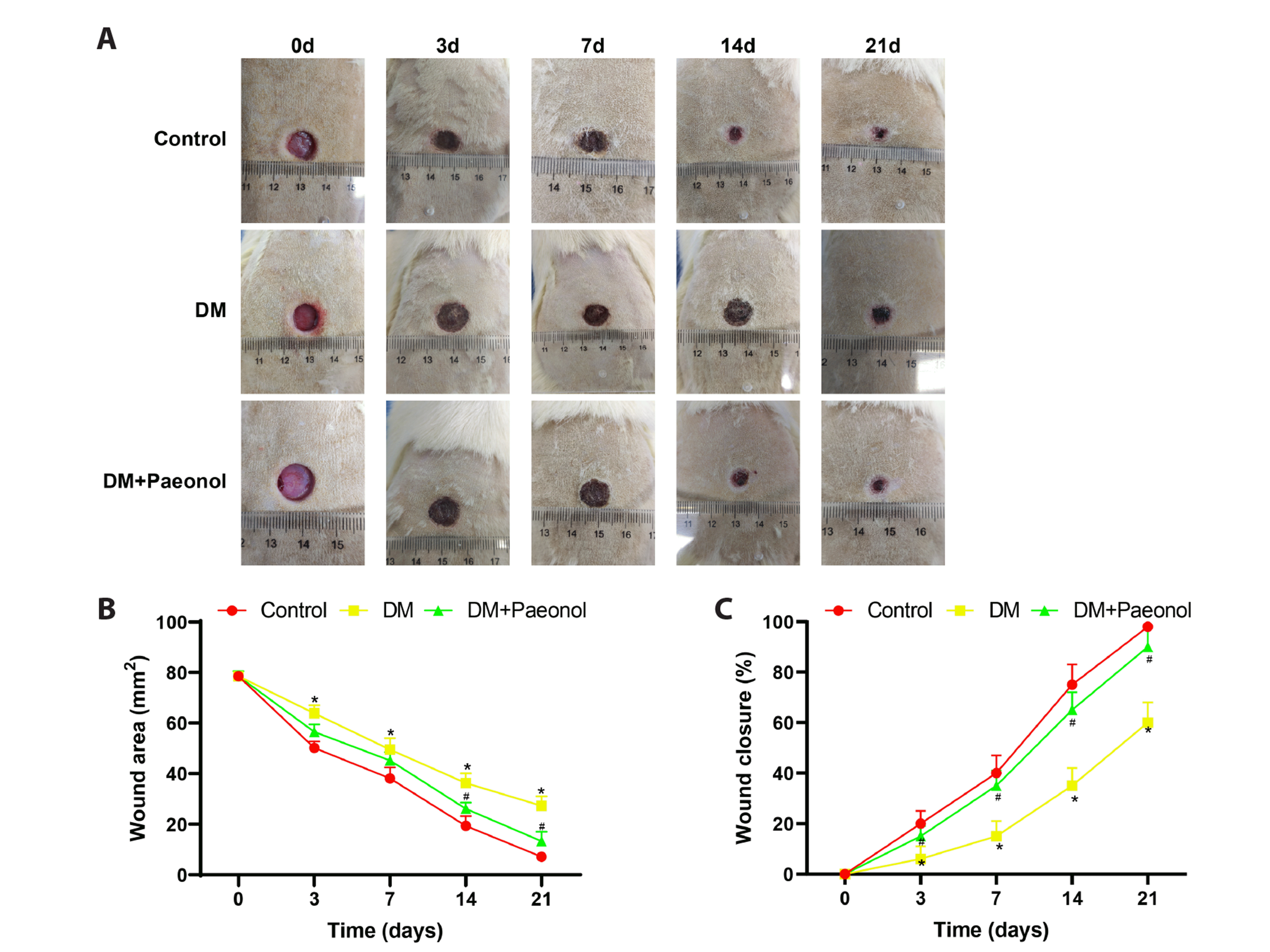
Paeonol promotes wound healing in diabetic rats
Paeonol has been reported to exert therapeutic effects on diseases including DM at a dosage of 50 mg/kg [21,26]. Therefore, we chose to investigate the effect of 50 mg/kg paeonol on wound healing in rats with STZ-induced DM. The wound healing of the diabetic rats was delayed in comparison with that of the control rats. The wound of the diabetic rats failed to heal on day 21 and the remaining wound area on days 3, 7, 14 and 21 was larger than that of the control rats (Fig. 1A, B, p < 0.05). Therefore, the wound closure rate of the diabetic rats was lower than that of the control rats (Fig. 1C, p < 0.05). After paeonol treatment, the wound healing efficiency of the diabetic rats increased and the wound was almost fully healed on day 21. Moreover, the remaining wound area was significantly reduced and the wound closure rate was elevated on days 3, 7, 14 and 21 (Fig. 1, p < 0.05). From the above, paeonol could promote wound healing in diabetic rats.
Paeonol reduces inflammation and promotes collagen deposition in diabetic ulcers
Next, we evaluated the effect of paeonol on wound healing in ulcerated DM rats using histological staining and immunohistochemistry methods. H&E staining was used for observation of inflammatory cells in the ulcerated sites. The diabetic rats showed inflammatory cell infiltration, redness, and swelling, while these symptoms were alleviated by paeonol treatment (Fig. 2A). Masson staining for detecting collagen fibers in the ulcerated sites revealed that the diabetic rats had fewer collagen fibers than the control rats. The collagen deposition in the ulcerated site of the diabetic rats was augmented after paeonol treatment (Fig. 2B, p < 0.05). Immunohistochemistry was utilized to detect the expression of the proliferation marker Ki67 and angiogenic factors CD31 and VEGF in the ulcerated sites. Compared with the control rats, the diabetic rats showed lower expression of Ki67, CD31 and VEGF; however, the expression of these proteins was elevated by paeonol treatment (Fig. 2C, D, p < 0.05). Taken together, these results indicated that paeonol accelerated wound healing in diabetic rats by reducing inflammation and promoting collagen deposition.

Fig. 2
Paeonol reduces inflammation and promotes collagen deposition in diabetic ulcers.
Ulcerated DM rats were treated with or without paeonol. (A) H&E assessment of the pathological characteristics of the ulcers. (B) Masson staining for revealing collagen deposition in the ulcers. Immunohistochemical detection of the expression of Ki67 (C), CD31 and VEGF (D). The data were expressed as mean ± standard deviation. Each group had 5 rats. Scale bar = 50 μm. *p < 0.05, **p < 0.01, ***p < 0.001, compared with the control group. #p < 0.05, ###p < 0.001, compared with the DM group. DM, diabetes mellitus; VEGF, vascular endothelial growth factor; IHC, immunohistochemistry.
![]()
Paeonol promotes M2 macrophage polarization in diabetic ulcers
Paeonol was found to promote wound healing in diabetic rats, but the mechanism was still unclear. Given the role of macrophage polarization in wound healing, we further detected the expression of proteins associated with M1 and M2 macrophages on day 21. The western blots showed that the expression of M1 macrophage-related inflammatory factors IL-1β and TNF-α increased and that of M2 macrophage-related inflammatory factors IL-4 and IL-10 decreased in the ulcerated site of diabetic rats; however, the expression trends of these inflammatory factors were reversed by paeonol treatment (Fig. 3A, p < 0.05). Furthermore, western blot was used to detect the expression of the M1 macrophage markers CD86 and CD80 and the M2 macrophage marker CD206. Compared with the control rats, the diabetic rats showed higher CD86 and CD80 expression and lower CD206 expression. Paeonol treatment decreased the expression of CD86 and CD80 and increased that of CD206 in the ulcerated DM rats (Fig. 3B, Supplementary Fig. 2A, p < 0.05). In addition, immunofluorescence staining was used to detect the expression of the M1 macrophage marker iNOS and the M2 macrophage marker arginase 1. The ulcerated DM rats showed higher iNOS expression and lower arginase 1 expression than the control rats, while paeonol treatment decreased the expression of iNOS and increased that of arginase 1 (Fig. 3C, D). The above results indicated that paeonol inhibited M1 macrophage polarization and activated M2 macrophages in ulcerated DM rats.
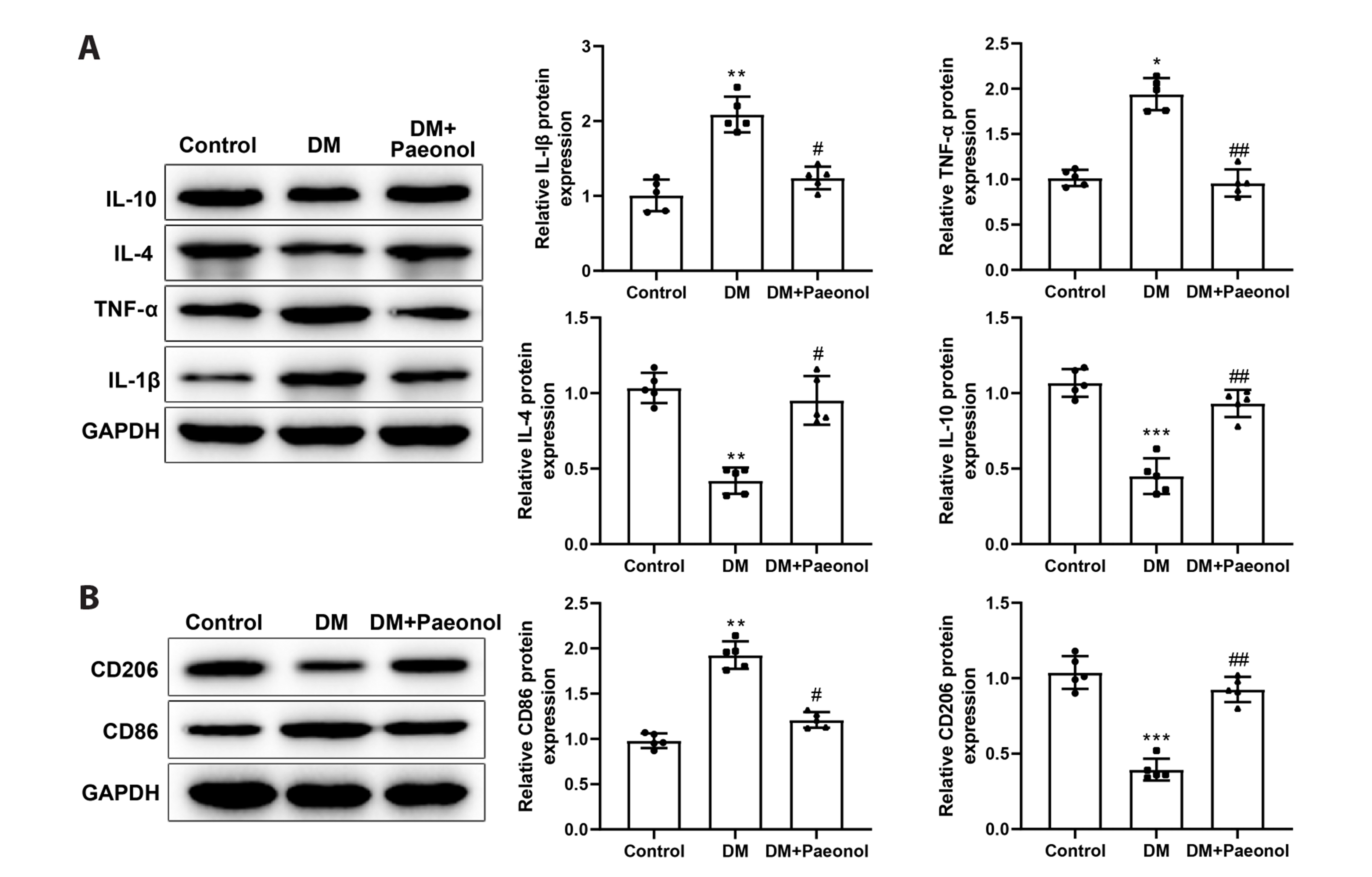
Fig. 3
Paeonol promotes M2 macrophage polarization in diabetic ulcers.
Ulcerated DM rats were treated with or without paeonol. (A, B) Western blot was used to detect the expression of inflammatory factors IL-1β, TNF-α, IL-4 and IL-10 and macrophage markers CD86 and CD206 in the ulcers on the 21st day. (C, D) Immunofluorescence was used to detect the expression of iNOS and arginase 1 in the ulcers. Scale bar = 50 μm. The data were expressed as mean ± standard deviation. Each group had 5 rats. *p < 0.05, **p < 0.01, ***p < 0.001, compared with the control group. #p < 0.05, ##p < 0.01, compared with the DM group. DM, diabetes mellitus; IL, interleukin; TNF-α, tumor necrosis factor α.
![]()
![]()
Paeonol improves the repair of high glucose-induced cell damage by modulating macrophage polarization
In order to validate the regulation of paeonol on macrophage polarization in vitro, we first detect the viability of paeonol-treated RAW264.7 macrophages using the MTS method. Different concentrations of paeonol had no impact on the viability of RAW264.7 cells, indicating that paeonol treatment did not induce cytotoxicity to macrophages (Fig. 4A). LPS + IFN-γ treatment and LPS + IL-4 treatment were used to induce M1 and M2 polarization of RAW264.7 cells, respectively. The PCR analysis demonstrated that LPS + IFN-γ treatment and LPS + IL-4 treatment increased the expression of the M1 marker iNOS and the expression of the M2 marker arginase 1, respectively; paeonol treatment reduced iNOS expression in RAW264.7 macrophages treated with LPS + IFN-γ and increased arginase 1 expression in RAW264.7 macrophages treated with LPS + IL-4 (Fig. 4B, p < 0.05). The immunofluorescence detection further suggested that paeonol could promote M2 macrophage polarization and inhibit M1 macrophage polarization (Fig. 4C). Next, we co-cultured high glucose-treated mouse skin fibroblasts with M1 or M2-polarized macrophages treated with or without paeonol. Western blot was used to detect the expression of inflammatory factors (IL-1β, TNF-α, IL-4, and IL-10), angiogenic factors (CD31 and VEGFA), and collagen I/III in the supernatant of mouse skin fibroblasts. High glucose treatment increased the expression of IL-1β and TNF-α and decreased that of IL-4, IL-10, CD31, VEGFA, collagen I, and collagen III (Fig. 4D, E, p < 0.05). Co-culture with M1 macrophages further elevated the expression of IL-1β and TNF-α and reduced that of IL-4, IL-10, CD31, VEGFA, collagen I, and collagen III, while co-culture with M2 macrophages brought about opposite effects (Fig. 4D, E, p < 0.05). Paeonol treatment reduced the expression of IL-1β and TNF-α and elevated that of IL-4, IL-10, CD31, VEGFA, collagen I, and collagen III in the supernatant of high glucose-treated fibroblasts co-cultured with either M1 or M2 macrophages (Fig. 4D, E, p < 0.05). Western blot experiments showed that the expression of IL-1β, TNF-α, IL-4, IL-10, CD31, VEGF, collagen I, and collagen III in mouse fibroblasts treated with paeonol alone were not significantly different from those in blank control cells (Supplementary Fig. 2B, C). The above data suggested that paeonol ameliorated the repair of high glucose-induced damage of fibroblasts by promoting M2 macrophage polarization.
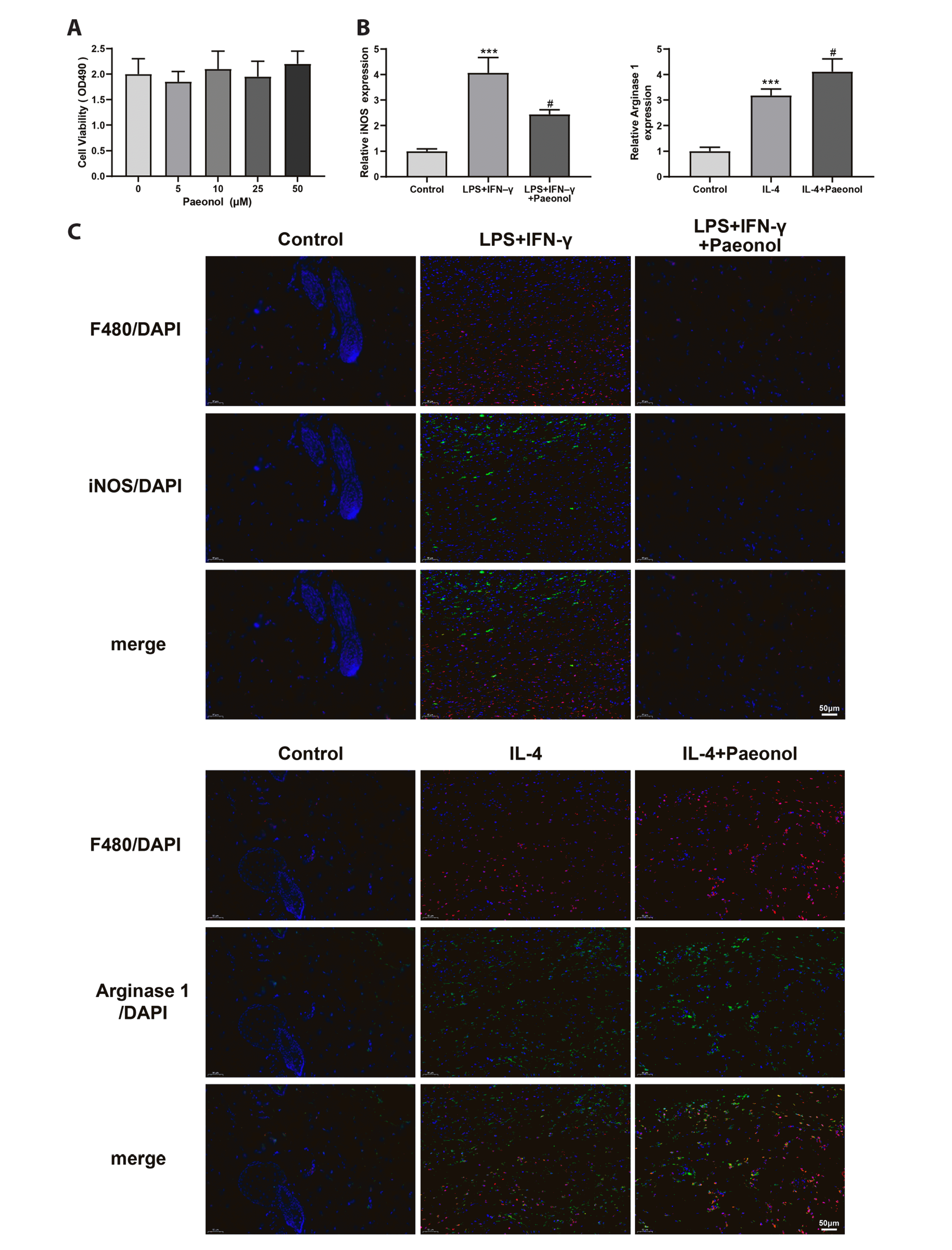
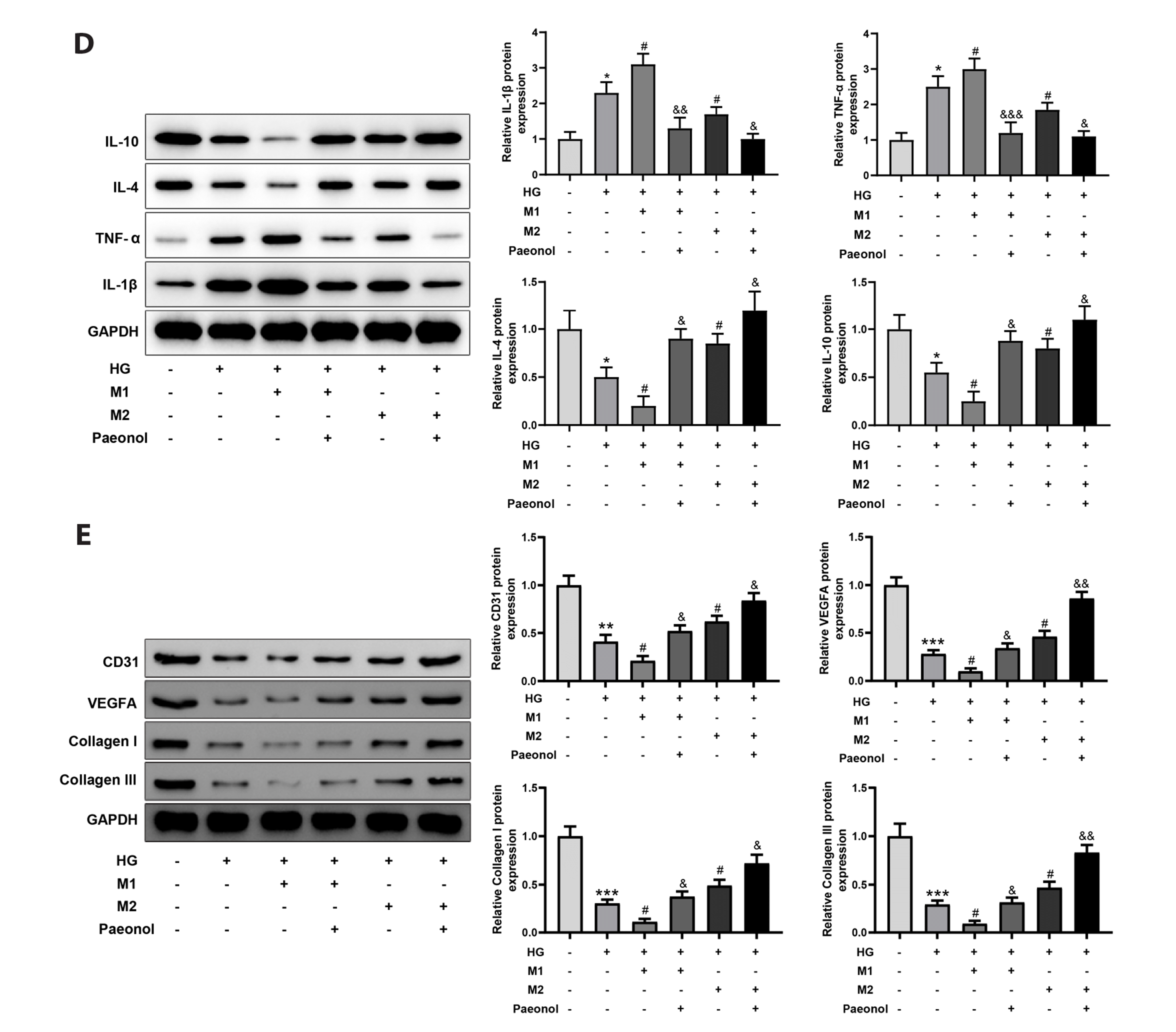
Fig. 4
Paeonol improves the repair of high glucose-induced cell damage by modulating macrophage polarization.
(A) MTS kit was used to assess the effect of paeonol treatment (0, 5, 10, 25, and 50 μM) on macrophage viability. RAW264.7 cells were polarized into M1 and M2 macrophages under LPS + IFN-γ treatment and IL-4 treatment, respectively. Then M1 or M2 macrophages were treated with paeonol and RT-qPCR (B) and immunofluorescence (C) was used to detect the expression of the M1 marker iNOS and the M2 marker arginase 1. Next, HG-treated mouse skin fibroblasts were co-cultured with M1 or M2-polarized macrophages treated with or without paeonol. (D, E) Western blot was used to detect the expression of inflammatory factors (IL-1β, TNF-α, IL-4, and IL-10), angiogenic factors (CD31 and VEGFA), and collagen I/III in the supernatant of mouse skin fibroblasts. Scale bar = 50 μm. The data were presented as mean ± standard deviation of three independent replicate experiments. *p < 0.05, compared with the control group. *p < 0.05, **p < 0.01, ***p < 0.001, compared with the control group. #p < 0.05, compared with the HG, LPS + IFN-γ, or LPS + IL-4 group. &p < 0.05, &&p < 0.01, &&&p < 0.001, compared with the HG + M1 or HG + M2 group. HG, high glucose; LPS, lipopolysaccharide; IFN, interferon; IL, interleukin; TNF-α, tumor necrosis factor α; RT-qPCR, Reverse transcription-quantitative polymerase chain reaction.
![]()
![]()
DISCUSSION
Globally, about 20% of individuals with diabetes develop diabetic wounds, with leg and foot being most often affected [27]. Diabetic ulcerations constitute a major health problem because of their resistance to healing which results from a constellation of both intrinsic and extrinsic factors regulating inflammation, tissue hypoxia, and extracellular matrix [28]. This study demonstrated that the delayed wound healing in diabetic rats was partially attributed to an imbalance of macrophage polarization. Paeonol, an anti-inflammatory agent, facilitated wound repair in diabetic rats and restored fibroblast function by promoting M2 macrophage polarization.
As macrophages play a critical role in wound healing, many new strategies for promoting diabetic wound healing are based on modulating macrophage polarization. For example, exosomes derived from melatonin-pretreated mesenchymal stem cells ameliorated diabetic wound healing by increasing the ratio of M2/M1 macrophages through activation of PTEN/Akt signaling pathway [29]. Insulin supplementation reduced inflammatory responses in diabetic wounds by promoting transition of macrophages from M1 to M2 phenotype through activation of Akt/Rac-1 signaling pathway and upregulation of PPAR-γ [30]. Molecules such as IL-25, FOXM1 and microRNA-145a-5p also improved diabetic wound healing by stimulating M2 macrophage polarization [31-33].
In this study, paeonol increased the healing efficiency and closure rate of back ulcers in diabetic rats. Although the effect of paeonol on diabetic wound healing has not been reported before, paeonol has shown great potential as a drug for treating other diabetic complications. Firstly, paeonol could alleviate diabetes itself by stimulating glucose uptake and decreasing lipid accumulation, which was associated with increased phosphorylation of Akt [34]. Paeonol promoted Opa1-dependent mitochondrial fusion and reduced oxidative stress in diabetic cardiomyopathy by activating the CK2α/Jak2/Stat3 pathway [35]. Paeonol impeded the progression of diabetic renal fibrosis by upregulating Sirt1 and activating its downstream Nrf2/ARE pathway [36]. From the perspective of neuroprotection, paeonol has been shown to reduce neurological pain, neuronal dysfunction, and oxidative stress in diabetic rats [37]. Moreover, paeonol suppressed oxidative stress, inflammatory response, and apoptosis of high glucose and palmitic acid-treated human umbilical vein endothelial cells by increasing the expression of Sirt1, which might have implications for macrovascular complications in diabetic cardiopathy [26].
The regulation of paeonol on macrophage function has been reported by several studies. For instance, paeonol reduced IL-1β production in LPS-treated mouse J774A.1 macrophages by blocking NLRP3 inflammasome and NF-κB signaling pathways [38]. Paeonol relieved acute pancreatitis by inhibiting M1 macrophage polarization and reactive oxygen species generation through inactivation of NLRP3 inflammasome [39]. The NLRP3 inflammasome is a critical component of the innate immune system, which mediates the activation of caspase-1 and the secretion of proinflammatory cytokines IL-1β and IL-18 in response to microbial infection and cellular damage [40]. Activation of the NLRP3 inflammasome sustains the inflammatory response in diabetic wounds [41]. The suppression of NLRP3 inflammasome activation can boost M2 macrophage polarization to accelerate skin wound healing [42]. Therefore, paeonol may facilitate diabetic wound healing by stimulating M2 macrophage polarization through inactivation of NLRP3 inflammasome. In this study, paeonol treatment decreased inflammatory cell infiltration and expression of IL-1β, TNF-α and iNOS and increased collagen fibers and expression of IL-4, IL-10, Ki67, CD31, VEGF, collagen I/III, and arginase 1 in the ulcerated site of diabetic rats, indicating that paeonol may accelerate wound healing by promoting collagen deposition, angiogenesis, and M2 macrophage polarization. In vitro, paeonol inhibited the M1 polarization of RAW264.7 macrophages and promoted the transition to M2 macrophages. Moreover, paeonol reduced the expression of IL-1β and TNF-α and increased that of IL-4, IL-10, CD31, VEGF, and collagen I/III in the supernatant of high glucose-treated fibroblasts co-cultured with either M1 or M2-polarized macrophages.
In summary, paeonol may serve as a new drug for diabetic wound repair by promoting M2 macrophage polarization. However, the pharmacological effect of paeonol has not been investigated in clinical settings and the molecules directly targeted by paeonol in diabetic wounds are unknown. These issues must be solved to promote the clinical application of paeonol in diabetic wound repair.
 XML Download