Journal List > Acute Crit Care > v.38(1) > 1516083672

Kayumov, Habimana, Kim, Obiweluozor, Jeong, and Cho: Extracorporeal circulation models in small animals: beyond the limits of preclinical research

Abstract

Extracorporeal membrane oxygenation (ECMO) use has remarkably increased in recent years. Although ECMO has become essential for patients with refractory cardiac and respiratory failure, extracorporeal circulation (ECC) is associated with significant complications. Small-animal models of ECC have been developed and widely used to better understand ECC-induced pathophysiology. This review article summarizes the development of small-animal ECC models, including the animal species, circuit configuration, priming, perioperative procedures, cannulation, and future perspectives of small-animal ECMO models.

INTRODUCTION

Extracorporeal life support (ECLS) is essential in critical care, supplying adequate oxygen and blood flow to vital organs for failing hearts or lungs [1-3]. ECLS or extracorporeal membrane oxygenation (ECMO) is an advanced version of cardiopulmonary bypass (CPB) that is customized to provide heart and/or lung support for longer durations (days to weeks) [4]. The main purpose of ECMO is to allow time to recover or to serve as a bridge to heart or lung transplantation. Unlike CPB, ECMO can be categorized based on the target of support. Many combinations exist, but historically, the main divisions are veno-venous (VV) ECMO for respiratory support [5] and veno-arterial (VA) ECMO for cardiac or cardiopulmonary support [6].
Extracorporeal circulation (ECC) induces systemic inflammatory response syndrome (SIRS) in the body, which often limits its potential benefits or causes failure to reach the targeted treatment [1,2,7]. This is often triggered by contact between blood and the foreign surface of the extracorporeal circuit [2,8], which leads to the activation of the complement system, resulting in the activation of inflammatory cells such as neutrophils and monocytes and the release of inflammatory mediators [8]. Although our understanding of the pathophysiology of ECC has advanced, its current limitations, such as SIRS and coagulopathy, restrict its long-term applications [9]. Despite improvements in medical engineering, ECC is still associated with significant risks and complications [2,9,10]. Thus, the development of reproducible animal models of ECC is increasingly important for improving its safety and outcomes.
Animal models of ECC have been developed in various sciences to understand the pathophysiology of various conditions [11] and to validate novel interventions [10,12]. Animal models of ECLS have mainly been established in large animals. However, these models are costly in terms of budgetary requirements, other resources, and personnel. Small animals have received more attraction, both due to the need for a lower budget and less personnel [13] and because they enable cellular or even molecular/genetic studies.
The first small-animal ECC model was reported by Popovic et al. in 1968 [14]. The study included 70 male Sprague-Dawley rats undergoing hypothermic CPB. They applied 70 minutes of CPB circulation at 14 ℃ and reported physiological parameters, such as heart rate, respiratory rate, oxygen blood oxygen content, and hematocrit, during CPB. Later, in 1983, Alexander et al. [15] performed 6 hours of partial CPB in 30 rats. The development of ECMO devices in the early 21st century increased the importance of small-animal models. Since then, interest in establishing small-animal ECC models has grown.
To better understand the available small-animal models of ECC, we summarized existing models between 1968 and 2020. The aim of this review is to discuss the strategies and current limitations of extant models and to focus on specific targets to improve the reproducibility of small-animal ECC models.

MODEL ESTABLISHMENT

Study Selection

We searched PubMed, Scopus, and Medline from 1968 to 2020, and two clinicians screened the articles to identify those that included small-animal ECC models. Studies were excluded if there were no information regarding the ECC model or surgical procedures, if the models were in animals other than rodents (rabbits were excluded), and if the same procedure was repeated by other researchers. Detailed data were obtained in the following major categories: the main theory, the purpose of the study, the species, animals’ weights, groups, the method of selection and inclusion, the number of animals, perioperative techniques (including animal preparation, sedation, respiratory support, and catheterization), the type of ECC, the duration of ECC, priming volume, and priming content.
In total, 23 studies were included: 19 studies with rats and four with mice. An overview of the included studies is shown in Table 1 [1,2,7,10,12-30]. Nineteen studies applied the VA type of ECC, whereas VV ECC was performed in two studies and arterio-arterial was performed in one study. One study conducted both VA and VV ECC models in rats [13]. The median duration of ECC varied from 30 minutes to 6 hours.

Species and weight of models

The most commonly used weight of rats was 300–500 g. The weight of the animals can be used to indicate normal cardiac output, thereby making it possible to manage the desired ECC flow. When establishing an ECC model, the weight is an essential determinant in adjusting priming volume. For mice, the weights were 25–35 g in all studies [2,26,27,29,31].

Circuit configuration

The circuit configuration of the experimental ECLS model is compatible with the circuit in clinical settings, with elements including cannulas, tubes, a reservoir, an oxygenator, and a pump. Silicone-based drainage and perfusion cannulas that allow the desired laminar blood flow are used for arterial or venous cannulation. Additional components (e.g., a reservoir or infusion line) can be optionally added to the circuit [13]. It is logical to set the circuit capacity to be as low as possible to minimize the contact of blood with the foreign surface of the circuit [32]. A large amount of priming volume may cause severe hemodilution of the animal blood undergoing ECC unless allogenic blood is used for priming [13].
In experimental research, a commercially available polyethylene-based oxygenator is used [8]. Until small-animal membrane oxygenators became commercially available, bubble oxygenators were commonly used in all ECC models [18,33,34]. However, bubble oxygenators require a larger priming volume and allow intimate contact between blood and gas, which induces damage to erythromyeloid reticuloendothelial cells [15]. A micro-membrane oxygenator for small animals requires a small priming volume (3–4 ml), with a sufficient gas exchange surface, and many investigators have successfully established ECC models with a priming volume of less than 10 ml [1,35]. Membrane oxygenators are designed to prevent direct contact between blood and gases and allow sufficient gas exchange through micro-pores [36,37]. Finally, the small-animal ECC circuit contains a single peristaltic pump that drains blood and passes it through the oxygenator to the arterial line [13].

Priming

Previously, a large amount of allogenic blood was required to fill the priming circuit [16,23]. However, using allogenic blood as a priming solution restrained immunological and physiological studies, as it may lead to profound pathophysiologic reactions. Crystalloids and colloids are now the most commonly used solutions to fill ECC circuits [1,2,7,8,10-13,17-20,22,24,25]. Hetastarch, hydroxyethyl starch-sterile, and Gelofusine are the most often used colloids, while Plasma, A-Lyte solution, Ringer solution, Saline, and sodium bicarbonate are the most commonly applied crystalloids.
Other medications such as heparin and mannitol can also be included in the priming solution, depending on the strategy and preferences of the investigator [18-21,24,25,27]. In our previous study, we introduced a new mixture of priming solution using 50% albumin and 50% saline, which successfully maintained a few hours of ECMO run and improved the animals’ survival postoperatively [13].
Applying less priming solution may be associated with improved outcomes. The average amount of priming solution for 400–500 g rats, which are commonly used by researchers, ranges between 10 and 16 ml [10,12-14,17,19,20,22,24,25,38]. In mouse models, the successfully applied priming amount varied between 0.4 and 0.85 ml in previously published papers [2,27,29,31].

Perioperative procedures

Surgical procedures are usually performed under general anesthesia. Choosing an appropriate anesthetic and administration method depends on the investigator’s preference. The most common methods of administration are intraperitoneal injection and inhalation anesthesia [1,2,7,10,12,13,17-22,24,25,27,29,31,38]. Ketamine, sodium pentobarbital, fentanyl, pancuronium, and isoflurane are generally used for anesthesia induction. A repeated dose of the same anesthetic or isoflurane inhalation can be used for maintenance [7,10,17,20,21]. The selection of a correct anesthetic and its dosage is of the utmost importance because an overdose and/or incorrect administration may directly alter the cardiovascular system, leading to adverse outcomes and death. Light but adequate anesthesia is essential. In our previous study, we used inhalation isoflurane alone (both induction and continuous) [13,30]. Animals should be placed on a heating pad to manipulate body temperature throughout the experiment. The surgical field is shaved and cleaned with antiseptics.

Cannulation

Surgery can be performed under the aid of a microscope (mouse) or surgical loupe (rats) according to researchers’ preference. The incision site is determined based on the type of ECC and other additional interventions. The neck and groin areas are commonly applied surgical areas of peripheral cannulation. Cannulation sites can differ depending on the type of ECC and the surgeon’s preference. In VA ECC, venous blood is drained from the right atrium through the right jugular vein or femoral veins, and oxygenated blood is perfused to the circulatory system through the right carotid artery or femoral artery as well as the tail artery. In VV ECC, as our previous study introduced, blood is drained from the right jugular vein and perfused to the left jugular vein. Theoretically, blood can be drained from the right femoral vein to the left femoral vein. The design and proper placement of the venous cannula result in adequate drainage flow. Several researchers have used modified multi-orifice 16-gauge angiocatheters as drainage cannulas [10,12,17,19,22,24,25,38]. However, the sharp end of the angiocatheter may damage the vessel or heart, making it dangerous to apply, especially for early-career researchers [13]. We recently introduced a drainage cannula that has a blunt end with eight additional side holes on the side of the cannula tip. This new cannula has resulted in safer cannulation and an increased flow rate [13]. Moreover, Natanov et al. [29] recently reported the application of a double-lumen silicone catheter for VV-ECMO in a mouse model that minimized surgical trauma and eventually improved experimental outcomes. The size of the angiocatheter for perfusion can be chosen depending on the size of the arterial vessel. The most widely used perfusion cannulas are 20–24 gauge angiocatheters [1,2,7,10,13,17-23,38].
Furthermore, successful ECC models with open-chest techniques and abdominal vessel cannulation have been reported [1], but the invasiveness of these procedures may result in bleeding and poor postoperative survival. Intravenous injection of 1,000 IU/kg heparin should be administered either prior to or immediately after cannulation to prevent blood clot formation during ECC. A repeated dose of heparin administration is recommended if ECC is sustained for more than 2 hours.

FUTURE PERSPECTIVES OF SMALL-ANIMAL ECMO MODELS: AFFORDABLE IN VIVO STUDIES

Small-animal models of ECMO are essential for understanding the mechanics and energetics of ECMO and testing novel extracorporeal interventions. ECMO research is valuable for overcoming complications related to ECMO and patient factors. Molecular studies are more feasible to perform in small animals in various disease models for ECMO (e.g., acute and chronic heart failure, respiratory failure, or septic shock). ECMO can be applied to a wide range of genetically modified and commercially available mice to investigate molecular levels during ECC (Table 2, Figure 1).

CONCLUSIONS

Although the use of ECMO is exponentially increasing, there is much more to learn about the effect of ECMO on cellular and molecular levels. To better understand the in vivo mechanisms of ECMO from diverse future perspectives, small-animal ECMO research may allow us to conduct affordable in vivo animal research.

HIGHLIGHTS

▪ This review article summarizes the development of small-animal extracorporeal circulation models, including animal species, circuit configuration, priming, perioperative procedures, cannulation, and future perspectives of small-animal extracorporeal membrane oxygenation models.

Notes

CONFLICT OF INTEREST

Hwa Jin Cho Dong-Ick Shin is an editorial board member of the journal but was not involved in the peer reviewer selection, evaluation, or decision process of this article. No other potential conflicts of interest relevant to this article were reported.

FUNDING

None.

AUTHOR CONTRIBUTIONS

Conceptualization: HJC, DK, FOO, ISJ. Data curation: MK, RH. Formal analysis: ISJ. Funding acquisition: HJC, DK, ISJ. Methodology: MK, RH. Project administration: MK, ISJ. Visualization: HJC, RH, ISJ. Writing–original draft: MK, HJC. Writing–review & editing: all authors.

ACKNOWLEDGMENTS

This study was supported by Chonnam National University Hospital (BCRE22118, BCRI22025. BCRI19045, BCRI123051) and National research foundation (NRF-2019R1D1A3A03103899 and NRF-2020R1F1A1073921) to In Seok Jeong and Hwa Jin Cho.

REFERENCES

1. Ali AA, Downey P, Singh G, Qi W, George I, Takayama H, et al. Rat model of veno-arterial extracorporeal membrane oxygenation. J Transl Med. 2014; 12:37.
2. Luo S, Tang M, Du L, Gong L, Xu J, Chen Y, et al. A novel minimal invasive mouse model of extracorporeal circulation. Mediators Inflamm. 2015; 2015:412319.
3. Tramm R, Ilic D, Davies AR, Pellegrino VA, Romero L, Hodgson C. Extracorporeal membrane oxygenation for critically ill adults. Cochrane Database Syst Rev. 2015; 1:CD010381.
4. Bateman RM, Sharpe MD, Jagger JE, Ellis CG, Solé-Violán J, López-Rodríguez M, et al. 36th International Symposium on Intensive Care and Emergency Medicine : Brussels, Belgium. 15-18 March 2016. Crit Care. 2016; 20(Suppl 2):94.
5. Abrams D, Bacchetta M, Brodie D. When the momentum has gone: what will be the role of extracorporeal lung support in the future? Curr Opin Crit Care. 2018; 24:23–8.
6. Raleigh L, Ha R, Hill C. Extracorporeal membrane oxygenation applications in cardiac critical care. Semin Cardiothorac Vasc Anesth. 2015; 19:342–52.
7. Chang RW, Luo CM, Yu HY, Chen YS, Wang CH. Investigation of the pathophysiology of cardiopulmonary bypass using rodent extracorporeal life support model. BMC Cardiovasc Disord. 2017; 17:123.
8. Cresce GD, Walpoth BH, Mugnai D, Innocente F, Rungatscher A, Luciani GB, et al. Validation of a rat model of cardiopulmonary bypass with a new miniaturized hollow fiber oxygenator. ASAIO J. 2008; 54:514–8.
9. Giridharan GA, Lee TJ, Ising M, Sobieski MA, Koenig SC, Gray LA, et al. Miniaturization of mechanical circulatory support systems. Artif Organs. 2012; 36:731–9.
10. Bianchini EP, Sebestyen A, Abache T, Bourti Y, Fontayne A, Richard V, et al. Inactivated antithombin as anticoagulant reversal in a rat model of cardiopulmonary bypass: a potent and potentially safer alternative to protamine. Br J Haematol. 2018; 180:715–20.
11. Yuan L, Su D, Liu X, Lu H, Li Y, Tong S. Cerebral blood flow changes during rat cardiopulmonary bypass and deep hypothermic circulatory arrest model: a preliminary study. Annu Int Conf IEEE Eng Med Biol Soc. 2013; 2013:1807–10.
12. Rungatscher A, Linardi D, Tessari M, Menon T, Luciani GB, Mazzucco A, et al. Levosimendan is superior to epinephrine in improving myocardial function after cardiopulmonary bypass with deep hypothermic circulatory arrest in rats. J Thorac Cardiovasc Surg. 2012; 143:209–14.
13. Cho HJ, Kayumov M, Kim D, Lee K, Onyekachi FO, Jeung KW, et al. Acute immune response in venoarterial and venovenous extracorporeal membrane oxygenation models of rats. ASAIO J. 2021; 67:546–53.
14. Popovic P, Horecky J, Popovic VP. Hypothermic cardiopulmonary bypass in white rats. Ann Surg. 1968; 168:298–301.
15. Alexander B, Al Ani HR. Prolonged partial cardiopulmonary bypass in rats. J Surg Res. 1983; 35:28–34.
16. Grocott HP, Mackensen GB, Newman MF, Warner DS. Neurological injury during cardiopulmonary bypass in the rat. Perfusion. 2001; 16:75–81.
17. Dong GH, Xu B, Wang CT, Qian JJ, Liu H, Huang G, et al. A rat model of cardiopulmonary bypass with excellent survival. J Surg Res. 2005; 123:171–5.
18. Ordodi VL, Paunescu V, Ionac M, Sandesc D, Mic AA, Tatu CA, et al. Artificial device for extracorporeal blood oxygenation in rats. Artif Organs. 2008; 32:66–70.
19. Huang H, Yin R, Zhu J, Feng X, Wang C, Sheng Y, et al. Protective effects of melatonin and N-acetylcysteine on hepatic injury in a rat cardiopulmonary bypass model. J Surg Res. 2007; 142:153–61.
20. Jungwirth B, Kellermann K, Blobner M, Schmehl W, Kochs EF, Mackensen GB. Cerebral air emboli differentially alter outcome after cardiopulmonary bypass in rats compared with normal circulation. Anesthesiology. 2007; 107:768–75.
21. Qing M, Shim JK, Grocott HP, Sheng H, Mathew JP, Mackensen GB. The effect of blood pressure on cerebral outcome in a rat model of cerebral air embolism during cardiopulmonary bypass. J Thorac Cardiovasc Surg. 2011; 142:424–9.
22. Waterbury T, Clark TJ, Niles S, Farivar RS. Rat model of cardiopulmonary bypass for deep hypothermic circulatory arrest. J Thorac Cardiovasc Surg. 2011; 141:1549–51.
23. Mackensen GB, Sato Y, Nellgård B, Pineda J, Newman MF, Warner DS, et al. Cardiopulmonary bypass induces neurologic and neurocognitive dysfunction in the rat. Anesthesiology. 2001; 95:1485–91.
24. Fujii Y, Shirai M, Inamori S, Shimouchi A, Sonobe T, Tsuchimochi H, et al. Insufflation of hydrogen gas restrains the inflammatory response of cardiopulmonary bypass in a rat model. Artif Organs. 2013; 37:136–41.
25. Fujii Y, Shirai M, Tsuchimochi H, Pearson JT, Takewa Y, Tatsumi E, et al. Hyperoxic condition promotes an inflammatory response during cardiopulmonary bypass in a rat model. Artif Organs. 2013; 37:1034–40.
26. Du Q, Shen Y, Yu J, Huang S, Pan S. Extracorporeal membrane oxygenation (EMCO) is an optimal method to cure the pneumonia caused by endotoxin in mice. Int J Clin Exp Pathol. 2016; 9:10796–802.
27. Madrahimov N, Boyle EC, Gueler F, Goecke T, Knöfel AK, Irkha V, et al. Novel mouse model of cardiopulmonary bypass. Eur J Cardiothorac Surg. 2018; 53:186–93.
28. Xie XJ, Tao KY, Tang ML, Du L, An Q, Lin K, et al. Establishment and evaluation of extracorporeal circulation model in rats. Sichuan Da Xue Xue Bao Yi Xue Ban. 2012; 43:770–4.
29. Natanov R, Khalikov A, Gueler F, Maus U, Boyle EC, Haverich A, et al. Four hours of veno-venous extracorporeal membrane oxygenation using bi-caval cannulation affects kidney function and induces moderate lung damage in a mouse model. Intensive Care Med Exp. 2019; 7:72.
30. Kayumov M, Kim D, Raman S, MacLaren G, Jeong IS, Cho HJ. Combined effects of sepsis and extracorporeal membrane oxygenation on left ventricular performance in a murine model. Sci Rep. 2022; 12:22181.
31. Madrahimov N, Natanov R, Boyle EC, Goecke T, Knöfel AK, Irkha V, et al. Cardiopulmonary bypass in a mouse model: a novel approach. J Vis Exp. 2017; (127):56017.
32. Riera J, Argudo E, Ruiz-Rodríguez JC, Ferrer R. Extracorporeal membrane oxygenation for adults with refractory septic shock. ASAIO J. 2019; 65:760–8.
33. Gaylor JD. Membrane oxygenators: current developments in design and application. J Biomed Eng. 1988; 10:541–7.
34. Clark RE, Beauchamp RA, Magrath RA, Brooks JD, Ferguson TB, Weldon CS. Comparison of bubble and membrane oxygenators in short and long perfusions. J Thorac Cardiovasc Surg. 1979; 78:655–66.
35. Berner M, Clément D, Stadelmann M, Kistler M, Boone Y, Carrel TP, et al. Development of an ultra mini-oxygenator for use in low-volume, buffer-perfused preparations. Int J Artif Organs. 2012; 35:308–15.
36. Iwahashi H, Yuri K, Nosé Y. Development of the oxygenator: past, present, and future. J Artif Organs. 2004; 7:111–20.
37. Obstals F, Vorobii M, Riedel T, de Los Santos Pereira A, Bruns M, Singh S, et al. Improving hemocompatibility of membranes for extracorporeal membrane oxygenators by grafting nonthrombogenic polymer brushes. Macromol Biosci. 2018; 18:1700359.
38. Lebreton G, Tamion F, Bessou JP, Doguet F. Cardiopulmonary bypass model in the rat: a new minimal invasive model with a low flow volume. Interact Cardiovasc Thorac Surg. 2012; 14:642–4.
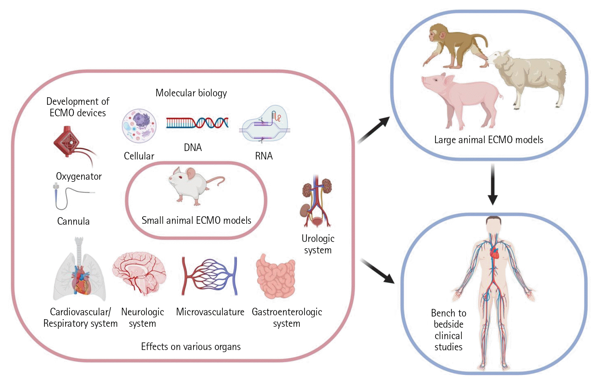
Figure 1.
Common experimental extracorporeal membrane oxygenation (ECMO) models. Created with BioRender.com.
acc-2023-00381f1.tif
Table 1.
Description of previously published studies included in the review
Study Year Species ECC type Duration of ECC (min) Drainage catheter Perfusion catheter Priming volume (ml) Purpose of study
Popovic et al. [14] 1968 Rat VA/CPB 70 PE 200 tubing (ID: 1.4 mm) PE 90 tubing (0.86 mm) 13.7 Model development
Alexander et al. [15] 1983 Rat VA/CPB 360 NA NA 8.7 Device development
Grocott et al. [16 2001 Rat VA/CPB 60 Dual-stage venous cannula (4.5 Fr) Angiocatheter (20G) 45 Neuropathophysiology
Dong et al. [17] 2005 Rat VA/CPB 60 Angiocatheter (16G) Angiocatheter (22G) 16 Model establishment
Ordodi et al. [18] 2008 Rat AA/CPB 120 Angiocatheter (24G) Angiocatheter (24G) 20 Device development
Huang et al. [19] 2007 Rat VA/CPB 60 Angiocatheter (16G) Angiocatheter (22G) 10 Pathophysiology
Jungwirth et al. [20] 2007 Rat VA/CPB 90 Dual-stage venous cannula (4.5 Fr) Angiocatheter (20G) 16 Neuropathophysiology
Qing et al. [21] 2011 Rat VA/CPB 90 Dual-stage venous cannula (4.5 Fr) Angiocatheter (20G) 18 Neuropathophysiology
Waterbury et al. [22] 2011 Rat VA/CPB 80 Angiocatheter (16G) Angiocatheter (20G) 12 CPB and DHCA development
Rungatscher et al. [12] 2012 Rat VA/CPB 90 Angiocatheter (16G) Angiocatheter (18G) 10.5 Pharmacology
Mackensen et al. [23] 2001 Rat VA/CPB 150 Angiocatheter (16G) Angiocatheter (22G) 10 Model establishment
Fujii et al. [24] 2013 Rat VA/CPB 60 Angiocatheter (16G) Polyethylene tubing (NA) 15 Pharmacology
Fujii et al. [25] 2013 Rat VA/CPB 120 Angiocatheter (16G) Polyethylene tubing (NA) 15 CPB pathophysiology
Ali et al. [1] 2014 Rat VA/ECMO 10 Angiocatheter (20G) Angiocatheter (24G) 8 Model establishment
Luo et al. [2] 2015 Mouse VA/ECC 30 Angiocatheter (24G) Angiocatheter (24G) 0.4 ECC-induced SIRS
Du et al. [26] 2016 Mouse NA NA NA NA NA ECMO treatment in pneumonia
Chang et al. [7] 2017 Rat VA/CPB, ECLS 30 Angiocatheter (14G) Angiocatheter (20G) 20 CPB pathophysiology
Bianchini et al. [10] 2018 Rat VA/CPB 60 Angiocatheter (16G) Angiocatheter (22G) 10 Pharmacology
Madrahimov et al. [27] 2018 Mouse VA/CPB 90 Polyurethane tube (2 Fr) Angiocatheter (27G) 0.85 Model establishment
Xie et al. [28] 2012 Rat VA 120 Trocar (24G) Trocar (24G) NA Platelet therapy with CPB
Natanov et al. [29] 2019 Mouse VV/ECMO 240 Double-lumen silicone catheter (2 Fr) Double-lumen silicone-based catheter (2 Fr) 0.5 VV-ECMO induced pathophysiology
Cho et al. [13] 2021 Rat VA+VV/ECMO 120 Modified neonatal feeding tube (5 Fr) Angiocatheter (24G) 14 Immunology
Kayumov et al. [30] 2022 Rat VA/ECMO 120 Modified neonatal feeding tube (5 Fr) Angiocatheter (24G) 14 Septic shock, pressure-volume change

ECC: extracorporeal circulation; VA: veno-arterial; CPB: cardiopulmonary bypass; NA: not applicable; AA: arterio-arterial; DHCA: deep hypothermic circulatory arrest; ECMO: extracorporeal membrane oxygenation; SIRS: systemic inflammatory response syndrome; ECLS: extracorporeal life support; VV: veno-venous.

Table 2.
Current and future perspectives on ECMO research
ECMO research
ECMO-related mechanics LV unloading
ECMO-related energetics Effect of ECC on ATP production in mitochondria
ECMO-related complications Understand pathophysiology to prevent complications.
Provide advanced care to patients that decrease life-threatening events.
Neuro-pathophysiology Post-cardiac arrest ischemic brain injury in patients on ECMO or ECPR
ECMO-related mechanical complications in the brain
The effect on the brain microcirculation
The effect on the neurologic performance
Development of ECMO devices Hemocompatible and biocompatible ECMO devices
Cause fewer mechanical complications.
Cause less vascular (endothelial) reaction.
Protect end-organs functions.
Adjunctive treatments in ECMO Mesenchymal stem cells during ECMO
Mechanical adjunctive treatment in ECMO
Pharmacodynamics and pharmacokinetics The effects of the circuit/oxygenator/pump on various medications

ECMO: extracorporeal membrane oxygenation; LV: left ventricle; ECC: extracorporeal circulation; ATP: adenosine triphosphate; ECPR: extracorporeal cardiopulmonary resuscitation.

TOOLS
Similar articles