

 PDF
PDF Citation
 Print
Print


Introduction
Inverted papilloma in paranasal sinus is known to accompany malignant changes in about 10% [1], and therefore is required complete surgical resection to achieve tumor control. Since the introduction of endoscopy, many studies have reported that endoscopic approach showed similar or even better results compared to open. Currently, endoscopic sinus surgery is being performed as the first treatment option for inverted papilloma. However, despite advances of endoscopic technology, surgery for lesion originating frontal sinus is still considered a difficult field due to the complex anatomical structure. According to a literature report, 1%-16% of inverted papilloma occur in the frontal sinus [2]. Therefore, it may be necessary to use an external approach for complete resection of the lesion. Among them, osteoplastic flap (OPF) surgery has been frequently used since the mid-1960s [3]. While this method facilitates access to the frontal sinus, the possibility of postoperative complications such as numbness of surgical site, alopecia, injury of frontal branch of facial nerve is well known [4]. Mini OPF has been introduced as an alternative procedure, which is less invasive. We report a case of successful treatment with mini OPF for recurrent inverted papilloma originating from the frontal sinus.
Go to : 
Case
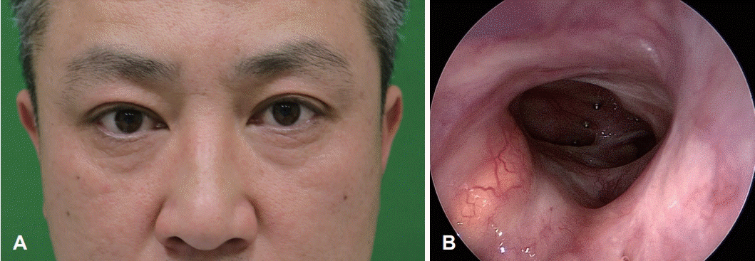
A 42-year-old male was referred with progressive downward deviation and proptosis of the right orbit, and diplopia during upward gaze that had occurred a month ago (Fig. 1A). He had no medical past history, except for smoking and drinking. On ophthalmologic examination, there were 6 mm exophthalmos, ophthalmoplegia and elevated intraocular pressure of right side, but no abnormalities in visual acuity. Several polypoid masses with mucopurulent discharge were found in the right middle meatus by nasal endoscope (Fig. 1B), which were confirmed as inverted papilloma on biopsy. CT showed a 3 cm sized well defined unilocular cystic mass in the right superior intraorbital extraconal space. This lesion was accompanied by bony erosion on the right orbital roof and compression of the eyeball and extraocular muscle, but no invasion. And soft tissue density was present in right ostiomeatal unit, ethmoid and frontal sinus (Fig. 2A and B). MRI revealed a non-enhancing right superior extraconal mass arising from right frontal sinus, showing low signal intensity in T1 weighted images and high signal intensity in T2 weighted images. And lobular soft tissue masses were observed in frontal and ethmoid sinuses, which is heterogeneously enhanced with low and intermediate signal intensity on T1 and T2 weighted images, respectively (Fig. 2C and D).
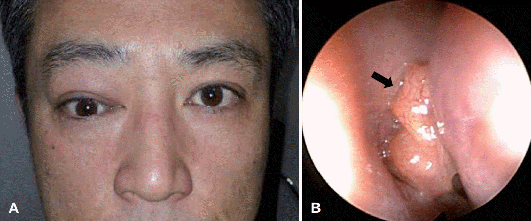 | Fig. 1.Preoperative profile and endoscopic finding. A: Frontal view shows downward deviation and proptosis of the right orbit. B: Several polypoid masses with mucopurulent discharge in the right middle meatus are shown in nasal endoscopy (arrow).
|
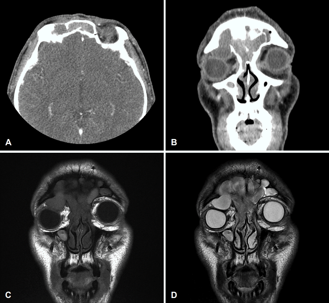 | Fig. 2.Preoperative paranasal sinus CT and MRI. Axial (A) and coronal (B) images of CT scan show well defined unilocular cystic mass in the right superior intaorbital extraconal space and soft tissue density in frontal sinus. T1-weighted (C) and T2-weighted (D) MRI show non-enhancing right superior extraconal mass with low signal intensity, which is presumed to be a mucocele. And heterogeneously enhancing lesion in frontoethmoid sinuses have low and intermediate signal intensity with possible bony erosion, respectively.
|
It was judged to be a lesion combined with mucocele and inverted papilloma, and endoscopic removal through the modified Lothrop procedure was planned first. The origin of inverted papilloma identified during surgery was the frontal sinus intersinus septum and posterior wall. The surrounding mucosa including the origin site was removed as completely as possible. The lesion suspected of being mucocele was resolved through marsupialization. The ophthalmic discomfort was improved after the surgery. The difference of exophthalmos between the right and left eyes was reduced to 1.5 mm. The biopsy result was inverted papilloma with mild to moderate dysplasia.
During follow-up without evidence of recurrence or complications, polypoid masses considered to be recurrent were observed in the right frontal sinus on nasal endoscopy at 20 months after the operation (Fig. 3A). CT and MRI showed soft tissue density in the superior and posterior wall of frontal sinus (Fig. 3B and C), and revision surgery via left frontal sinus external approach was planned. Initially, an attempt was made to remove the tumor through an endoscopic approach, but it was incomplete. Therefore, an incision was performed along the medial side under the left eyebrows for access to the frontal sinus for removal of the remaining lesion. After finding and preserving supratrochlear nerve, the mini OPF was lifted into a 12×8 mm rectangular shape (Fig. 4A). Papillomatous lesions observed in the superior, posterior wall and supraorbital area of right frontal sinus were removed using microdebrider, curette, malleable suction monopolar cautery and drill. The OPF was fixed with a mini plate and incision site was closed with suture (Fig. 4B). The biopsy result revealed invered papilloma. After surgery, satisfactory cosmetic results were obtained without any specific complications (Fig. 5A). For 3 years postoperatively, the patient has been undergoing follow-up with no recurrence on nasal endoscopy and imaging study (Fig. 5B).
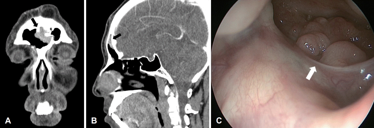 | Fig. 3.Paranasal sinus CT and endoscopic finding at postoperative 20 months. A and B: Coronal (A) and sagittal (B) images of CT scan show soft tissue density in superior posterior wall of frontal sinus (black arrows). C: Nasal endoscopy shows multiple polypoid masses were observed in superior and posterior wall of the right fontal sinus (white arrow).
|
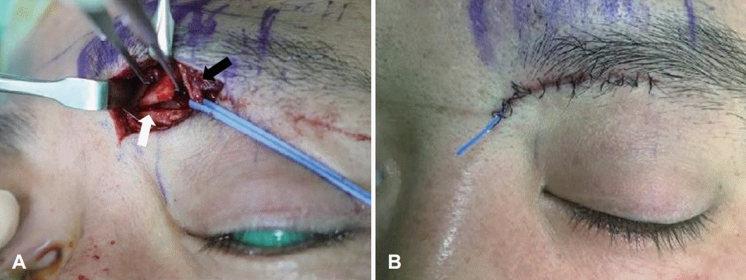 | Fig. 4.Intraopeative findings of mini osteoplastic flap. A: An incision is made along inferior margin of left eyebrow. The frontal sinus is exposed when the mini osteoplastic flap (white arrow) is removed while identifying supratrochlear neurovascular bundles (black arrow). B: The incision site is closed after removal of remaining lesions in frontal sinus.
|
This study was approved by Inje Universtiy Haeundae Paik Hospital Institutional Review Board (2023-05-031).
Go to :
Discussion
The number of inverted papillomas occurring in the frontal sinus is relatively small, but the risk of recurrence is greater than that of other sinuses [1]. In addition, since there is a risk of malignant transformation into squamous cell carcinoma, it is important to completely resection once the lesion is identified.
With the development of endoscopy and surgical instruments, the endoscopic approach is widely used as a standard treatment for inverted papilloma surgery. In the case of inverted papilloma originating from the frontal sinus, it has been successfully treated with a modified endoscopic Lothrop procedure and its usefulness has been reported [5]. However, when the primary site is located on the superior, lateral or anterior wall of the frontal sinus, complete resection may be difficult with only an endoscopic nasal approach [6,7]. So an external approach such as OPF or frontal sinus trephination is needed for complete removal of tumor in such case. In this case, frontal sinus trephination was considered; however, the lesion was found to be diffuse multifocal on the posterior wall, requiring a larger working space. Additionally, there was some recurrent inverted papilloma in the supraorbital area in the right frontal sinus, which was not detected in preoperative imaging and was therefore removed.
Frontal sinus obliteration via an OPF was first described by Hoffman in 1904 [8]. Since fat obliteration was introduced in 1956 [9], OPF has continued to be highly valued by many experienced frontal sinus surgeons. It is considered the gold standard for surgical treatment of chronic frontal sinusitis, especially when alternative surgical procedures have failed. Traditional OPF is a method of accessing the bilateral frontal sinuses through bicoronal incision, which has potential for postoperative complications such as cosmetic problems, cerebrospinal fluid leakage, and forehead numbness [10]. Recently, unilateral OPF has been reported, and a surgical technique that preserves the contralateral frontal sinus has also been developed. This technique allows for extensive removal of inverted papilloma invading the frontal sinus on the affected side without obliterating the frontal sinus, following a bicoronal incision [11,12]. To reduce postoperative complications, mini OPF using an extended Lynch incision was developed [4]. Compared to conventional OPF, mini OPF has a relatively short recovery period after surgery and is less invasive. In mini OPF, a Lynch incision is made under the eyebrow on the lesion side, and then an additional incision is made toward the nasion. At this time, attention should be paid not to damage to the supratrochlear nerve bundles. After lifting the periosteum, make a bone incision along the upper orbital edge and fracture the upper medial side to expose the frontal sinus. After removing the lesion in the frontal sinus, return the flap to its original position, fix it with a plate, and perform skin closure.
In the past, it was common to perform frontal sinus obliteration with autologous fat tissue, but recently methods that do not obliterate frontal sinuses have been tried. If frontal sinus obliteration is not performed, the possibility of complications such as sinusitis is lowered because drainage of mucus can occur through the preserved natural ostium of frontal sinus. In addition, it is easy to follow up the lesion with nasal endoscope after surgery. And it also shows better cosmetic results as no scars are formed on the donor site for obtaining fat tissue [13]. In this case, following the endoscopic modified Lothrop procedure, the frontal sinus natural ostium was widely open, eliminating the need for obliteration. Additionally, obliteration was not performed for the purpose of monitoring the recurrence of the lesion.
The authors considered mini OPF as a method to perform complete removal with minimal complications in recurrent inverted papilloma that invaded the superior wall of frontal sinus. Additionally, it was confirmed that the aeration of the frontal sinus was maintained after surgery because the frontal sinus was not occluded.
In conclusion, we report a successful case of mini OPF surgery for recurred frontal sinus inverted papilloma. Endoscopic surgery has been widely used as a standard treatment for frontal sinus inverted papilloma. Mini-OPF may be an optional treatment on the case of recurred, endoscopically unreached, frontal sinus inverted papilloma.
Go to :
 XML Download