

 PDF
PDF Citation
 Print
Print


서 론
소엽성 모세혈관종(lobular capillary hemangioma, LCH)과 비소엽성 모세혈관종(non-LCH)은 피부와 점막에서 발생하는 폴립 모양의 양성 혈관성 병변이다[1]. 명확한 병인은 밝혀진 바 없으나, 일반적으로 만성적인 약한 국소 자극, 외상성 손상, 호르몬 요인 또는 특정 종류의 약물과 같은 다양한 자극에 반응하여 발생한다고 알려져 있으며 구강, 비강 점막 등에서 발생한다[1)].소엽성 모세혈관종이 후두에서 발견된 사례는 국내외로 극히 드물며[2-5], 실제로 저자들이 조사한 문헌 중 기관 삽관이나 후두 손상, 흡연력 없이 양측성으로 소엽성 혹은 비소엽성 모세혈관종이 발견된 사례는 보고된 바가 없었다. 최근 저자들은 3개월 전 코로나 바이러스 질병(coronavirus disease 2019, COVID-19) real-time reverse transcription-polymerase chain reaction (RT-PCR) 검사에서 확진 판정을 받고 3주 전부터 목 이물감을 호소하는 환자에서 후두 육아종으로 오인하여 수술하였지만 소엽성 모세혈관종과 비소엽성 모세혈관종으로 진단된 증례를 치험하였기에 문헌 고찰과 함께 보고하고자 한다.
Go to : 
증 례
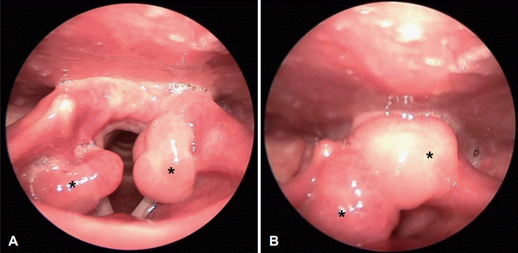
45세 여자 환자가 내원 3주 전부터 시작된 목소리 변화와 목 이물감, 호흡 불편감을 주소로 일차 의료기관에서 의뢰되었다. 기저질환은 없고, 3개월 전 COVID-19 RT-PCR 검사에서 양성으로 확인되었으며, 흡연력은 없고, 음주력도 미미하였다. 사이클로스포린 등의 면역억제제를 포함한 특이 약물 복용력도 없었다. 내원 3개월 전 COVID-19 감염 당시 인후통과 목 이물감이 동반되었으나 내원 당시에는 인후통이나 기침, 콧물, 가래 등의 다른 인후두 증상은 없었고 목 이물감과 호흡 불편감이 동반되었다. 또한 후두 외상을 당한 병력이나 기관삽관을 시행한 과거력 역시 없었다. 경성 후두 내시경 검사에서 양측 피열부에 육아종성 병변으로 의심되는 약 10 mm 가량의 종물이 관찰되었다(Fig. 1). 그 외 성대의 경계와 움직임 및 피열후두개주름과 후두개 등 다른 해부학적 구조는 모두 정상소견으로 확인되었다.
COVID-19 감염 후 후두 육아종이 보고된 바 있고, 병변의 위치 및 외관적으로 육아종의 가능성이 높아 이에 대한 치료를 계획하였다. 음성치료를 동반한 양성자 펌프 억제제(proton pump inhibitor) 치료를 고려하였으나, 환자의 목 이물감 및 자극으로 인한 호흡 불편감이 심하여 보존적 치료 대신 진단과 치료를 위한 CO2 레이저 후두 미세 수술을 계획하였다.
전신마취 후 레이저 튜브로 기관 삽관을 시행하였으며, 현수 후두경을 걸어 시야를 확보하고, 400 mm 렌즈를 부착한 수술용 현미경을 이용하였으며 CO2 레이저를 미세조정기(micromanipulator)에 연결하여 병변을 절제하였다. 양측 종물은 모두 약 2-3 mm의 자유연을 확보하여 일괄절제(en block resection)를 시행하였으며, 에피네프린 거즈를 위치시켜 지혈하고 수술을 종료하였다. 크기는 약 0.8 cm로 관찰되었으며 조직검사를 의뢰하였다. 술후 1일에 후두내시경 소견에서 수술 부위의 이상 소견은 없었고, 목소리는 정상적으로 회복되었으며, 환자는 음성 안정 및 주의 사항 교육 후 퇴원하였다. 술후 1주째 후두내시경 검사에서 수술 부위 점막의 정상 치유 과정이 관찰되었고, 술후 3개월에 시행한 후두 내시경 검사에서도 재발의 소견은 없었다(Fig. 2). 수술 후 확인된 조직병리학적 검사상 좌측 종물은 소엽성 모세혈관종으로, 우측 종물은 비소엽성 모세혈관종으로 최종 진단되었다(Fig. 3).
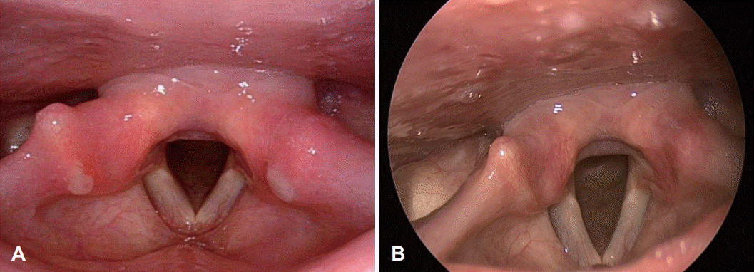 | Fig. 2.The rigid laryngoscopic image of both arytenoid that taken after surgery. A: At eighth postoperative day. It appears to be a normal wound healing process. B: Three-month after surgery, there was no evidence of recurrence and the arytenoid mucosa recovered well.
|
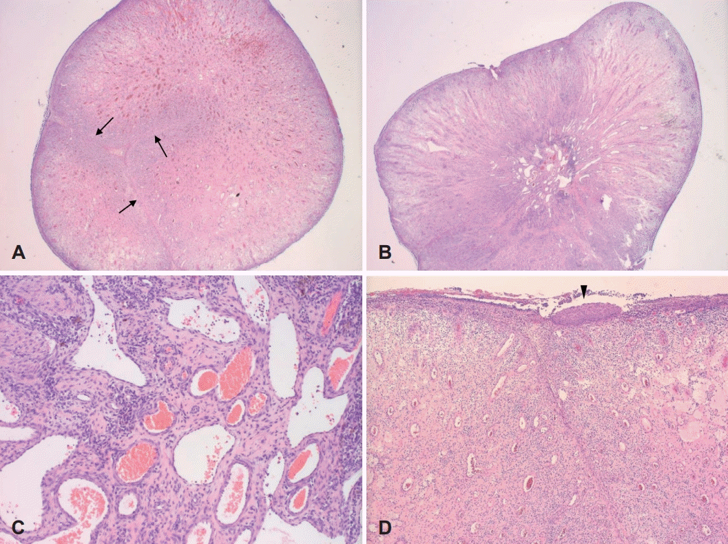 | Fig. 3.Histopathologic finding with hematoxylin and eosin staining showed the typical pattern of pyogenic granuloma. The left arytenoid nodular mass, 0.8 cm-sized, shows congested small vessels aggregation with thin connective tissue septae (arrows), suggesting lobular capillary hemangioma (LCH) type of pyogenic granuloma (A, ×12.5) and the right arytenoid mass, 1.0 cm-sized, reveals central branching vessels and many elongated vessels; suggesting non-LCH type of pyogenic granuloma (B, ×12.5). In high power view, small vessels with or without vascular wall aggregation with stromal hemosiderin-laden macrophages and stromal fibroblastic proliferation are observed (C, ×100). Diffuse surface ulceration with focal remnant squamous epithelium (arrowhead) and underlying edematous stroma (D, ×40).
|
Go to :
고 찰
성문 후부에 발생하는 종물은 피열연골의 성대돌기에 발생하는 육아종이 가장 흔하며, 원인에 따라 접촉성 육아종과 기관 삽관 후 발생하는 삽관 육아종이 대표적이다[6]. 그 외에도 드물게 암종의 발생이나 소엽성 모세혈관종도 보고된 바 있다. 따라서 다양한 질환과의 감별이 필요하며 생검 소견이 진단의 확립에 결정적인 역할을 한다[1].
후두 육아종은 경계가 명확하고 구형 및 난원형인 경우에는 악성일 가능성이 매우 낮아 보존적 치료를 먼저 시도하며, 이에는 양성자 펌프 억제제를 통한 인후두 역류 방지와 음성 치료 등이 포함된다[6]. 보존적 치료에 실패하거나 육아종의 위험인자가 없을 경우 또는 병변의 위치가 일반적이지 않을 때는 후두 미세 수술을 시행하게 된다[6]. 반면 소엽성 및 비소엽성 모세혈관종의 치료 원칙은 작고 무증상일 때는 정기적인 추적 관찰이 권장되나 병변이 크거나 증상이 있는 경우 제거가 필요하다[1]. 절제 생검을 원칙으로 하며, 제거 과정에서 현저한 합병증이 예상되는 경우에는 절개 생검이라도 시행해야 한다. 최근에는 Nd:YAG 레이저, CO2 레이저 등을 이용한 절제가 사용되고 있으며, 수술 후 예후가 좋고 합병증이 적은 것으로 알려져 있다[7]. 본 예에서는 기관삽관 등의 육아종을 유발할 원인인자가 명확하지 않은 점, 환자의 불편감 등을 고려하여 CO2 레이저를 이용한 국소 절제를 시행하였으며, 3개월간의 추적 검사에서 재발하지 않았다.
소엽성 모세혈관종과 비소엽성 모세혈관종은 피부, 입술, 혀, 구강 점막, 비강 등에서 발견할 수 있는 양성종양이며, 후두에는 비교적 드물게 발생한다[1,3]. 최초에는 병변이 화농성 유기체에 의해 발생한다고 생각되어 화농성 육아종(pyogenic granuloma)으로 불렸으나, 이는 잘못된 용어(misnomer)이며, Epivatianos 등은 이를 조직병리학적으로 분류하여 특징을 비교하여 소엽성 모세혈관종과 비소엽성 모세혈관종으로 분류하였다[1,8].
조직병리학적으로 소엽성 모세혈관종은 표면적으로 부종, 모세혈관 확장 또는 염증성 과립 조직 반응 등의 변화를 겪지 않지만 소엽 응집체로 이루어진 모세혈관증식 및 각 소엽을 구분짓는 점액체 또는 섬유성 결합 조직의 격벽을 특징으로 한다[9]. 반면 비소엽성 모세혈관종은 과립 조직과 유사한 고도의 혈관 증식을 특징으로 한다[9].
소엽성, 비소엽성 모세혈관종이 발생하는 명확한 기전은 밝혀지지 않았으나 임신, 경구 피임약과 연관성이 밝혀져 호르몬 변화가 영향을 미칠 것으로 추정되고 있다[10]. 또한 몇몇 연구에 따르면 국소 외상[1] 혹은 사이클로스포린과 같은 일부 약물[11]도 원인이 될 수 있다고 알려져 있다. 일반적으로 몇주 안에 최대 크기에 도달하며, 시간이 지나도 크기가 감소하지 않는다. 종양의 크기는 0.5 cm에서 4 cm에 이르며, 평균 지름은 1.1 cm이다[1]. 본 증례에서 역시 정확한 발생 시기는 알 수 없으나 종양의 크기가 증가하며 증상을 일으킨 것으로 보인다.
이 증례의 환자는 3개월 전 COVID-19 감염이 있었고, COVID-19 감염 이후 나타날 수 있는 대표적인 이비인후과적 후두 합병증으로는 성문 후방 협착(posterior glottic stenosis), 후두 부종(laryngeal edema), 육아종(granuloma) 등이 있다[12]. 그러나 COVID-19 감염 후 발생한 소엽성 혹은 비소엽성 모세혈관종은 국내와 국외를 비롯하여 보고된 바 없다. 이에 저자들은 COVID-19 감염에 의한 발생에 대해서도 추측을 하였으나 인과관계 입증에는 어려움이 있었다. 본 증례에서 발생한 소엽성 및 비소엽성 모세혈관종은 COVID-19 감염에 의한 직접적인 점막 변화의 경과이기보다는 감염 후 발생하는 일반적인 감기 증상 및 이로 인한 후두 자극 등이 원인일 것으로 생각된다.
저자들은 상기도 감염 외 특이 병력 없이 목 이물감과 목소리 변화를 주소로 내원한 환자에서 내시경적 검사상 육아종의 소견을 보였으나 수술적 절제를 통해서 얻은 조직병리검사상 소엽성 및 비소엽성 모세혈관종으로 진단하였다. 후두에서 양측성 종물이 관찰될 경우 육아종의 가능성이 가장 높으나, 본 증례에서 보듯이 소엽성 혹은 비소엽성 모세혈관종의 가능성도 염두에 둘 필요가 있다는 점에서 임상적 의의가 있어 본 증례를 보고하는 바이다.
Go to :
 XML Download