Journal List > Korean J Anesthesiol > v.76(4) > 1516083627

Flaviano, Bettinelli, Assandri, Muhammad, Benigni, Cappelleri, Mariano, Lorini, and Bugada: Erector spinae plane versus fascia iliaca block after total hip arthroplasty: a randomized clinical trial comparing analgesic effectiveness and motor block

Abstract

Background

Ultrasound-guided supra-inguinal fascia iliaca block (FIB) provides effective analgesia after total hip arthroplasty (THA) but is complicated by high rates of motor block. The erector spinae plane block (ESPB) is a promising motor-sparing technique. In this study, we tested the analgesic superiority of the FIB over ESPB and associated motor impairment.

Methods

In this randomized, observer-blinded clinical trial, patients scheduled for THA under spinal anesthesia were randomly assigned to preoperatively receive either the ultrasound-guided FIB or ESPB. The primary outcome was morphine consumption 24 h after surgery. The secondary outcomes were pain scores, assessment of sensory and motor block, incidence of postoperative nausea and vomiting and other complications, and development of chronic post-surgical pain.

Results

A total of 60 patients completed the study. No statistically significant differences in morphine consumption at 24 h (P = 0.676) or pain scores were seen at any time point. The FIB produced more reliable sensory block in the femoral nerve (P = 0.001) and lateral femoral cutaneous nerve (P = 0.018) distributions. However, quadriceps motor strength was better preserved in the ESPB group than in the FIB group (P = 0.002). No differences in hip adduction motor strength (P = 0.253), side effects, or incidence of chronic pain were seen between the groups.

Conclusions

ESPBs may be a promising alternative to FIBs for postoperative analgesia after THA. The ESPB and FIB offer similar opioid-sparing benefits in the first 24 h after surgery; however, ESPBs result in less quadriceps motor impairment.

Introduction

Regional anesthesia techniques are a recommended component of multimodal pain management after total hip arthroplasty (THA) [1,2]. The hip joint has complex innervation [3] involving branches of the femoral nerve (FN), obturator nerve (ON), and sciatic nerve (SN), while the lateral femoral cutaneous nerve (LFCN) innervates the skin of the lateral side of the thigh [4].
The fascia iliaca block (FIB) is an established and effective technique, especially when ultrasound (US) guidance and proximal approaches are used. Recent studies have demonstrated that US-guided FIBs using the supra-inguinal approach is associated with improved pain control and significant opioid-sparing effects after THA [5,6].
However, concerns still exist regarding the routine use of FIBs. Similar to the lumbar plexus block, the FIB is associated with motor block [6], which can complicate early rehabilitation by delaying immediate postoperative mobilization. Therefore, alternative regional anesthesia techniques with motor-sparing strategies are worth investigating in the setting of enhanced recovery and fast-track surgery.
The erector spinae plane block (ESPB) involves the injection of local anesthetics in the fascial plane between the transverse processes and erector spinae muscles. Despite some controversy, this new approach has generated tremendous interest and may be applicable to a wide variety of surgeries. The use of the ESPB at L4 for hip surgery has been previously described [7,8], and a retrospective study has demonstrated improved ambulation with comparable analgesia when low thoracic continuous ESPBs replaced continuous FIBs within a well-established clinical pathway for patients undergoing THA [9].
However, to date, no randomized clinical trial has compared the ESPB with the FIB in patients undergoing THA. Therefore, we designed this study to test the analgesic superiority of the FIB over the ESPB in terms of 24-h morphine consumption, while also evaluating motor impairment as a secondary outcome.

Materials and Methods

This single-center randomized clinical trial was reported according to the Consolidated Standards of Reporting Trials (CONSORT) guidelines [10] and approved by the Institutional Review Board (2019-0191) of ASST Papa Giovanni XXIII – Bergamo on December 30, 2019 and registered on ClinicalTrials.gov (NCT04574154). Our study was conducted in accordance of the Ethical Principles for Medical Research Involving Human Subjects, as outlined in the Helsinki Declaration.
Patients aged ≥ 18 years who were undergoing primary unilateral THA using a posterolateral approach and provided informed consent met the inclusion criteria. The exclusion criteria were as follows: allergy to any drug used in the study, contraindications to spinal anesthesia and/or the use of a general anesthetic, kidney failure (glomerular filtration rate < 50 ml/min), epilepsy, psychiatric disease, pre-existing neurologic deficits or neuropathies, pregnancy, pre-existing alcohol/opioid use disorder, revision or emergency surgery, planned postoperative admission to an intensive care unit, and refusal to provide informed consent. Randomization was performed using a computer-generated random number table to allocate patients to one of the two study groups.
Preoperatively, an investigator blinded to the group allocation performed a sensory assessment of the FN, ON, and LFCN in response to cold (ice) in the anterior, medial, and lateral aspects of the thigh. The motor functions of the FN and ON were tested using the method previously described by Neal [11]. Briefly, to test FN motor function, the investigator supported the knee under the popliteal fossa and the patient was asked to extend the knee. To test the motor function of the ON, the leg was abducted and the patient was asked to adduct the leg toward the midline. Demographic data were collected, and patients were assigned a unique identification code.
On the day of surgery, a sealed envelope with group allocation was opened by a team member who was not involved in the data collection.
In the FIB group, an ultrasound-guided longitudinal supra-inguinal FIB was performed on the ipsilateral side of the surgery according to the technique described by Desmet et al. [6]. Briefly, a linear 6–13 MHz ultrasound probe (HFL38xi; SONOSITE Fujifilm S-Nerve®, USA) was placed in the sagittal plane to obtain an image of the anterior superior iliac spine. By sliding medially, the fascia iliaca and sartorius, iliopsoas, and internal oblique muscles were identified, using the “bow-tie sign.” A 22 gauge (G), 50-mm needle (SonoPlex®, Pajunk Medizintechnologie, Germany) was introduced 1 cm cephalad to the inguinal ligament using an in-plane approach. The fascia iliaca was separated from the iliacus muscle using saline to create a space where the needle tip was further advanced. The deep circumflex artery was identified superficial to the fascia iliaca, lifted up by saline injection, and used as a marker of successful penetration of the fascia iliaca. A total volume of 40 ml of ropivacaine 0.5% was injected. Correct placement was defined as the spread of the local anesthetic cranial to the point where the iliac muscle travels under the abdominal wall muscles (Fig. 1). If correct spread was not immediately visualized, the injection was stopped and the needle was repositioned.
In the ESPB group, an ultrasound-guided ESPB was performed on the ipsilateral side of the surgery using a previously described technique [7]. Moving cephalad from the sacrum, we identified the L5, L4, and L3 transverse processes and erector spinae muscles posteriorly. A 100-cm, 21 G needle (Sonoplex® Pajunk Medizintechnologie, Germany) was directed in-plane and the needle tip was positioned anterior to the erector spinae muscle at the corner of the transverse process. After the initial saline injection, dissection of the plane was observed by injecting the same volume and concentration of the local anesthetic solution that was used in the FIB group. Correct placement was defined as the spread of local anesthetic cranially and caudally from the injection point, dissecting the plane between the transverse processes and erector spinae muscles (Fig. 1). As in the FIB group, the injection was stopped, and the needle was repositioned if the correct spread was not immediately visualized.
After 20 min, all the patients’ blocks were tested by a blinded observer for sensory and motor block using the same cold test described previously for preoperative testing.
Spinal anesthesia was performed at L2–L3 with 2.2 ml of isobaric bupivacaine 0.5% without adjuvants in all patients, regardless of allocation group, after either the FIB or ESPB had been performed and tested. All patients also received dexamethasone (4 mg), ibuprofen (400 mg), and tranexamic acid (10 mg/kg) intravenously before entering the operating room according to our institutional protocol for THA.
For postoperative analgesia after surgery, all patients received scheduled ibuprofen (400 mg) and paracetamol (1,000 mg) every 8 h, until discharge from hospital. Rescue analgesia was provided by patient-controlled analgesia (PCA) with intravenous morphine (no basal rate; bolus 1 mg, lockout 10 minutes, maximum dose 20 mg in 4 h), which was started on arrival to the post-anesthesia care unit (PACU). In the PACU, postoperative nausea and vomiting (PONV) was assessed and, if indicated, treated with metoclopramide 10 mg or ondansetron 4 mg intravenously. Potential block-related complications were also recorded.
At 6, 24, and 48 h postoperatively, an independent investigator (blinded to allocation group) evaluated cumulative morphine consumption, pain scores at rest and with movement using the numeric rating scale (NRS; ranging from 0–10 with 0 = no pain and 10 = worst pain imaginable), and PONV and any other complications. After 6 h, the patients were tested again. After confirming resolution of spinal anesthesia, sensory testing to cold in the territories of the FN, ON, and LFCN was performed using ice, while motor function was tested by knee flexion and hip adduction. This additional evaluation was performed with the intention of confirming the preoperative block assessment, since the block may not have been fully established before surgery. A successful sensory and/or motor block was defined as positive sensory/motor testing at either 20 min or 6 h.
The occurrence of chronic postsurgical pain (CPSP) was assessed one year after surgery using the Pain-Detect Questionnaire. According to the International Association for the Study of Pain, severe CPSP was defined as an NRS > 5 [12].

Statistical analysis

The primary outcome of the study was total morphine consumption (mg) 24 h postoperatively. Secondary outcomes were the following: total morphine consumption (mg) at 6 and 48 h; block success defined as sensory and motor block in the territories of the LFCN, FN, and ON; NRS pain scores; incidence of PONV; and incidence of other adverse events.
We calculated the sample size using G*Power (3.1) based on previously published data. In the study by Desmet et al. [6], the mean ± standard deviation (SD) morphine consumption at 24 h was 10.25 ± 1.64 mg in patients receiving FIB, while Tulgar et al. [13] reported the consumption of tramadol as 13 ± 5 mg after ESPB (equivalence: 100 mg tramadol = 10 mg of morphine). This difference in morphine consumption corresponded to an estimated effect size of 0.74. Given this effect size, 30 patients per group were needed to test the superiority of the FIB over the ESPB, with 80% power and an α of 0.05 using a 2-sided 2-sample t test. To adjust for possible drop-outs, 64 total patients were included.
According to the central limit theorem, we assumed that the distribution of our sample was normal. Data are therefore presented as the mean ± SD for continuous variables and as the proportion (%) for categorical variables. Standard hypothesis tests (2-sided t-tests) were performed to analyze the baseline characteristics and outcome parameters. For multiple testing, repeated-measures analysis of variance with post-hoc Tukey’s honest significant difference correction was applied. Categorical data were assessed using frequency tables and χ2 or Fisher’s exact tests (for cell counts < 5). Statistical significance was set at P < 0.05. All statistical analyses were performed using SPSS Software (version 28.0, IBM Corp., USA).

Results

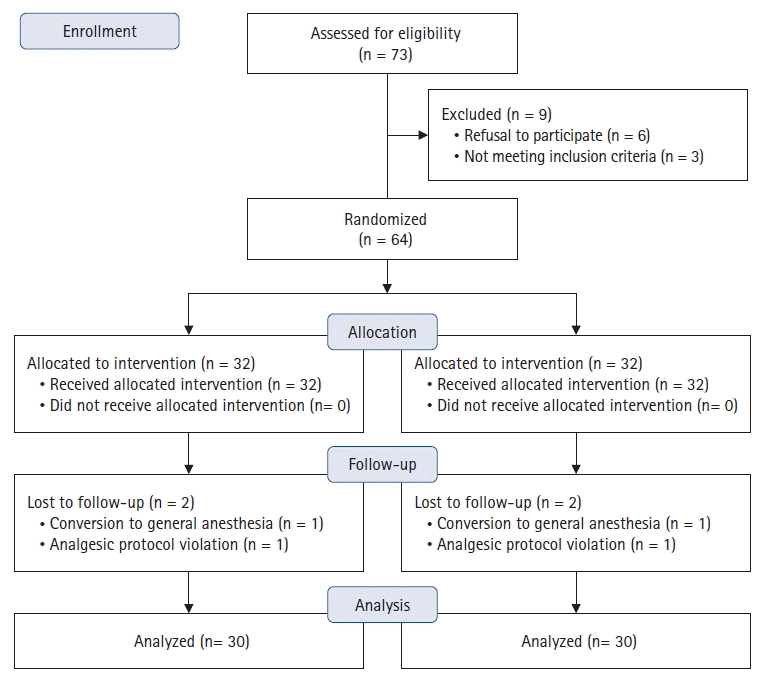
From January 2020 to May 2021, a total of 73 patients were assessed for eligibility, and 64 were enrolled (Fig. 2). The baseline characteristics are summarized in Table 1. All study participants developed a sensory block in the expected distribution prior to surgery. The postoperative clinical outcomes are summarized in Table 2.
Regarding the primary outcome, no statistically significant differences were observed in morphine consumption at 24 h (P = 0.676) or 48 h (P = 0.654). Pain values expressed using the NRS were not statistically significantly different at any time point (at rest or during movement) (Fig. 3).
Statistically significant differences, however, were observed in sensory testing with the FIB, which, produced a more reliable sensory block for the FN (P = 0.001) and LFCN (P = 0.018) compared to the ESPB. No significant differences were observed in the ON (P = 0.091). However, the quadriceps motor block was greater with the FIB than with the ESPB (P = 0.002). No significant differences were observed in hip adduction (P = 0.253).
No differences were observed between the groups in terms of CPSP incidence and severe CPSP, and none of the patients reported developing chronic neuropathic pain. Additionally, side effects were not different between the groups, and no complications were reported.

Discussion

The results of this randomized, observer-blinded study showed that the FIB, while producing a more reliable sensory block of the femoral and LFCN, is not superior to the ESPB in terms of pain management after THA using a posterolateral approach. However, the patients who receive an FIB experienced more of a motor block that remained for several hours after surgery. Thus, the ESPB may represent an acceptable motor-sparing regional analgesic alternative to the FIB within a multimodal pain management regimen in this surgical population.
Our randomized controlled trial compared the FIB with the ESPB in the THA surgical population. While the FIB is known to decrease pain and morphine consumption after THA, it is often associated with motor impairment of the quadriceps [6]. A previous retrospective study has shown that replacing FIB catheters with a continuous ESPB results in increased early ambulation with comparable analgesia in patients undergoing elective primary THA for the first two days after surgery [9]. This is the first report to suggest that the ESPB could be an important analgesic alternative to the FIB for enhancing surgical recovery after THA.
Our prospective study confirmed this hypothesis by demonstrating that the FIB does not clearly provide superior analgesia at rest or with movement in the first 48 h, and morphine consumption is not significantly different between the two techniques at any time point. Thus, replacing the FIB with the ESPB is unlikely to deprive patients of effective analgesia, but may offer significant advantages in the form of reduced motor impairment in the immediate postoperative period.
As the trend towards outpatient THA has already started occurring in some countries, interest in motor-sparing techniques is growing [14]. Certain patients may successfully undergo same-day THA with careful patient selection and consistent use of perioperative strategies specifically aimed at ultra-rapid recovery and early mobilization.
Regardless of the anesthetic and analgesic techniques employed, falls after any surgery are a critical concern. Despite its low incidence, falls are considered a serious adverse outcome of joint replacement. Multiple risk factors have been identified, and the role of regional blocks continues to be debated [15]. However, minimizing quadriceps impairment after joint replacement can only improve patient safety given the inherent risk of postoperative falls in this population. Thus, regional blocks that can provide effective analgesia without motor compromise are critical in the current era of rapid recovery and safe patient discharge.
In our population, the incidence of motor block, measured by quadriceps impairment/knee extension, was significantly lower with the ESPB, while no differences were observed in motor function during hip adduction. Hip adduction is controlled by the lumbar plexus (adductor longus, adductor brevis, and pectineus muscles) and sacral plexus (adductor magnus muscle). Therefore, blocking the lumbar plexus branches alone may not invariably reduce adduction strength. Studies on the FIB have reported different degrees of motor block, and hip adduction has always been difficult to test because of difficulties in ascertaining the relative contribution of each nerve and muscle group [1618]. Thus, the clinical relevance of adductor motor impairment in the postoperative recovery of THA patients remains difficult to quantify.
Additionally, the exact mechanism of action of the ESPB continues to be debated. In theory, the anterior spread of the injectate may reach the paravertebral/epidural space and intervertebral foramen [19,20]. However, multiple structures exist that form a barrier to local anesthetic spread in this region, which may differ at the thoracic and lumbar levels.
At the lumbar level, the main structures limiting the anterior diffusion of injectate are the middle thoracolumbar fascia (three sublayers attached to the lateral edges of transverse processes), anterior thoracolumbar fascia (passes between the psoas and quadratus lumborum muscles and attaches to the inner/anterior surfaces of the transverse processes), and intertransverse ligaments and muscles [21]. Cadaver studies suggest that no active spread to the lumbar plexus roots occurs, and injectate spread is typically limited to the dorsal rami [22,23]. However, discrepancies frequently exist between cadavers and living human studies, and the migration of local anesthetics has already been documented in surgical patients. In the specific setting of hip surgery, a case series has shown active deposition of dye at the L3–L5 roots in the psoas compartment after an L4 ESPB [7]. Our study provides further support for this finding as the patients in the ESPB group in our study experienced similar analgesia and opioid consumption as those in the FIB group while exhibiting reduced nerve conduction in the lumbar plexus territory (Fig. 4). As suggested by Tulgar et al. [21], we speculate that the needle tip position (deep to the intertransverse ligament and middle thoracolumbar fascia at the corner of the transverse process) and higher volumes of local anesthetic may promote anterior spread of injectate similar to that of other blocks referred to as ‘paravertebral by proxy’ (Fig. 4) [24]. Variations in the technique used to perform the ESPB and other ‘proxy’ techniques may produce differential injectate spread and clinical effects [20].
Finally, in a study using lidocaine for bilateral ESPB, systemic absorption of local anesthetics was proposed as a contributor to the analgesic effect of the ESPB [25] and may account for the additional pain relief beyond the local effect. Systemically administered local anesthetics have been described as an effective mode of analgesia and reduced hyperalgesia [26,27], and these benefits may translate into fascial plane blocks such as the ESPB. Further studies are needed to differentiate and appropriately attribute the local and systemic contributions of local anesthetic injections to fascial plane blocks. However, this may be inconsequential in the clinical setting of acute pain management.
Postoperative pain in patients undergoing THA has been reported to peak at 8 h and progressively decrease thereafter [28]. Most studies evaluate pain at discrete time points, such as at 12 or 24 h after surgery, which may be considered late in the pain trajectory and may leave gaps in assessment during the early period of intense pain [28]. Our study focused on this early interval from 0 h to 8 h after surgery when both the FIB and ESPB may provide effective analgesia, and indeed, no differences in pain scores or morphine consumption were found.
Since a wide variety of single-injection nerve and fascial plane blocks have been effective for perioperative analgesia in patients undergoing THA [28], the motor block effect is a major concern because it is associated with slower recovery and early mobility. The anterior quadratus lumborum block [29,30] and the pericapsular nerve group block have been shown to provide adequate pain relief comparable to that of the FIB, with reduced motor quadriceps impairment [31,32]. Our study suggests that the ESPB may be another motor-sparing regional analgesic option; however, additional studies comparing the ESPB with alternate approaches are needed to determine the optimal technique.
The prevalence of chronic pain after THA has been reported to be in the range of 27%–38%, with 6%–12% reporting moderate to severe pain and 1%–2% reporting a neuropathic component [33]. None of our patients reported neuropathic pain; however, the cumulative incidence of CPSP in our population was 20%, with no difference between the groups, although our study was underpowered for this outcome. CPSP is a complex phenomenon, and socioeconomic and psychological factors play a major role beyond the effectiveness of postoperative analgesia [34]. It is not surprising that a single treatment would fail to prevent chronic pain development. Pain in the early postoperative period is a known risk factor for CPSP [35]. Postoperative pain was well controlled in our study population in both groups, reinforcing that multimodal analgesia, including regional anesthesia techniques, is pivotal to reduce acute pain and minimize the risk of CPSP.
This study had several limitations. First, a type II error is always possible with a negative study, as it may be underpowered to show a difference in the primary outcome despite an a priori sample size calculation. Second, this was a single-center study, and findings at our institutional population may not be generalizable to other populations. Third, the physicians performing the blocks are experts in regional anesthesia; therefore, the comparison of these two block techniques may be limited to centers with expertise in regional anesthesia. Additionally, all patients in our study underwent THA using a posterolateral approach, which may be appropriate for the ESPB [36]; however, whether these results are generalizable to studies using an anterior approach is not certain. Finally, our study was conducted in a setting of enhanced recovery and multimodal analgesia, thus, these results may differ in patients who do not receive this level of perioperative care.
In conclusion, the ESPB may be a feasible alternative to the FIB as a regional analgesic technique within a multimodal pain management regimen for patients undergoing THA. The ESPB provides analgesia and opioid-sparing effects similar to those of the FIB with superior preservation of motor function. Further comparative effectiveness studies are required to evaluate alternative regional analgesic techniques and to determine the optimal motor-sparing block for hip surgery.

Notes

Funding

None.

Conflicts of Interest

No potential conflict of interest relevant to this article was reported.

Data Availability

All data generated or analyzed during this study are included in this published article.

Author Contributions

Edoardo Flaviano (Conceptualization; Investigation; Writing – original draft; Writing – review & editing)

Silvia Bettinelli (Investigation; Writing – review & editing)

Maddalena Assandri (Data curation; Investigation; Writing – review & editing)

Hassam Muhammad (Data curation; Investigation; Writing – review & editing)

Alberto Benigni (Conceptualization; Investigation; Writing – review & editing)

Gianluca Cappelleri (Conceptualization; Writing – original draft; Writing – review & editing)

Edward Rivera Mariano (Writing – original draft; Writing – review & editing)

Luca Ferdinando Lorini (Conceptualization; Project administration; Writing – original draft; Writing – review & editing)

Dario Bugada (Conceptualization; Data curation; Formal analysis; Investigation; Methodology; Project administration; Supervision; Validation; Writing – original draft; Writing – review & editing)

References

1. Memtsoudis SG, Cozowicz C, Bekeris J, Bekere D, Liu J, Soffin EM, et al. Peripheral nerve block anesthesia/analgesia for patients undergoing primary hip and knee arthroplasty: recommendations from the International Consensus on Anesthesia-Related Outcomes after Surgery (ICAROS) group based on a systematic review and meta-analysis of current literature. Reg Anesth Pain Med. 2021; 46:971–85.
2. Anger M, Valovska T, Beloeil H, Lirk P, Joshi GP, Van de Velde M, et al. PROSPECT guideline for total hip arthroplasty: a systematic review and procedure-specific postoperative pain management recommendations. Anaesthesia. 2021; 76:1082–97.
3. Bugada D, Bellini V, Lorini LF, Mariano ER. Update on selective regional analgesia for hip surgery patients. Anesthesiol Clin. 2018; 36:403–15.
4. Birnbaum K, Prescher A, Hessler S, Heller KD. The sensory innervation of the hip joint--an anatomical study. Surg Radiol Anat. 1997; 19:371–5.
5. Carella M, Beck F, Piette N, Denys S, Kurth W, Lecoq JP, et al. Effect of suprainguinal fascia iliaca compartment block on postoperative opioid consumption and functional recovery in posterolateral-approached total hip arthroplasty: a single-blind randomized controlled trial. Reg Anesth Pain Med 2022. Advance Access published on Jun 15, 2022. doi: 10.1136/rapm-2021-103427.
6. Desmet M, Vermeylen K, Van Herreweghe I, Carlier L, Soetens F, Lambrecht S, et al. A longitudinal supra-inguinal fascia iliaca compartment block reduces morphine consumption after total hip arthroplasty. Reg Anesth Pain Med. 2017; 42:327–33.
7. Tulgar S, Senturk O. Ultrasound guided Erector Spinae Plane block at L-4 transverse process level provides effective postoperative analgesia for total hip arthroplasty. J Clin Anesth. 2018; 44:68.
8. Bugada D, Zarcone AG, Manini M, Lorini LF. Continuous Erector Spinae Block at lumbar level (L4) for prolonged postoperative analgesia after hip surgery. J Clin Anesth. 2019; 52:24–5.
9. Xu L, Leng JC, Elsharkawy H, Hunter OO, Harrison TK, Vokach-Brodsky L, et al. Replacement of fascia iliaca catheters with continuous erector spinae plane blocks within a clinical pathway facilitates early ambulation after total hip arthroplasty. Pain Med. 2020; 21:2423–9.
10. Schulz KF, Altman DG, Moher D; CONSORT Group. CONSORT 2010 statement: updated guidelines for reporting parallel group randomised trials. BMJ. 2010; 340:c332.
11. Neal JM. Assessment of lower extremity nerve block: reprise of the Four P's acronym. Reg Anesth Pain Med. 2002; 27:618–20.
12. Schug SA, Lavand'homme P, Barke A, Korwisi B, Rief W, Treede RD; IASP Taskforce for the Classification of Chronic Pain. The IASP classification of chronic pain for ICD-11: chronic postsurgical or posttraumatic pain. Pain. 2019; 160:45–52.
13. Tulgar S, Kose HC, Selvi O, Senturk O, Thomas DT, Ermis MN, et al. Comparison of ultrasound-guided lumbar erector spinae plane block and transmuscular quadratus lumborum block for postoperative analgesia in hip and proximal femur surgery: a prospective randomized feasibility study. Anesth Essays Res. 2018; 12:825–31.
14. Wainwright TW, Memtsoudis SG, Kehlet H. Fast-track hip and knee arthroplasty...how fast? Br J Anaesth. 2021; 126:348–9.
15. Memtsoudis SG, Danninger T, Rasul R, Poeran J, Gerner P, Stundner O, et al. Inpatient falls after total knee arthroplasty: the role of anesthesia type and peripheral nerve blocks. Anesthesiology. 2014; 120:551–63.
16. Bendtsen TF, Pedersen EM, Moriggl B, Hebbard P, Ivanusic J, Børglum J, et al. Suprainguinal fascia iliaca block: does it block the obturator nerve? Reg Anesth Pain Med. 2021; 46:832.
17. Bendtsen TF, Pedersen EM, Moriggl B, Hebbard P, Ivanusic J, Børglum J, et al. Anatomical considerations for obturator nerve block with fascia iliaca compartment block. Reg Anesth Pain Med. 2021; 46:806–12.
18. Weller RS. Does fascia iliaca block result in obturator block? Reg Anesth Pain Med. 2009; 34:524.
19. Bonvicini D, Boscolo-Berto R, De Cassai A, Negrello M, Macchi V, Tiberio I, et al. Anatomical basis of erector spinae plane block: a dissection and histotopographic pilot study. J Anesth. 2021; 35:102–11.
20. Chin KJ, Lirk P, Hollmann MW, Schwarz SK. Mechanisms of action of fascial plane blocks: a narrative review. Reg Anesth Pain Med. 2021; 46:618–28.
21. Tulgar S, Ahiskalioglu A, Aydin ME, Jadon A, Forero M, Gürkan Y. Lumbar erector spinae plane block: a miracle or self-persuasion? Reg Anesth Pain Med. 2021; 46:638–9.
22. Elsharkawy H, Bajracharya GR, El-Boghdadly K, Drake RL, Mariano ER. Comparing two posterior quadratus lumborum block approaches with low thoracic erector spinae plane block: an anatomic study. Reg Anesth Pain Med 2019. Advance Access published on Mar 28, 2019. doi: 10.1136/rapm-2018-100147.
23. Ivanusic J, Konishi Y, Barrington MJ. A cadaveric study investigating the mechanism of action of erector spinae blockade. Reg Anesth Pain Med. 2018; 43:567–71.
24. Costache I, Pawa A, Abdallah FW. Paravertebral by proxy - time to redefine the paravertebral block. Anaesthesia. 2018; 73:1185–8.
25. De Cassai A, Bonanno C, Padrini R, Geraldini F, Boscolo A, Navalesi P, et al. Pharmacokinetics of lidocaine after bilateral ESP block. Reg Anesth Pain Med. 2021; 46:86–9.
26. Challapalli V, Tremont-Lukats IW, McNicol ED, Lau J, Carr DB. Systemic administration of local anesthetic agents to relieve neuropathic pain. Cochrane Database Syst Rev. 2019; 2019.
27. Haller Y, Gantenbein AR, Willimann P, Spahn DR, Maurer K. Systemic ropivacaine diminishes pain sensitization processes: a randomized, double-blinded, placebo-controlled, crossover study in healthy volunteers. Pain Ther. 2014; 3:45–58.
28. Panzenbeck P, von Keudell A, Joshi GP, Xu CX, Vlassakov K, Schreiber KL, et al. Procedure-specific acute pain trajectory after elective total hip arthroplasty: systematic review and data synthesis. Br J Anaesth. 2021; 127:110–32.
29. Kukreja P, MacBeth L, Sturdivant A, Morgan CJ, Ghanem E, Kalagara H, et al. Anterior quadratus lumborum block analgesia for total hip arthroplasty: a randomized, controlled study. Reg Anesth Pain Med 2019. Advance Access published on Oct 25, 2019. doi: 10.1136/rapm-2019-100804.
30. Polania Gutierrez JJ, Ben-David B, Rest C, Grajales MT, Khetarpal SK. Quadratus lumborum block type 3 versus lumbar plexus block in hip replacement surgery: a randomized, prospective, non-inferiority study. Reg Anesth Pain Med. 2021; 46:111–7.
31. Aliste J, Layera S, Bravo D, Jara Á, Muñoz G, Barrientos C, et al. Randomized comparison between pericapsular nerve group (PENG) block and suprainguinal fascia iliaca block for total hip arthroplasty. Reg Anesth Pain Med. 2021; 46:874–8.
32. Pascarella G, Costa F, Del Buono R, Pulitanò R, Strumia A, Piliego C, et al. Impact of the pericapsular nerve group (PENG) block on postoperative analgesia and functional recovery following total hip arthroplasty: a randomised, observer-masked, controlled trial. Anaesthesia. 2021; 76:1492–8.
33. Nikolajsen L, Brandsborg B, Lucht U, Jensen TS, Kehlet H. Chronic pain following total hip arthroplasty: a nationwide questionnaire study. Acta Anaesthesiol Scand. 2006; 50:495–500.
34. Grosu I, de Kock M. New concepts in acute pain management: strategies to prevent chronic postsurgical pain, opioid-induced hyperalgesia, and outcome measures. Anesthesiol Clin. 2011; 29:311–27.
35. Fletcher D, Stamer UM, Pogatzki-Zahn E, Zaslansky R, Tanase NV, Perruchoud C, et al. Chronic postsurgical pain in Europe: An observational study. Eur J Anaesthesiol. 2015; 32:725–34.
36. Zhang J, He Y, Wang S, Chen Z, Zhang Y, Gao Y, et al. The erector spinae plane block causes only cutaneous sensory loss on ipsilateral posterior thorax: a prospective observational volunteer study. BMC Anesthesiol. 2020; 20:88.
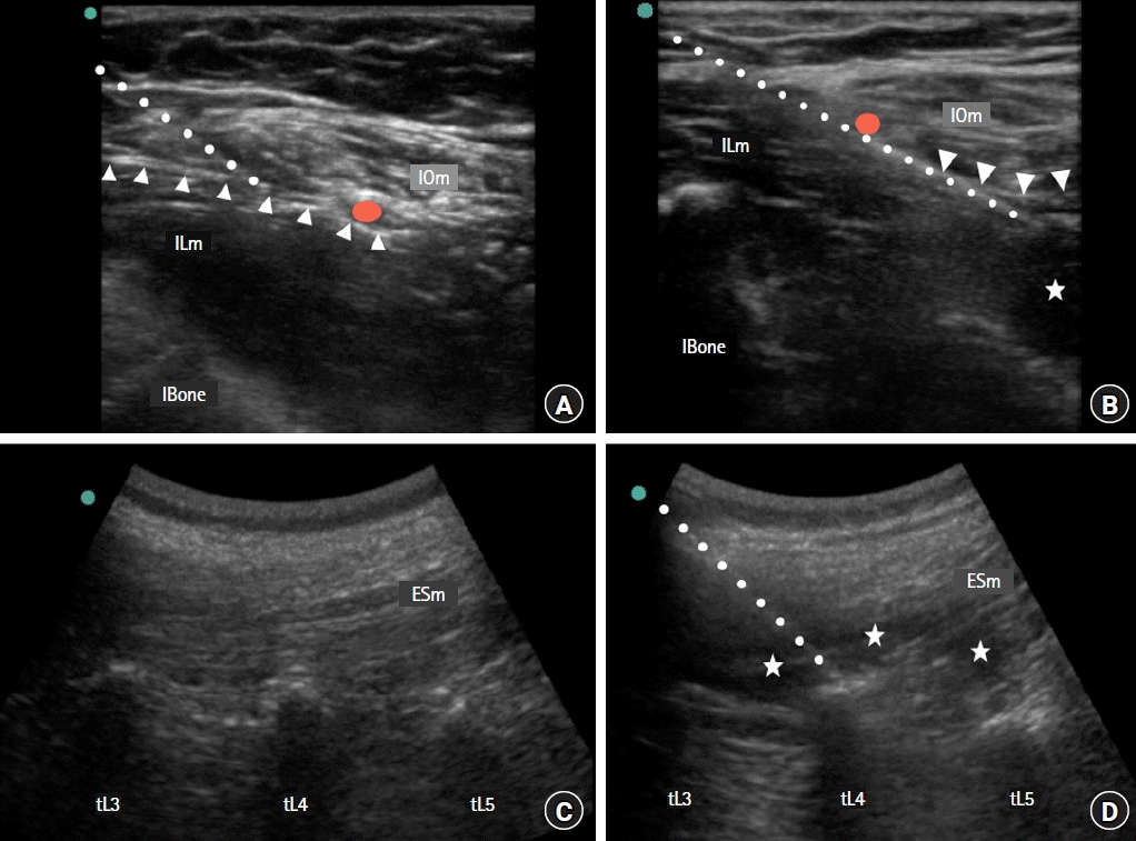
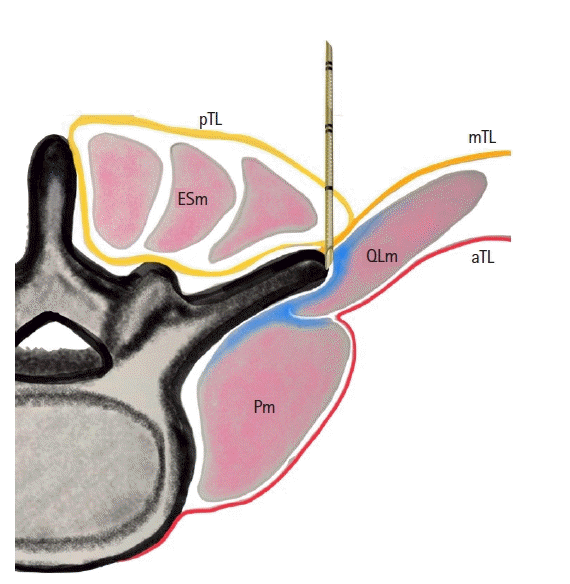
Fig. 1.
Ultrasound scans. (A) Fascia iliaca block pre-injection and (B) post-injection, (C) erector spinae plane block pre-injection and (D) post-injection. The red circle indicates the deep circumflex artery, the triangular dotted line indicates the fascia iliaca, the stars indicate the local anesthetic, and the dotted line indicates the needle path. tL3, tL4, and tL5 indicate the transverse process of the third, fourth, and fifth lumbar vertebra, respectively. IOm: internal oblique muscle, ILm: iliacus muscle, IBone: iliac bone, ESm: erector spinae muscle.
kja-22669f1.tif
Fig. 2.
Consolidated Standards of Reporting Trials (CONSORT) patient flowchart.
kja-22669f2.tif
Fig. 3.
Pain values at rest and during movement in the first 48 h. NRS: numeric rating scale at rest, mNRS: numeric rating scale during movement, FIB: fascia iliaca block, ESPB: erector spinae plane block.
kja-22669f3.tif
Fig. 4.
Proposed mechanism of local anesthetic (blue) diffusion. The orange line indicates the middle thoraco-lumbar fascia (mTL), the red line indicates the anterior thoraco-lumbar fascia (aTL), and the yellow line indicates the posterior thoraco-lumbar fascia (pTL). ESm: erector spinae muscle, QLm: quadratus lumborum muscle, Pm: psoas muscle.
kja-22669f4.tif
Table 1.
Baseline Patient Characteristics
Variable FIB group (n = 30) ESPB group (n = 30)
Sex (F) 20 (66.7) 12 (40)
Age 71.4 ± 10.1 69.3 ± 12
ASA
 I 3 (6.7) 3 (10)
 II 20 (60) 20 (66.7)
 III 10 (33.3) 7 (23.3)
BMI 26 ± 3.8 28 ± 4.3
Duration of surgery (min) 80 ± 16.8 78 ± 14
Preoperative sensory assessment*
 Anterior thigh 30 (100) 30 (100)
 Medial thigh 30 (100) 30 (100)
 Lateral thigh 30 (100) 30 (100)

Values are presented as number (%) or mean ± SD. FIB: fascia iliaca block, ESPB: erector spinae plane block, ASA: American Society of Anesthesiologists, BMI: body mass index. *Baseline preoperative sensory assessment: intact preserved sensory discrimination.

Table 2.
Postoperative Clinical Outcomes and Block Features in terms of Sensory and Motor Testing
Variable FIB group (n = 30) ESPB group (n = 30) P value Cramer’s V/95% CI for the difference in means
Morphine consumption
 6 h 1.4 ± 1.6 2.1 ± 3.6 0.424 [−2.5, 0.8]
 24 h 6.7 ± 5.5 7.4 ± 6.8 0.676 [−3.9, 2.5]
 48 h 9.3 ± 7.7 10.6 ± 10.6 0.654 [−7.3, 4.7]
PONV 3 (10) 0 (0) 0.149 0.23
Urinary retention 5 (16.7) 5 (16.7) 0.953 0.01
Others* 0 (0) 1 (0.03) 0.830 0.13
Sensory block
 FN 27 (93.3) 16 (53.3) 0.001 0.45
 ON 17 (58.6) 11 (36.7) 0.091 0.22
 LFCN 19 (63.3) 11 (36.7) 0.018 0.31
Motor block
 Knee extension 15 (53.6) 4 (13.3) 0.002 0.42
 Hip adduction 7 (25.9) 4 (13.3) 0.253 0.15
CPSP at 1 year
 CPSP 3 (13.6) 6 (26.1) 0.297 0.16
Severe CPSP
 NRS > 5 0 (0) 2 (8.7) 0.157 0.21

Values are presented as number (%) or mean ± SD. FIB: fascia iliaca block, ESPB: erector spinae plane block, PONV: postoperative nausea and vomiting, FN: femoral nerve, ON: obturator nerve, LFCN: lateral femoral cutaneous nerve, CPSP: chronic post-surgical pain, NRS: numeric rating scale. *Delirium and agitation.

TOOLS
Similar articles