

 PDF
PDF Citation
 Print
Print


Introduction
Airway management in children is often challenging owing to their unique anatomical and physiological features. The safe apnea time to maintain appropriate oxygen saturation in children is shorter than that of adults, and desaturation with bradycardia frequently occurs especially in children with difficult airways. Therefore, various techniques have been introduced to extend the time available for airway management and for successful intubation in children with expected or unexpected difficult airway [1]. Freehand fiberoptic intubation, fiberoptic-guided intubation through a supraglottic airway device (SAD), or intubation using a videolaryngoscope are the recommended techniques for difficult pediatric airway management [2]. However, although equipment available for difficult airway management is increasing, their utilization without sufficient training and skills in emergent airway management conditions is concerning.
The SAD is a more useful device for securing the airway compared to endotracheal intubation [3], and can be used as an intubation conduit [4]. Fiberoptic-guided intubation via an SAD has the advantage of ventilation and continuous oxygenation during the procedure [1,5]. Moreover, this technique demonstrated a higher success rate and lower incidence of hypoxia compared to indirect intubation using a videolaryngoscope in infants with difficult airways [6]. Therefore, this technique can maximize the safety of children and the success rate of intubation in children with both expected and unexpected difficult airways, who are prone to hypoxia and subsequent bradycardia during apnea [1,5]. Therefore, resident training of fiberoptic-guided intubation via an SAD is necessary for safe airway management during pediatric anesthesia. Since trainees undergo rotations of several subspecialties of anesthesia during residency and spend only limited time in the pediatric anesthesia department, providing adequate training for this technique is challenging.
The cumulative sum test, which allows monitoring individual medical performance during the learning process by determining when a predefined acceptable level of performance has reached, has been used in various studies on learning of certain procedures [7–10]. Limited information exists on the amount of training required for several advanced airway techniques, including fiberoptic-guided intubation through an SAD in pediatric patients.
We hypothesized that anesthesiology residents would be able to gain proficiency in fiberoptic-guided intubation via an SAD in a pediatric manikin after repeated trials, and planned a prospective study. Our primary aim was to obtain the learning curves of individual residents for the procedure, and our secondary aim was to analyze the success rates and procedure times upon trial accumulation.
Go to : 
Materials and Methods
Study design and population
This study was designed as a single-armed, prospective study. The study protocol was approved by the Institutional Review Board of Seoul National University Hospital (No: 2006-160-1135, approval date: 07/20/2020). This study was registered prior to participant enrolment at http://clinicaltrials.gov (publication date: 07/21/2020). Candidates were recruited from August 2020 to July 2021. We followed the recommendations of the Standards for QUality Improvement Reporting Excellence in Education publication guidelines for educational improvement (SQUIRE-EDU) [11].
Medical doctors under residency training in anesthesiology and pain medicine from a single tertiary hospital in Seoul, Korea, were requested to participate and enrolled in the study after obtaining informed consent. Inclusion criteria were 30 or more pediatric SAD insertion experiences and 30 or more fiberoptic bronchoscopy (FOB) manipulations. Exclusion criteria were prior experience with fiberoptic-guided tracheal intubation through an SAD to patients or manikin and refusal to participate in the study.
Training materials
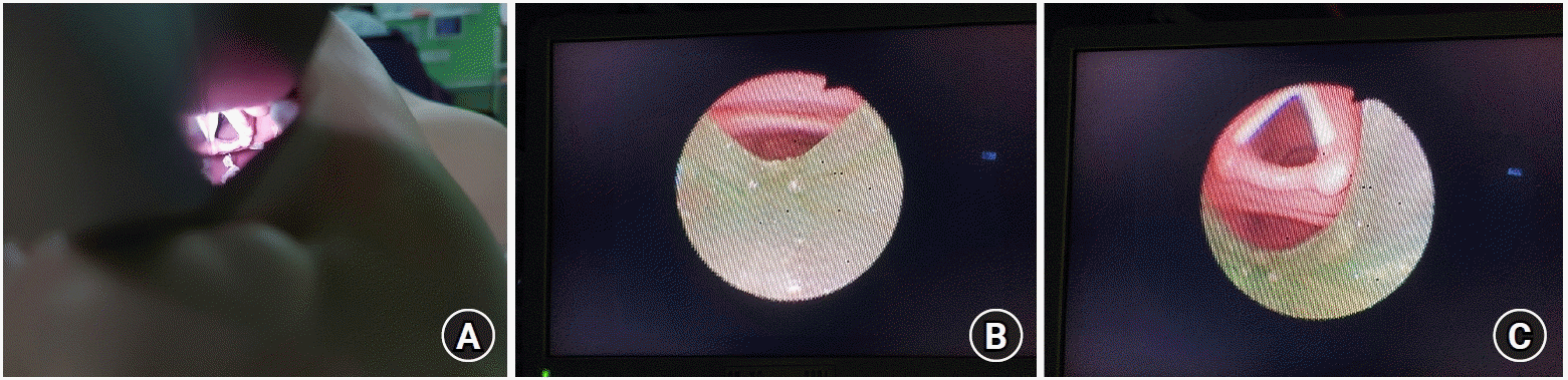
A manikin designed for pediatric intubation training (Pediatric Intub Trainer Torso 255-00001TM, Laerdal Medical Korea LLC, Korea) was prepared. When visualized using a direct laryngoscope with curved blade #2, the Cormack-Lehane grade was II. An SAD of AuraGain™ #2 (Ambu®, Denmark), a cuffless endotracheal tube (ETT) with an inner diameter of 5.0 mm (Shiley™ oral/nasal ETT, Medtronic Korea Co., Ltd., Korea), and an FOB with a diameter of 3.8 mm (LF-GPTM, Olympus Korea, Korea) were used. AuraGainTM was chosen owing to its frequent use and high success rate as an intubation conduit [12,13]. The glottis of the manikin visualized from a direct laryngoscope or an FOB are shown in Fig. 1. Upon visualization from the point approximately 1 cm proximal to the distal orifice of the SAD, the vocal cords of the manikin could be observed with appropriate positioning of the SAD, which has a score of 4 as previously recommended by Brimacombe [14]. However, anterior flexion of the bronchoscope was necessary.
Training protocol
Each ‘trial’ consisted of four steps, which are described in detail in Table 1. Before beginning the experiment, two investigators explained the entire procedure with a demonstration, followed by a discussion with question and answer session. Thereafter, the participants started a series of trials for the procedure. During the experiment, no real-time feedback was provided for self-reflection. A minimum of eight trials were performed by each participant; further details are provided in the ‘cumulative sum analysis’ section.
Table 1.
Stepwise Discrimination of the Fiberoptic-guided Endotracheal Intubation through Ambu® AuraGainTM SAD to a Pediatric Manikin
![]()
At the end of the training, the participants provided self-assessment for understanding and confidence in proficiency before and after the training using a 10-point numeric scale. Moreover, the overall satisfaction with the training was surveyed using a 10-point numeric scale.
Procedure evaluation
For each step, every repositioning, withdrawal, or advancement maneuver of any device owing to difficulty in advancement or poor vision were counted as an ‘attempt.’ For each trial, the number of ‘attempts’ for each step, procedure times for each step, and overall procedure time were recorded. If the number of ‘attempts’ for any step exceeded five, failure was declared for the trial, and the next trial was commenced from the beginning [15]. If the participant succeeded in every step of the trial, the trial was declared successful. For failed trials, the procedure time was recorded until the end of the 5th attempt of the failed step.
Cumulative sum analysis
We set our primary outcome as the learning curve of each participant and employed the cumulative sum analysis to construct them [9]. In adults, the success rate of fiberoptic-guided intubation via an SAD is reportedly above 90% [12,16,17]. In children, the success rate for vocal cord identification via a bronchoscope through an SAD is approximately 80% [18,19]. Considering that our participants did not possess any prior experience, we set the acceptable failure rate as 20% and unacceptable failure rate as 40% that was twice the acceptable rate. Both the alpha and beta error rate were set as 0.1, in the conventional method [20]. With these assumptions, the parameters for the cumulative sum analysis were calculated as shown in Table 2. Three values of h0, h1, and s were used for plotting the learning curve for each participant. The cumulative sum score started from zero at the beginning that decreased by s for each success and increased by 1 - s for each failure in the trials. When the sum reached h0 that is the lower boundary limit, the acquisition of proficiency was declared. However, when the sum reached h1 that is the upper boundary limit, a failure in acquiring proficiency was declared. We did not limit the number of trials or total training time, and the training was completed when the cumulative sum score of the participant reached either h0 or h1. Additional trials up to the 15th trial were allowed if the participant wished to continue with the practice even after acquiring proficiency. Since eight consecutive successes without failure were the minimum required number of trials, each participant performed at least eight trials.
Table 2.
Calculation of Variables in the Cumulative Sum Method
![]()
Statistical analysis
Changes in the procedure times for each step according to the number of trials were analyzed using a linear mixed-effects model with the number of trials as a fixed effect and participant as a random effect to account for the correlation between the multiple measurements within a participant. The procedure times were log-transformed owing to the skewed distributions with large values to improve normality. The relationship between the trial numbers and log-transformed procedure times were analyzed assuming a linear or piecewise linear effect that assumed a combination of intervals with linear slopes. The points of the slope change were determined as the points at which the linear mixed-effects model had the lowest Akaike and Bayesian information criteria. The model with a single point of slope change was selected for each of the four steps and total time. Since the times were log-transformed, the ratio of the cumulative geometric means was obtained for each increment in the trial number. The procedure time for failed trials was also included in the regression, since the procedure time reflects the apnea time of patients in actual clinical settings regardless of success or failure. Self-assessment of proficiency before and after the training was compared using the Wilcoxon signed-rank test. For comparison of the means, student t-test or paired t-test was used for the parametric data and Mann–Whitney U test for the nonparametric data. Normality tests for variables were performed using the Kolmogorov–Smirnov test for data of less than 30 participants and Shapiro–Wilk test for data with more than 30 participant. Statistical analyses were performed using SPSS® version 22 (IBM®, USA) and SAS® version 9.4 (SAS Institute, USA).
Go to :
Results
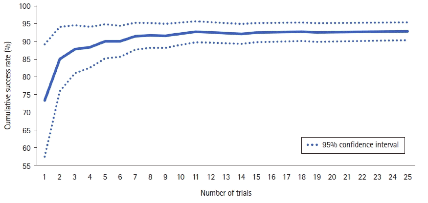
A total of 30 trainees were enrolled; no one dropped out from the study. The mean ± standard deviation (SD) duration of anesthesiology training of the participants before the practice was 28.0 ± 11.3 months. Twelve (40%) of them were males and 18 (60%) were females. Overall, the participants performed 404 trials for the procedure, recording 375 (92.8%) successful trials. At the end of the practice session, all the participants succeeded in acquiring proficiency. The median (interquartile range) number of trials required to acquire proficiency was 8 (8–12). One participant crossed h1 in the cumulative sum analysis but eventually succeeded in crossing h0. Fig. 2 shows the cumulative sum lines of the participants for the procedure. Fig. 3 shows the cumulative success rate of participants as the number of trials increased.
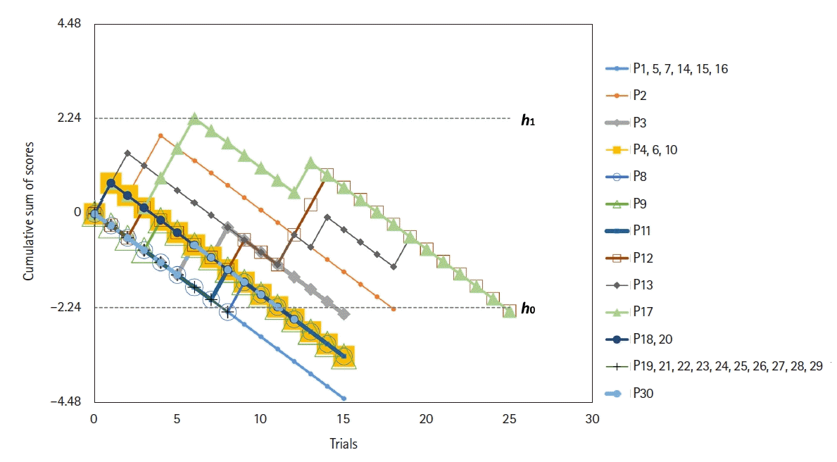 | Fig. 2.Cumulative sum lines of the participants for the practice. Lines h1 and h0 represent the upper and lower boundary limits, respectively. When the line of an individual crosses h0, we declared that the individual gained proficiency. At the end of the practice, all the participants succeeded in acquiring proficiency within 25 times of trial.
|
Among the 29 failed trials, 25 (86.2%) were due to failure in step 4, while only four (13.8%) were in step 3, and none in step 1 or 2. After the end of the practice session, all the participants recorded a failure rate of 20% or lower that was regarded as satisfactory. A summary of the individual results is presented in Table 3.
Table 3.
Summary of Individual Results of Practice
![]()
The mean ± standard deviation (SD) procedure time for each trial was 71.3 ± 50.7 s overall; successful trials showed a significantly shorter time compared to failed trials (median values 52.0 vs. 185.8 s; P < 0.001). Significant differences in the procedure time between the successful and failed trials were also seen in steps 3 (median values 13.4 vs. 122.2 s; P = 0.001) and 4 (median values 17.0 vs. 130.8 s; P < 0.001). The mean procedure time for step 4 (34.2 s) accounted for 48.0% of the mean total elapsed time (71.3 s).
As the trials were repeated, the procedure time gradually decreased for every step. Trends in the elapsed times for each step and total time fit into the piecewise linear effect model that indicates the presence of two phases of rapid decrease and slow decrease in the procedure times. A total of 1,851 measurements from the 371 trials were included in the regression models. Data from the 16th or later trials were excluded from the piecewise linear mixed-effects model since the data were from only four trainees who failed three or more times until the 15th trial. The slope of the decrease between the 1st and 2nd trial was 32.2% (95% CI [21.2, 41.7], P < 0.001), while it was 4.6% (95% CI [3.5, 5.6], P < 0.001) between the 2nd and 15th trial. For the individual steps, the decrease in the procedure times slowed down following the 5th trial for steps 1 and 3, after the 2nd trial for steps 2 and 4. The detailed data for each step are shown in Table 4. Fig. 4 shows a scatter plot of the total procedure time against the trial numbers.
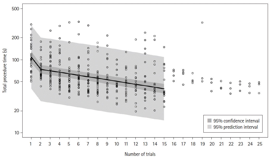 | Fig. 4.Scatter plots with regression lines of the total procedure time. The points of slope change are at trial 2. Lines for the 95% CI in the 95% prediction interval were not drawn from the 15th trial since the piecewise linear mixed-effects modeling was performed only until the 15th trial.
|
Table 4.
Changes in Procedure Times for Each Step and Total Practice
![]()
For steps 1 to 3, the success rates of the first attempt were 99.5%, 97.3%, and 91.6%, respectively. However, only 65.8% trials were successful in step 4 in the first attempt. The distribution of the number of attempts for each step is listed in Table 5.
Table 5.
Number of Attempts for Each Step
![]()
The median (Q1, Q3) of the self-assessment scores before and after the practice on a 10-point numeric scale were 2 (2, 5) and 8 (7.5, 9), respectively, with a statistically significant increase following practice (P < 0.001). The median (Q1, Q3) satisfaction score of the participants on a 10-point numeric scale was 10 (9.5, 10).
Go to :
Discussion
This is the first study to evaluate the learning curve of fiberoptic-guided intubation using an SAD in a pediatric manikin. The cumulative sum test is a simple method for the evaluation of learning a procedure and easily provides the number of trials required to obtain proficiency for a specific procedure after serial monitoring of the procedures, with the provided acceptable and unacceptable failure rates [7,9]. Upon using this method, we observed that all the participating residents became proficient in fiberoptic-guided intubation via an AuraGainTM SAD using a pediatric manikin in 25 trials.
The use of SADs has been emphasized in both expected and unexpected difficult mask ventilation and intubation, and allows continuous oxygenation and ventilation while serving as a conduit during fiberoptic-guided intubation [21]. Fiberoptic-guided intubation via an SAD is particularly beneficial, especially in children with specific anomalies, such as Pierre Robin Sequence or Treacher–Collins syndrome, since the SAD can overcome anatomic airway obstruction and optimize the laryngeal view using an FOB [6]. The success rate of this technique is reportedly similar to videolaryngoscopy in children with difficult airways [6]. However, the first attempt had a success rate of approximately 50–60% [6], indicating the need for sufficient practice prior to clinical application. None of the candidates who were eligible for screening had experienced fiberoptic-guided intubation via an SAD during their residency. The strength of our study is that we present the achievement of proficiency in a reasonable number of trials for this procedure, without awaiting a challenging case.
During the experiment, the time taken for the procedure demonstrated a decreasing trend upon repetition of the trials. The decline in the procedure time became slower following the 5th trial for Steps 1 and 3, 2nd trial for Steps 2 and 4, and overall. These findings demonstrate a significant improvement in the procedure time following the number of experiences for each step. In the first trial of step 2, some participants spent a long time visualizing the vocal cords since the bronchoscope required to be anteriorly flexed. After learning this in the first experience, the participants easily identified the vocal cords from the second trial onward; thus, a marked decrease in the procedure time for step 2 in the 2nd trial was recorded. The slow decline in the procedure time for step 3 following the 5th trial implies that advancement of the bronchoscope into the trachea required approximately five times of experience to gain dexterity.
In addition, the participants struggled for a long time in step 4 and demonstrated the highest failure rate. The ETT did not easily pass the glottis over the bronchoscope owing to collision with the arytenoid cartilages or the posterior wall of the glottis [22]. Turning the bevel to face the ground reportedly facilitates advancing the ETT over the bronchoscope by minimizing impingement and reducing the gap between the ETT and the bronchoscope [22]. However, the participants achieved a significant improvement in the procedure time at the second trial. Since the participants were instructed to rotate the ETT to adjust the direction of the bevel during practice when resistance was felt, we speculated that most of them achieved self-reflection following the first trial. Certain strategies exist that facilitate advancing the ETT over the bronchoscope, such as using a smaller-sized ETT or a Parker Flex-TipTM tube [23] to reduce the gap between the bronchoscope and the ETT, or using a reinforced tube that can change its direction easily [24]. However, we did not change the size or type of the ETT for the purpose of training and maintaining consistency of the data.
Consequently, the overall success rate of fiberoptic-guided intubation via AuraGainTM was 92.8% in our study that was similar to previous results in adults [12,16,17] and pediatric patients [13]. In pediatric data, the success rate of intubation via AuraGainTM was reported to be 100%; however, the proficiency of the practitioner was not specified [13]. As shown in Fig. 3, the cumulative success rate of participants rapidly increased from the 2nd trial, and exceeded 90% in the 5th trial. Considering the cumulative rate to include failures in earlier trials in which the participants were not accustomed to the procedure, our success rate is satisfactory. In addition, the mean procedure time of 71.3 s in our study was shorter than 96 s from previous pediatric data [13]. Since children have low oxygen reservoirs, reducing the procedure time is as important as increasing the success rate in clinical situations. Although we cannot directly compare the results obtained using a manikin with those from real patients, our results are satisfactory, and we believe our residents would reduce the time of intubation via SADs in real clinical situations.
Our study has certain limitations. First, since the practice session was conducted on a single manikin rather than patients, we cannot ensure the same level of proficiency of the procedure for patients in clinical settings. Further studies on practice with actual patients are warranted. Second, in many situations, this procedure is actually completed by removing the SAD, while keeping the ETT in situ. In the instruction step before the practice, removing the SAD was also demonstrated with the corresponding explanation. Since the decision to remove the SAD varies based on clinical situations, we did not plan to evaluate the procedure time or success rate for that step in our study. However, as removal of SAD is actually performed under various circumstances, including that step would have been reflected in the procedure time in reality. Third, we did not record or strictly control the direction of the bevel for each attempt in step 4, except for the instruction before the start of the practice. Fourth, performing this study in a smaller manikin with a smaller FOB would be more instructive, since younger children present with difficult airways and suffer more complications related to ventilation. Finally, further studies using other types of SAD, such as air-Q laryngeal airway, are necessary, since it could affect the success rate and the number of trials to achieve competency in residents.
In conclusion, anesthesiology residents obtained proficiency within 25 times of practice for fiberoptic-guided intubation in pediatric manikins through an AuraGainTM SAD with an acceptable success rate and procedure time. The procedure time markedly decreased following the first experience, indicating that this airway management skill is a relatively simple technique without a high entry barrier. Further studies should focus on the effect of dummy training in improving the success rate in actual clinical situations and learning curve of fiberoptic-guided intubation via an SAD in children with difficult airways.
Go to :
 XML Download