Journal List > Korean J Anesthesiol > v.76(4) > 1516083619

Cesur, Aksu, Güler, and Kuş: Modified pectoral nerve block versus bi-level erector spinae plane block for postoperative analgesia after radical mastectomy surgery: a prospective, randomized, controlled trial

Abstract

Background

Regional anesthesia techniques constitute an important part of successful analgesia strategies in the perioperative care of patients undergoing breast surgery. The advent of ultrasound-guided regional anesthesia has led to the development of fascial plane blocks. The large array of blocks available for postoperative analgesia in breast surgery has increased the accessibility of regional anesthesia but has also created a dilemma of choice. This study compared the analgesic efficacy of the ultrasound-guided modified pectoral nerve (PECS) block and erector spinae plane block (ESPB) in patients undergoing radical mastectomy.

Methods

Seventy women were enrolled in this prospective, double-blind, randomized control trial. After exclusion, 67 female patients who underwent radical mastectomy were finally analyzed. Ultrasound-guided PECS blocks and ESPBs were performed with 30 ml 0.25% bupivacaine. Postoperative morphine and pain scores were compared between the groups.

Results

Postoperative total morphine consumption in the first 24 h was significantly higher in the PECS group (P < 0.001). The ESPB group exhibited significantly reduced morphine consumption at all postoperative time points. Numeric rating scale scores were lower in the ESPB group at 6, 12, and 24 h postoperatively at rest and when coughing.

Conclusions

Ultrasound-guided bi-level ESPBs provided better postoperative analgesia than PECS blocks after radical mastectomy surgery.

Introduction

Breast cancer is the most prevalent cancer diagnosed in women worldwide [1]. Over one-third of patients who undergo breast cancer surgery experience acute postoperative pain, which may lead to the development of chronic postsurgical pain [2,3]. Although breast surgeries can significantly improve quality of life and can be curative in cases of malignancy, acute and chronic postoperative pain may be severe and cause functional impairment [4]. Therefore, postoperative pain remains a burden for patients and a challenge for surgeons and anesthesiologists [5,6].
The innervation of the breast is complex and thus multiple nerves and interfascial planes are available to target [7]. Various analgesic techniques, such as paravertebral blocks, thoracic epidural anesthesia, pectoral nerve blocks, serratus anterior plane blocks, and erector spinae plane blocks (ESPBs), have been used to reduce postoperative pain [7,8].
The modified pectoral nerve (PECS) block was first described by Blanco et al. [9] and consists of a PEC I injection plus a second injection under the pectoralis minor muscle at the level of the fourth rib in the anterior axillary line. The PECS block has been shown to provide safe and effective postoperative analgesia in breast surgery [1012]. The ultrasound-guided ESPB was first described by Forero et al. [13] for the effective management of thoracic wall neuropathic pain in an outpatient setting. The ESPB has since been used as a regional anesthetic technique for thoracic, breast, thoracolumbar spine, and abdominal surgeries [8,14,15].
The primary aim of this study was to compare the analgesic effects of the ultrasound-guided modified PECS block and bi-level ESPB in terms of morphine consumption in the first 24 h postoperatively in patients undergoing breast surgery with axillary lymph node dissection.

Materials and Methods

Study design

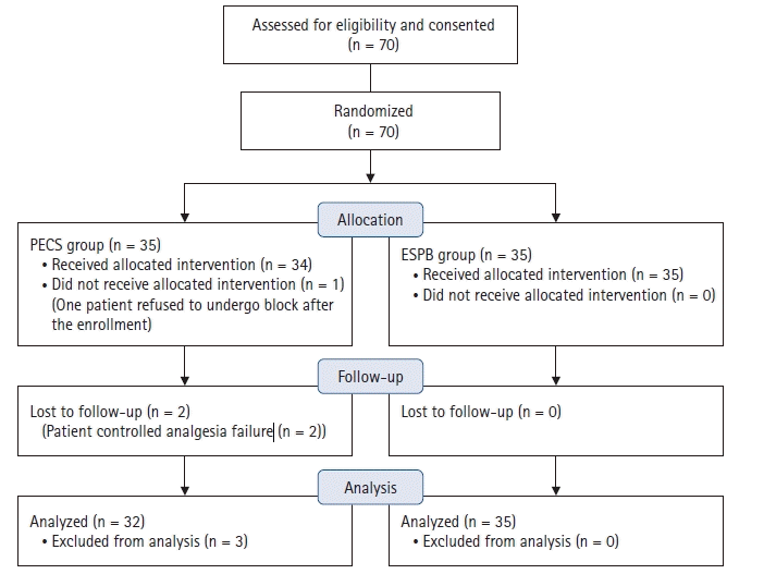
This prospective, randomized, controlled, double-blinded trial was approved by the Kocaeli University Ethical Committee (KIA 2018/487). The trial was registered at ClinicalTrials.gov (NCT04689581) and written informed consent was obtained from all participants. This study was conducted in accordance with the ethical principles of the Helsinki Declaration 2013. A Consolidated Standards of Reporting Trials (CONSORT) flow diagram was used for patient enrollment and allocation (Fig. 1). Female patients aged between 18 and 70 years with American Society of Anesthesiologists (ASA) physical status scores I–III scheduled for elective unilateral radical mastectomy surgery with lymph node dissection between January 2020 and January 2021 were included in the study. Exclusion criteria were morbid obesity defined as a body mass index > 40 kg/m2, infection of the skin at the site of the needle puncture area, coagulopathy, renal insufficiency (preoperative creatinine > 1.5 mg/dl), history of opioid abuse, pregnancy, allergy to the study medications (bupivacaine), inability to comprehend the numeric rating pain scoring system or patient-controlled analgesia (PCA) pump, and COVID-positivity.

Block procedures

The study included two groups of patients, those receiving a modified PECS block (PECS group) and those receiving a bi-level ESPB (ESPB group). Randomization was performed according to www.random.org, and patients were allocated using the sealed opaque envelope technique by a researcher (A.K.) who was not involved in the study. The same researcher (A.K.) assigned a random ID to each patient. Preoperatively, all patients were premedicated with midazolam 0.03 mg/kg and fentanyl 1 µg/kg. An experienced anesthesiologist (C.A.), blinded to the data collection until the end of the study, performed all blocks. In the preoperative block room, all patients received standard ASA monitoring (electrocardiogram, pulse oximetry, and non-invasive blood pressure) and oxygen supplementation via a nasal cannula. Blocks were performed approximately 30 min before the induction of general anesthesia. An Esaote MyLab US machine (Esaote, Italy) with a high-frequency linear probe (10–18 MHz) and a 21-gauge, 80-mm, insulated facet type needle (B Braun Sonoplex, Germany) were used for all blocks.
In the PECS group, blocks were performed with the patient in the supine position and the arm abducted to 90°. The ultrasound probe was placed obliquely over the lateral third of the clavicle. Once the axillary artery and vein were identified, the probe was moved laterally until the pectoralis major, minor, and serratus anterior muscles were identified in the same view. After skin infiltration with 1–2 ml 2% lidocaine, the needle was advanced, in the same plane as the probe, from the medial to lateral direction until the tip reached the plane between the pectoralis major and minor muscles, at which point 10 ml 0.25% bupivacaine was injected. Once the local anesthetic had spread to this plane, the needle was advanced to the fascial plane between the pectoralis minor and serratus anterior muscles, at which point an additional 20 ml 0.25% bupivacaine was administered (Fig. 2A).
In the ESPB group, blocks were performed unilaterally with the patient in the prone position. The ultrasound probe was placed 2–3 cm laterally from the midline at the level of the T4 spinous process using a sagittal approach. After identifying the erector spinae muscle, the needle was inserted in-plane from the cranial to caudal direction deep into the erector spinae muscle. The correct position of the needle tip was controlled with 1–2 ml of isotonic saline solution. Next, 15 ml 0.25% bupivacaine was administered at the T4 level. The same procedure was repeated for the second level (T2 spinous process). The local anesthetic spread in a fascial longitudinal pattern was visualized using ultrasound guidance at the two levels (Fig. 2B).
The sensory level of the block was assessed by another blinded observer (S.A.G.) with cold sensation every 5 min in each dermatomal distribution from T1 to T6. If the ice-cold sensation did not decrease in any segment up to 30 min, it was considered a block failure.

Intraoperative and postoperative management

All patients underwent standardized monitoring (pulse oximetry, electrocardiogram, and non-invasive blood pressure) in the operating room. General anesthesia was induced with an intravenous (IV) injection of fentanyl 1 µg/kg followed by propofol 2–3 mg/kg and rocuronium 0.6 mg/kg. Anesthesia was maintained with desflurane in combination with nitrous oxide in oxygen at a ratio of 2 : 1 to maintain a minimum alveolar concentration (MAC) between 1 and 1.3 in 2 L fresh gas flow. One investigator (S.C.) managed the anesthesia according to this protocol. The same surgical team conducted all surgeries; however, the surgeons did not perform local anesthetic wound infiltration. At the end of surgery, tramadol 1 mg/kg and paracetamol 15 mg/kg IV were administered to all patients. Ondansetron 0.1 mg/kg was also administered before completion of surgery. In the recovery room, all patients were given a PCA device containing morphine 0.5 mg/ml, set to deliver a 1-mg bolus dose of morphine, with an 8-min lockout time and 6-mg/h limit. A blinded pain nurse assessed postoperative pain using a numeric rating scale (NRS) ranging from 0 (no pain) to 10 (worst imaginable pain) at 1, 3, 6, 12, and 24 h postoperatively. The first rescue analgesia, IV diclofenac sodium 1.5 mg/kg, was administered in the case of an NRS score ≥ 4 at rest or while coughing. A bolus morphine dose (3 mg) was administered after 30 min if the NRS score remained ≥ 4.
Nausea and vomiting were assessed using four grades: grade 0, no nausea and vomiting; grade 1, mild nausea; grade 2, severe nausea or vomiting once; and grade 3, vomiting more than once. Ondansetron (0.1 mg/kg) was administered every 4 h postoperatively if the score was > 2 points.

Outcome measures

The primary outcome measure of the study was the total morphine consumption within the first 24 h after surgery. A pain nurse blinded to the study recorded the cumulative morphine consumption for each patient using the ID determined by A.K. at 1, 3, 6, 12, and 24 h postoperatively. The secondary outcome measures were the NRS scores at 1, 3, 6, 12, and 24 h postoperatively. The pain nurse also recorded the incidence of nausea and vomiting in the first 24 h. Potential block-related side effects and complications, such as hematoma or infection at the block site, were also recorded.

Statistical analysis

G*Power (version 3.0.10) was used to estimate the sample size (a priori). The total overall morphine consumption was used as the primary outcome variable. In a pilot study using 30 ml 0.25% bupivacaine, total morphine consumption in the first 24 h after surgery was 9.30 ± 3.24 mg for the PECS group (n = 7) and 6.18 ± 4.23 mg for the ESPB group (n = 6). Therefore, it was estimated that a sample size of 52 patients (26 per study group) would provide a power of 80%, with an α error of 0.05. We decided to include at least 35 patients per group due to possible patient drop outs.
All statistical analyses were performed using IBM SPSS for Windows (version 20.0; SPSS, USA). The Shapiro-Wilk test was used to assess the assumption of normality. Numerical variables are presented as the mean ± standard deviation (SD) or median (Q1, Q3), according to the normality of the data. Categorical variables are summarized as counts (percentages). Comparisons of numerical variables between groups were carried out using the independent samples t-test or Mann-Whitney U test, as appropriate. The association between two categorical variables was examined using the chi-square test. All statistical analyses were carried out with 5% significance, and a two-sided P value < 0.05 was considered statistically significant.

Results

Seventy women were enrolled in this study. One patient from the PECS group refused to undergo the block after enrollment, and two patients from the PECS group were lost during follow-up due to problems with the PCA device. Thus, data from 67 patients were included in the final analyses. No significant differences in demographic data were identified between the two groups (Table 1).
Total morphine consumption in the first 24 h postoperatively was significantly higher in the PECS group (P < 0.001) than in the ESPB group (PECS median, 11.0 mg; ESPB median, 5.0 mg), and morphine consumption was significantly lower at all postoperative time points in the ESPB group (Fig. 3, Table 2). Although rescue analgesia with diclofenac was administered in nine patients in the PECS group and one patient in the ESPB group, none of the patients in both groups require a morphine bolus. The postoperative rescue analgesia requirement was also significantly higher in the PECS group (P = 0.03).
The NRS scores both at rest and while coughing were similar at 1 and 3 h postoperatively. However, NRS scores were lower in the ESPB group than in the PECS group at 6, 12, and 24 h postoperatively both at rest and while coughing (Table 3).
Five patients in the PECS group and seven in the ESPB group experienced postoperative nausea; however, the difference was not statistically significant (P > 0.05).
No block failure or adverse events related to the block procedures, such as pneumothorax, hematoma, or infection at the block site, were observed in any patient.

Discussion

The findings of the present study show that the ultrasound-guided bi-level (T2–T4). ESPB reduced total morphine consumption in the first 24 h postoperatively significantly more than the modified PECS block. Postoperative pain scores were also significantly lower in the ESPB group.
PECS blocks are superficial interfascial plane blocks that access branches of the brachial plexus, which supplies innervation to the pectoral muscles and upper anterior thoracic wall [10]. Three recent meta-analyses of randomized controlled trials showed that the PECS block resulted in similar pain scores and opioid consumption to systemic analgesics and thoracic paravertebral blocks (TPVB) for breast surgery [1012].
Zhao et al. [16] performed a meta-analysis including 993 patients who received PECS blocks for radical mastectomies and recommended PECS-2 as an effective analgesia for early postsurgical pain. Kulhari et al. [17] compared the modified PECS block with the TPVB and reported that the PECS block was superior in terms of postoperative pain and opioid consumption in radical mastectomy. The PECS block may also be a safer alternative to the paravertebral block because it is easier to perform and is associated with a lower risk of pneumothorax. However, one of the most common complications of axillary dissections is trauma to the long thoracic nerve. In most cases, the nerve is stimulated electrically or mechanically for correct identification, but the nerve response has been blocked in more than one case after anesthetic injection, thus preventing identification and facilitating injury [18].
In 2016, Forero et al. [13] described the ultrasound-guided ESPB as a new regional technique in which the local anesthetic is injected into the interfascial plane between the transvers process of the vertebra and the erector spinae muscles. Several cadaveric studies have shown that local anesthetics reach both the ventral and dorsal rami of the spinal nerves, causing a sensory blockade over the anterolateral thorax. Although the debate is still ongoing, the weight of the available evidence suggests that this is the primary mechanism of action. Studies also support the assertion that the ESPB exhibits a significant analgesic effect in patients undergoing breast cancer surgery [19,20].
In a meta-analysis conducted by Leong et al. [21], ESPB was compared with no block in seven studies, the TPVB in four, and the PECS block in three. The analysis revealed that the ESPB reduced opioid consumption in the postoperative period more than systemic analgesics; however, no statistically significant difference was seen in the 24-h opioid consumption compared with the TPVB. Another meta-analysis conducted by Huang et al. [14] reported that the ESPB can be an effective alternative to the TPVB for multimodal analgesia management in thoracic and breast surgeries, which is consistent with the study conducted by Leong et al. Paraspinal facial plane blocks, such as the ESPB, are designed to provide local anesthetic spread into the paravertebral and intercostal spaces without the risks associated with inserting the needle tip into these spaces, hence the nickname paravertebral-by-proxy [22]. Given that the ESPB involves a simpler technique and is associated with a lower risk of serious complications, such as pneumothorax, this block could be a suitable alternative in thoracic and breast surgeries, as suggested by these meta-analyses.
Only three previous studies comparing the PECS block and ESPB have reported that the PECS block provided better pain management; however, the pain scores in the first 24 h were similar between the two blocks in these studies [21,2325]. Additionally, in the studies by Altıparmak et al. [23] and Gad et al. [24], all blocks were performed after the induction of anesthesia, and the PECS block was administered with the patient in the supine position, while the ESPB was administered using the lateral decubitus position. Studies have suggested that the different positions used in fascial plane blocks may affect the distribution of the local anesthetic [26]. The deep fascia, which is the target of interfascial plane blocks, is composed of multiple layers, and its dynamic properties depend on numerous factors [27]. One such factor is the mechanism by which muscle contraction creates stretch and movement in the fascia, allowing local anesthetics to spread to low-resistance areas through the pumping mechanism [28,29]. Second, the fascia may contain its own contractile elements that play a role in force generation [30]. Considering the interfascial plane blocks from this perspective, many unanswered questions remain. Although these authors reported that the patients were placed in the supine position as soon as possible after the block to eliminate the positional effect on the local anesthetic distribution, the lack of evaluation of dermatomal spread remains an important limitation. It should also be noted that fascial dynamics, and thus block effects, can be affected by anesthetic depth, muscle relaxation, and patient position.
Additionally, the ESPB was applied at the level of the T4 transverse process in all three studies.
PECS blocks may provide superior postoperative analgesic results owing to better coverage of the axilla and T2 dermatome. Aksu et al. [20] demonstrated the efficacy of the bi-level ESPB in breast surgery with axillary lymph node dissection. Therefore, the ESPB was employed as a bi-level procedure at the T2 and T4 transverse processes in the present study. The T2 level was selected as the second-level injection point because the origin of pain after axillary surgery involves the lateral cutaneous branch of T2 [31].
Finally, all three studies used more local anesthetic in the PECS block than in the ESPB block (30–35 ml vs. 20 ml, respectively). A previous meta-analysis investigating the analgesic efficacy of the ESPB in breast cancer surgery examined 12 randomized controlled studies involving 699 patients and reported an effective dose of the local anesthetic between 16 ml and 40 ml [32]. The same dose of the local anesthetic (30 ml) was applied for both the ESPB and PECS block in the present study in order to eliminate the potential effects of different doses. The study findings showed that the ultrasound-guided bi-level ESPB is an effective analgesic technique that can be used as part of a multimodal analgesia protocol in patients undergoing radical mastectomy.
The main limitation of this study was the lack of hemodynamic data recorded during the blocks. Second, patient satisfaction scores were not evaluated in this study. Third, the patients’ nausea and vomiting symptoms during the postoperative period may not have been correctly evaluated because each patient received routine ondansetron. It is difficult for doctors to blindly treat regional blocks performed using different methods. In a prospective, randomized, controlled, double-blinded trial, considering sedated patients as blinded may be biased.
We used 30 ml 0.25% bupivacaine for both blocks, and lower volumes might produce different results, especially for the bi-level ESPB.
Ultrasound-guided bi-level ESPBs provided better postoperative analgesia than PECS blocks after radical mastectomy. Future studies should investigate the optimum local anesthetic dose for interfascial plane blocks and factors affecting the spread of the local anesthetic.

Notes

Funding

None.

Conflicts of Interest

No potential conflict of interest relevant to this article was reported.

Data Availability

The datasets generated during and/or analyzed during the current study are not publicly available due to restrictions of the local ethical committee but are available from the corresponding author on reasonable request.

Author Contributions

Sevim Cesur (Data curation; Methodology; Project administration; Visualization; Writing – original draft; Writing – review & editing)

Can Aksu (Methodology; Project administration)

Sertaç Ata Güler (Data curation)

Alparslan Kuş (Methodology; Supervision)

References

1. DeSantis C, Ma J, Bryan L, Jemal A. Breast cancer statistics, 2013. CA Cancer J Clin. 2014; 64:52–62.
2. Wang L, Guyatt GH, Kennedy SA, Romerosa B, Kwon HY, Kaushal A, et al. Predictors of persistent pain after breast cancer surgery: a systematic review and meta-analysis of observational studies. CMAJ. 2016; 188:E352–61.
3. Andersen KG, Kehlet H. Persistant pain after breast cancer treatment: a critical review of risk factors and strategies for prevention. J Pain. 2011; 12:725–46.
4. Gan TJ. Poorly controlled postoperative pain: prevalence, consequences, and prevention. J Pain Res. 2017; 10:2287–98.
5. Turan M, Karaman Y, Karaman S, Uyar M, Gonullu M. Postoperative chronic pain after breast surgery with or without cancer: follow up 6 months. Eur J Anaesthesiol. 2014; 31:216.
6. Kulkarni AR, Pusic AL, Hamill JB, Kim HM, Qi J, Wilkins EG, et al. Factors associated with acute postoperative pain following breast reconstruction. JPRAS Open. 2017; 11:1–13. Erratum in: JPRAS Open 2021; 30: 170-1.
7. FitzGerald S, Odor PM, Barron A, Pawa A. Breast surgery and regional anaesthesia. Best Pract Res Clin Anaesthesiol. 2019; 33:95–110.
8. Urits I, Ostling PS, Novitch MB, Burns JC, Charipova K, Gress KL, et al. Truncal regional nerve blocks in clinical anesthesia practice. Best Pract Res Clin Anaesthesiol. 2019; 33:559–71.
9. Blanco R, Fajardo M, Parras Maldonado T. Ultrasound description of Pecs II (modified Pecs I): a novel approach to breast surgery. Rev Esp Anestesiol Reanim. 2012; 59:470–5.
10. Versyck B, van Geffen GJ, Chin KJ. Analgesic efficacy of the Pecs II block: a systematic review and meta-analysis. Anaesthesia. 2019; 74:663–73.
11. Hussain N, Brull R, McCartney CJ, Wong P, Kumar N, Essandoh M, et al. Pectoralis-II myofascial block and analgesia in breast cancer surgery: a systematic review and meta-analysis. Anesthesiology. 2019; 131:630–48.
12. Grape S, Jaunin E, El-Boghdadly K, Chan V, Albrecht E. Analgesic efficacy of PECS and serratus plane blocks after breast surgery: a systematic review, meta-analysis and trial sequential analysis. J Clin Anesth. 2020; 63:109744.
13. Forero M, Adhikary SD, Lopez H, Tsui C, Chin KJ. The erector spinae plane block: a novel analgesic technique in thoracic neuropathic pain. Reg Anesth Pain Med. 2016; 41:621–7.
14. Huang W, Wang W, Xie W, Chen Z, Liu Y. Erector spinae plane block for postoperative analgesia in breast and thoracic surgery: a systematic review and meta-analysis. J Clin Anesth. 2020; 66:109900.
15. Yayik AM, Cesur S, Ozturk F, Ahiskalioglu A, Ay AN, Celik EC, et al. Postoperative analgesic efficacy of the ultrasound-guided erector spinae plane block in patients undergoing lumbar spinal decompression surgery: a randomized controlled study. World Neurosurg. 2019; 126:e779–85.
16. Zhao J, Han F, Yang Y, Li H, Li Z. Pectoral nerve block in anesthesia for modified radical mastectomy: a meta-analysis based on randomized controlled trials. Medicine (Baltimore). 2019; 98:e15423.
17. Kulhari S, Bharti N, Bala I, Arora S, Singh G. Efficacy of pectoral nerve block versus thoracic paravertebral block for postoperative analgesia after radical mastectomy: a randomized controlled trial. Br J Anaesth. 2016; 117:382–6.
18. Miller B, Pawa A, Mariano ER. Problem with the PECS II block: the long thoracic nerve is collateral damage. Reg Anesth Pain Med 2019. Advance Access published on Apr 16, 2019. doi: 10.1136/rapm-2019-100559. Erratum in: Reg Anesth Pain Med 2019; 44: e3.
19. Gürkan Y, Aksu C, Kuş A, Yörükoğlu UH, Kılıç CT. Ultrasound guided erector spinae plane block reduces postoperative opioid consumption following breast surgery: a randomized controlled study. J Clin Anesth. 2018; 50:65–8.
20. Aksu C, Kuş A, Yörükoğlu HU, Tor Kılıç C, Gürkan Y. Analgesic effect of the bi-level injection erector spinae plane block after breast surgery: a randomized controlled trial. Agri. 2019; 31:132–7.
21. Leong RW, Tan ES, Wong SN, Tan KH, Liu CW. Efficacy of erector spinae plane block for analgesia in breast surgery: a systematic review and meta-analysis. Anaesthesia. 2021; 76:404–13.
22. Jüttner T, Werdehausen R, Hermanns H, Monaca E, Danzeisen O, Pannen BH, et al. The paravertebral lamina technique: a new regional anesthesia approach for breast surgery. J Clin Anesth. 2011; 23:443–50.
23. Altıparmak B, Korkmaz Toker M, Uysal Aİ, Turan M, Gümüş Demirbilek S. Comparison of the effects of modified pectoral nerve block and erector spinae plane block on postoperative opioid consumption and pain scores of patients after radical mastectomy surgery: a prospective, randomized, controlled trial. J Clin Anesth. 2019; 54:61–5.
24. Gad M, Abdelwahab K, Abdallah A, Abdelkhalek M, Abdelaziz M. Ultrasound-guided erector spinae plane block compared to modified pectoral plane block for modified radical mastectomy operations. Anesth Essays Res. 2019; 13:334–9.
25. Sinha C, Kumar A, Kumar A, Prasad C, Singh PK, Priya D. Pectoral nerve versus erector spinae block for breast surgeries: a randomised controlled trial. Indian J Anaesth. 2019; 63:617–22.
26. Ivanusic J, Konishi Y, Barrington MJ. A cadaveric study investigating the mechanism of action of erector spinae blockade. Reg Anesth Pain Med. 2018; 43:567–71.
27. Elsharkawy H, Pawa A, Mariano ER. Interfascial plane blocks: back to basics. Reg Anesth Pain Med. 2018; 43:341–6.
28. Lelean P. The migratory fascia hypothesis. J Bodyw Mov Ther. 2009; 13:304–10.
29. Stecco C, Gagey O, Belloni A, Pozzuoli A, Porzionato A, Macchi V, et al. Anatomy of the deep fascia of the upper limb. Second part: study of innervation. Morphologie. 2007; 91:38–43.
30. Condino S, Turini G, Parrini S, Stecco A, Busoni F, Ferrari V, et al. A semiautomatic method for in vivo three-dimensional quantitative analysis of fascial layers mobility based on 3D ultrasound scans. Int J Comput Assist Radiol Surg. 2015; 10:1721–35.
31. Woodworth GE, Ivie RM, Nelson SM, Walker CM, Maniker RB. Perioperative breast analgesia: a qualitative review of anatomy and regional techniques. Reg Anesth Pain Med. 2017; 42:609–31.
32. Hussain N, Brull R, Noble J, Weaver T, Essandoh M, McCartney CJ, et al. Statistically significant but clinically unimportant: a systematic review and meta-analysis of the analgesic benefits of erector spinae plane block following breast cancer surgery. Reg Anesth Pain Med. 2021; 46:3–12.
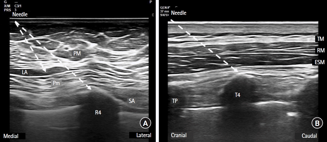
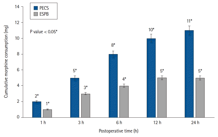
Fig. 1.
CONSORT flow diagram of the study. PECS: pectoral nerve block, ESPB: erector spinae plane block.
kja-22188f1.tif
Fig. 2.
Ultrasound image of the modified pectoral nerve block and erector spinae plane block. (A) Ultrasound image of the modified pectoral nerve block. (B) Ultrasound image of the erector spinae plane block. PM: pectoralis major muscle, Pm: pectoralis minor muscle, LA: local anesthetic, SA: serratus anterior muscle, R4: fourth rib, TM: trapezius muscle, RM: rhomboid muscle, ESM: erector spinae muscle, TP: transverse process, T4: 4th transverse process.
kja-22188f2.tif
Fig. 3.
Bar graph of morphine consumption according to time and group. PECS: modified pectoral nerve block, ESPB: erector spinae plane block. *Statistically significant difference between the PECS and ESPB groups. Values are presented as median (bars) with percentiles 25th–75th (error bars).
kja-22188f3.tif
Table 1.
Demographic and Characteristics of Patients
Variable PECS group (n = 32) ESPB group (n = 35) P value
Age (yr) 54.06 ± 10.52 51.05 ± 9.41 0.222
Height (cm) 160.96 ± 3.07 161.51 ± 3.83 0.526
Weight (kg) 71.28 ± 10.33 72.05 ± 12.21 0.880
BMI (kg/m2) 27.56 ± 4.26 27.74 ± 5.29 0.880
ASA status (I/II/III) 10 (31.3)/21 (65.6)/1 (3.1) 18 (51.4)/17 (48.6)/0 (0.0) 0.141
Duration of surgery (min) 87.34 ± 13.85 78.85 ± 10.78 0.007*

Values are presented as mean ± SD or number (%). BMI: body mass index, ASA: American Society of Anesthesiologists, PECS: modified pectoral nerve block, ESPB: erector spinae plane block. P values marked with the *symbol represents statistically significant difference.

Table 2.
Morphine Consumption at 1, 6, 12, and 24 Hours Postoperatively
Morphine consumption (mg) PECS group (n = 32) ESPB group (n = 35) P value
 1 h 2.0 (1.0, 3.0) 1.0 (1.0, 2.0) < 0.001
 3 h 5.0 (4.0, 6.7) 3.0 (2.0, 4.0) < 0.001
 6 h 8.0 (6.0, 10.0) 4.0 (3.0, 7.0) < 0.001
 12 h 10.0 (7.0, 13.0) 5.0 (4.0, 6.0) < 0.001
 24 h 11.0 (8.0, 15.0) 5.0 (4.0, 7.0) < 0.001

Values are presented as median (Q1, Q3). P values written in bold represents statistically significant difference. PECS: modified pectoral nerve block, ESPB: erector spinae plane block.

Table 3.
NRS Scores at rest and while Coughing at 1, 6, 12, and 24 Hours Postoperatively
NRS scores PECS group (n = 32) ESPB group (n = 35) P value*
NRS scores at rest
 1 h 3.0 (2.0, 4.0) 3.0 (2.0, 4.0) 0.942
 3 h 2.0 (2.0, 3.0) 2.0 (2.0, 3.0) 0.820
 6 h 2.0 (1.0, 2.0) 1.0 (0.0, 2.0) 0.003*
 12 h 1.0 (0.0, 2.0) 0.0 (0.0, 0.0) < 0.001*
 24 h 0.0 (0.0, 0.0) 0.0 (0.0, 0.0) 0.020*
NRS scores while coughing
 1 h 4.0 (2.0, 4.7) 4.0 (3.0, 6.0) 0.224
 3 h 2.5 (2.0, 4.0) 2.0 (2.0, 4.0) 0.744
 6 h 2.0 (2.0, 2.7) 2.0 (0.0, 2.0) 0.004*
 12 h 1.5 (0.0, 2.0) 0.0 (0.0, 0.0) < 0.001*
 24 h 0.0 (0.0, ;0.7) 0.0 (0.0, 0.0) 0.010*

Values are presented as median (Q1, Q3). NRS: numeric rating scale, PECS: modified pectoral nerve block, ESPB: erector spinae plane block. *P < 0.05 (Mann-Whitney U test). P values marked with the *symbol represents statistically significant difference.

TOOLS
Similar articles