

 PDF
PDF Citation
 Print
Print


INTRODUCTION
Endoscopic submucosal dissection (ESD) is a minimally invasive treatment for gastric neoplasms. ESD is now approved as the standard treatment for gastric dysplasia and early gastric cancer (EGC), having a negligible risk of lymph node metastasis. In contrast to surgical gastrectomy, ESD can provide functional and structural preservation of the stomach, even after complete and curative resection. However, the remaining stomach harbors the possibility of metachronous neoplasm recurrence.
A history of gastric cancer is one of the strongest risk factors for the development of gastric neoplasms. At diagnosis, patients with gastric cancer have synchronous gastric cancer in 5.4% to 5.8% of cases; this is particular common in older male patients.1,2 Even after surgery for gastric cancer, 3.0% to 5.4% of patients experience recurrence during follow-up.3,4 Following ESD for EGC, recurrence develops in completely preserved stomachs at a rate of approximately 3% annually.5,6
Low-grade dysplasia (LGD) and high-grade dysplasia (HGD) in the stomach are also significant risks for metachronous cancer development. The annual incidence of gastric cancer is 0.6% for mild-to-moderate dysplasia, and 6% for severe dysplasia within 5 years after diagnosis.7,8 Approximately 8.8% of LGD and 68.8% of HGD cases progress to gastric cancer during long-term follow-up.9
Metachronous recurrence is the principal reason for conducting surveillance following endoscopic resection. Male sex, intestinal metaplasia, and HGD are well-established risk factors for metachronous cancer recurrence.10 Helicobacter pylori infection is the sole amendable risk factor for metachronous recurrence.11 Proper surveillance can help to identify patients with recurrence in a timely manner. However, the clinical characteristics of metachronous gastric neoplasms (MGN) following ESD have not yet been thoroughly studied with respect to the various tiers of primary gastric neoplasms. In this study, we aimed to determine the characteristics of metachronous recurrence after ESD for gastric dysplasia and EGC.
Go to : 
METHODS
Patients
This retrospective study was conducted using electronic medical records. Patients diagnosed with gastric neoplasms and treated with ESD from January 2010 to December 2020 at The Catholic University of Korea, Yeouido St. Mary’s Hospital, Seoul, Korea were enrolled consecutively. The inclusion criteria were as follows: patients confirmed to have gastric dysplasia or EGC after ESD; resected EGC and dysplasia meeting the criteria for complete and curative resection; and follow-up for more than 1 year after ESD. The exclusion criteria were as follows: follow-up period of less than 1 year after ESD and final pathological results incompatible with curative ESD. To discriminate between synchronous and metachronous neoplasms, additional lesions discovered within 1 year after the diagnosis of the first lesion were judged as synchronous neoplasms and thus excluded from the analysis.
The clinical data analyzed in this study included age, sex, primary neoplasm histology, smoking status, alcohol use, presence of medical comorbidities, and H. pylori infection status.
Endoscopy procedures and follow-up
All ESD procedures were performed by two gastroenterologists (DYC and JIK) who had more than 500 ESD experiences and are certified as specialty board members of the Korean Society of Gastrointestinal Endoscopy. Narrow-band imaging and indigo-carmine chromoendoscopy were used to determine the extent of the lesion and define the border between the neoplasm and surrounding nonneoplastic epithelium. A glycerol solution containing epinephrine at a concentration of 1:10,000 was used to secure the submucosal layer. Circumferential incision and submucosal dissection were performed using a dual knife (Olympus) with or without insulated tip knives (FineMedics). After ESD, the patients were treated with a proton pump inhibitor for eight weeks to heal the iatrogenic ulcer. If the final histological results were compatible with curative resection, patients were monitored for ulcer healing and recurrence by periodic endoscopy. Participants with dysplasia underwent endoscopic surveillance at 2 to 3 months and then annually, and participants with EGC at 2 to 3 months, 6 months, 12 months, and then annually.
Pathological evaluation
The resected specimens were pinned, fixed on a cork plate, and immersed in a 10% formalin solution immediately after ESD completion. Specimens were sliced at a width of 2 mm for histological analysis. The Vienna classification system was used to diagnose gastric neoplasms. In this study, category 3, non-invasive low-grade adenoma/dysplasia was classified as LGD, while category 4.1 high-grade adenoma/dysplasia and 4.2 non-invasive carcinoma/carcinoma in situ were classified as HGD. Category 4.3 suspected invasive carcinoma and category 5 invasive neoplasia were classified as gastric cancer.
The background of atrophic gastritis and intestinal epithelial metaplasia was determined based on the condition of the adjacent nonneoplastic mucosa. Pathological diagnosis was determined based on the updated Sydney system.12 All pathological assessments were performed by an experienced pathologist. For determination of the presence of atrophic gastritis, serological values of pepsinogen I and pepsinogen II were preferred over endoscopic and histological findings.
Helicobacter pylori infection status
At the time of gastric cancer or dysplasia diagnosis, H. pylori infection status was evaluated. Giemsa staining was performed for the histological determination of infection. When H. pylori infection was confirmed, eradication therapy was initiated immediately. In most cases, H. pylori infection was confirmed at the time of gastric cancer or dysplasia diagnosis, and eradication was performed before ESD. In cases of additional confirmed infections after ESD, eradication regimens were administered after ESD. Successful eradication was determined using the urea breath test (HELIKIT; Isotechnika Inc.) or by histological evaluation.
Study outcomes
The primary outcome of this study was the development of a MGN after ESD for EGC and dysplasia. Dysplasia was stratified and analyzed by separating LGD and HGD. As a secondary outcome, the risk factors associated with MGN development were analyzed. The timing and histology of MGN were also analyzed.
Statistics
Baseline clinical characteristics are presented as mean±standard deviation. Categorical variables were compared and analyzed using the chi-squared test or Fisher exact test. For continuous variables, an independent t-test or Mann–Whitney analysis was used. To compare the incidence of recurrence and risk of MGN according to the primary histology of the neoplasms, an incidence curve was estimated using the Kaplan–Meier method, and hazard ratios and 95% confidence intervals (CIs) were calculated using the Cox proportional hazard regression model. Univariate and multivariate Cox proportional hazard regression analyses were performed to determine factors affecting the development of MGN. SPSS package (KoreaPlus Statistics Embedded on SPSS statistics 26 standard, Datasolution Inc.) was used for statistical analyses. A p-value <0.05 was regarded as statistically significant.
Ethical statements
This study was approved by the Institutional Research Ethics Committee of The Catholic University of Korea, Yeouido St. Mary’s Hospital (approval number: SC21RISI0110). A waiver of informed consent was obtained due to the use of retrospective data. This study was conducted in accordance with the principles of the Declaration of Helsinki.
Go to :
RESULTS
Baseline characteristics
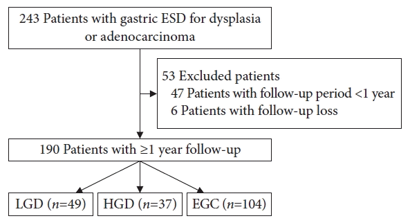
During the study period, 243 patients underwent ESD for dysplasia and EGC. Fifty-three patients were excluded from the analysis: six were lost during follow-up, and 47 had a follow-up period of less than 1 year. Thus, a total of 190 participants who met the inclusion criteria were analyzed.
The mean age was 64.4±10.4 years old. Of the participants, 140 were men, accounting for 73.7%. Regarding the primary neoplasm histology, 49 patients were diagnosed with LGD, 37 with HGD, and 104 with EGC (Fig. 1). None of the patients had multiple synchronous neoplasms. The baseline clinical characteristics were compared among the three histological groups (Table 1). Factors including age, sex, smoking, alcohol consumption, coexistence of medical comorbidities, use of antiplatelet drugs, and the presence of H. pylori infection did not differ among the groups. Regarding the pathological conditions of the primary neoplasms, factors including neoplasm size and location, presence of atrophic gastritis, and intestinal metaplasia did not differ among the groups.
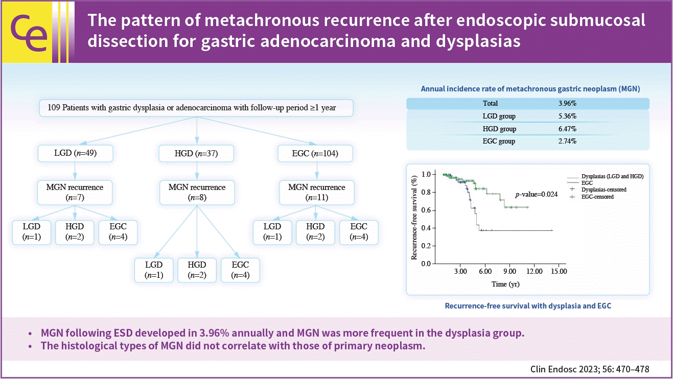
 | Fig. 1.Study design and patient disposition. ESD, endoscopic submucosal dissection; LGD, low-grade dysplasia; HGD, high-grade dysplasia; EGC, early gastric cancer.
|
Table 1.
Baseline characteristics of patients with gastric dysplasia and early gastric cancer
![]()
Development of MGNs
The mean follow-up duration was 3.45 years (standard deviation±2.25, median 2.88, minimum 1.02, maximum 14.28). The follow-up duration was 2.66±1.42 years in the LGD group, 3.34±2.39 years in the HGD group, and 3.86±2.43 years in the EGC group. The follow-up duration in the LGD group was significantly shorter than that in the HGD and EGC groups (p=0.008).
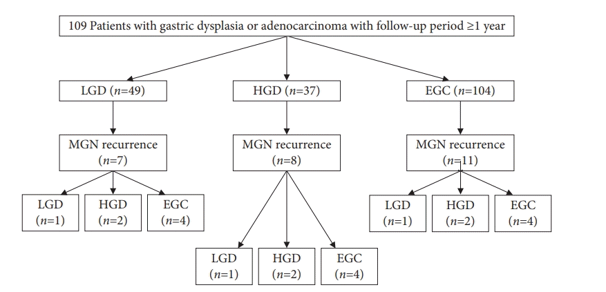
During the follow-up period, MGN was observed in 26 patients. There were no cases of multiple MGN lesions; they all occurred as a single lesion. The MGN locations differed from those of the primary lesions, and there were no cases of on-site recurrence. The MGN histology was LGD in six patients, HGD in six, and adenocarcinoma in 14. Histological diagnoses of MGN and primary neoplasm were independent of each other (p=0.458) (Fig. 2). The histology of the primary neoplasm did not predict MGN histology.
The overall annual incidence of MGN after ESD was 3.96% (Table 2). The incidence of MGN was 5.36% per year in the LGD group, 6.47% per year in the HGD group, and 2.74% per year in the EGC group. The incidences of MGN between the LGD and HGD groups did not differ (p=0.91). However, the differences in MGN incidences among the LGD, HGD, and EGC groups were significant (p=0.01 for the LGD-EGC comparison and p=0.0122 for the HGD-EGC comparison). For those with MGN development, the mean time interval from ESD to MGN was 4.1±1.8 years. The MGNs in the LGD group developed at mean interval of 4.2±0.9 years, those in the HGD group MGN at 3.6±1.3 years, and those in the EGC group MGN at 4.4±2.5 years, which was not significantly different among groups (p=0.67). Using the Kaplan–Meier model, the estimated mean MGN free survival time was 9.97 years (95% CI, 8.53–11.40) overall. The estimated mean MGN free survival time was shorter in the LGD group (4.95 years; 95% CI, 4.36–5.54) than those in the HGD and the EGC groups (8.73 years; 95% CI, 5.88–11.59 and 9.39 years; 95% CI, 8.38–10.40, respectively) (p=0.01) (Fig. 3A).
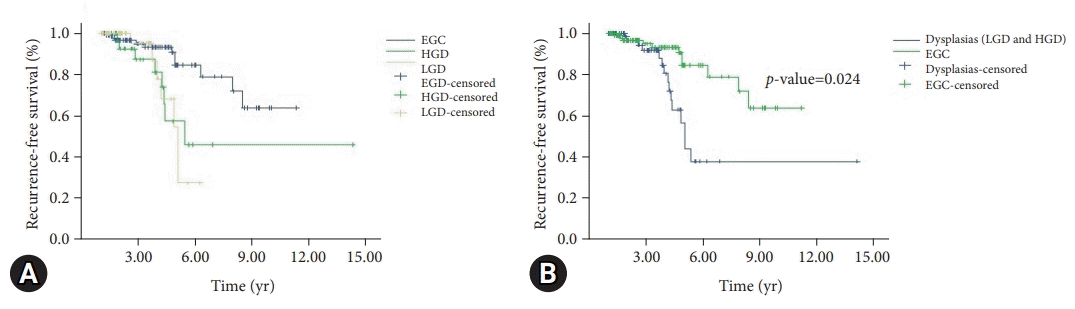 | Fig. 3.The outcomes of gastric endoscopic submucosal dissection. (A) Kaplan–Meier analysis of recurrence-free survival according to tumor grade (LGD, HGD, and EGC). (B) Kaplan–Meier analysis of recurrence-free survival with dysplasias and EGC. EGC, early gastric cancer; HGD, high-grade dysplasia; LGD, low-grade dysplasia.
|
Table 2.
The values for metachronous gastric neoplasm development according to histology of primary lesions
| LGD | HGD | EGC | p-value | |
|---|---|---|---|---|
| MGN development in total | 7/49 | 8/37 | 11/104 | |
| Annual incidence of MGN (%/yr) | 5.36 | 6.47 | 2.74 | 0.9091* for LGD-HGD |
| 0.0076* for LGD-EGC | ||||
| 0.0122* for HGD-EGC | ||||
| Time interval to MGN for observed subjects (mean±SD, yr) | 4.2±0.92 | 3.6±1.27 | 4.4±2.47 | 0.664 |
| MGN free survival (mean, 95% CI) | 4.95 (4.36–5.54) | 8.73 (5.88–11.59) | 9.39 (8.38–10.40) | 0.010 |
![]()
When the LGD and HGD groups were combined and reclassified as the dysplasia group, the estimated mean MGN free survival times for the dysplasia and EGC groups were 8.07 years (95% CI, 5.91–10.22) and 9.39 years (95% CI, 8.38–10.40), respectively, with that of the EGC group being significantly longer than that in the dysplasia group (p=0.024) (Fig. 3B). The risk of developing MGN during the follow-up period was significantly lower in the EGC group, with a risk of 0.305 (95% CI, 0.136–0.683), compared to the dysplasia group.
Treatment and risk factors of MGNs
Among the 26 patients with MGNs, 24 were treated with a second ESD procedure and two underwent a surgical gastrectomy. Both were adenocarcinomas, and the primary lesions were HGD and EGC, respectively.
There was no significant difference in baseline clinical characteristics between the MGN recurrence and non-recurrence groups (Tables 3, 4). The MGN incidence rate was 12.8% (5/39) in H. pylori-positive cases and 13.9% (21/151) in H. pylori-negative cases (p=0.860). No association was observed between the histological type of MGN and the presence of H. pylori infection.
Table 3.
The clinical characteristics of patients with and without metachronous gastric neoplasm
![]()
Table 4.
Risk factors for metachronous gastric neoplasm
![]()
Go to :
DISCUSSION
Since its introduction in the late 1990s, ESD has been proven to have clinical efficacy and safety as a standard treatment for EGC and dysplasia. The main advantage of ESD is its complete preservation of the stomach structure. However, in addition to the risk of unexplored lymph node metastasis, preserved stomachs also have the potential to retain metachronous neoplasms after ESD. Metachronous recurrence has been reported to have an annual rate of approximately 3.5% following ESD for EGC. H. pylori infection is a contributing factor to increased risk of recurrence.11 H. pylori eradication and scheduled surveillance EGD are recommended following ESD to monitor for the development of MGNs. However, regarding gastric dysplasia, a surveillance program following ESD has not yet been established for early detection of metachronous recurrence. In our study, the risk and frequency of metachronous recurrence were evaluated in patients with EGC and gastric dysplasia.
The enrolled participants were grouped according to primary neoplasm histology into LGD, HGD, and EGC groups. Risk factors known to contribute to tumorigenesis were compared between the three groups. These risk factors included age, sex, H. pylori infection, intestinal metaplasia, gastric atrophy, smoking, alcohol consumption, and comorbidities. Intestinal metaplasia was evaluated by endoscopic examination and confirmed by histological evaluation according to the updated Sydney system. The presence of atrophic gastritis was determined using the pepsinogen I and II ratios to avoid inter- and intra-observer variation in endoscopic evaluation and targeted biopsies. Contrary to the expected outcomes, baseline clinical properties, including age, did not differ among the groups. All three groups had mean age in the early 7th decade. These findings might be due to the small cohort size of this study in comparison to large cohorts in the literature; however, we can infer that the clinical background of patients with gastric neoplasms will be very similar regardless of histology.
Among the 190 patients, 26 presented with metachronous recurrences at an annual rate of 3.96%. MGN recurrence was more frequent in the LGD and HGD groups than that in the EGC group. In the analysis of the time of MGN occurrence, the mean time interval of MGN diagnosis from ESD was about 4.2 years, regardless of the histological diagnosis of the primary lesion. However, when the mean survival time before MGN occurrence was estimated using the Kaplan–Meier survival curve function, it was difficult to interpret the shorter MGN free mean survival time of LGD relative to those of HGD and EGC. The authors first acknowledge the possibility that there may have been bias errors in the analysis of the results because the number of participants in this study was relatively small and the observation period was not long enough. We could not find a scientifically appropriate explanation for these results. At present, it is not possible to determine whether this phenomenon occurred simply because of the short observation period, or because of other important reasons. However, based on this study, we intend to continue observing and investigating the causality of MGN timing. Between the MGN recurrence and non-recurrence groups, there were no significant differences in clinical characteristics such as age, male sex proportion, alcohol consumption and smoking, H. pylori infection, primary tumor size, presence of atrophy, and intestinal metaplasia. These findings seem to differ from other similarly designed studies in which factors including male sex, older age, and H. pylori infection contributed to metachronous recurrence.10,11,13 This is presumably because the risk produced by H. pylori infection was attenuated, as all patients with confirmed H. pylori infection in the study received eradication therapy before or after ESD. However, it is also possible that this result was due to the number of participants in the study not being large enough and the observation period not being long enough. Nevertheless, our study suggests that the contribution of well-known risk factors may be smaller than expected. In this study, the histology of the metachronous neoplasms was independent of the primary lesion. This suggests that metachronous cancer risk cannot be overlooked, even in LGD. Patients with LGD also require close surveillance, as do patients with EGC or HGD.
This study has several limitations. The first is the retrospective design of the protocol. This study was conducted as a retrospective review and analysis of an electronic medical database from a single institution. However, ESD for gastric neoplasms has been performed for over 20 years at our institution, and the clinical workup and process have been well established. The homogeneity of the study data and ESD performance are likely to be reliable. Second, the sample size was not sufficiently large. The number of enrolled participants was less than 200 in the final analysis. We reviewed and enrolled all the participants consecutively during the study period. Third, background histology, such as atrophy and intestinal metaplasia, was the investigators’ main interest as it is an influential factor in tumorigenesis. Although the updated Sydney system was employed to evaluate the presence of intestinal metaplasia, risk group stratification, such as Operative Link on Gastric Intestinal Metaplasia Assessment, could not be used. Regarding gastric atrophy, we used pepsinogen I and II ratios to avoid observer-dependent bias. However, we admit that serological diagnosis of atrophic gastritis is not completely accepted as a standard diagnostic tool.
In this study, we investigated the characteristics of MGN recurrence following gastric neoplasms, including LGD, HGD, and EGC. The annual global recurrence rate was 3.96%. Patients with dysplasia were more likely to develop MGN than those with EGC. The primary histology of gastric neoplasms did not predict the histological type of MGN. Surveillance endoscopy should be employed to monitor the recurrence of gastric neoplasms after endoscopic resection for gastric dysplasia and EGC.
Go to :
 XML Download