

 PDF
PDF Citation
 Print
Print


I. Introduction
A midline or median cleft lip is rare, with an incidence amongst cleft patients of only 0.43% to 0.73%1,2. It was initially presumed to arise from a failure of fusion between the medial nasal prominences; however, further study and observation in rats showed that it might be due to a failure of mesenchymal migration3.
In his widely adopted facial cleft classification, Tessier4 put a midline cleft of the upper lip in the 0 group, whereas the 5-type surgical classification proposed by Fearon5 has a midline cleft of the lip as type 1. The midline cleft is unique because it is the only type with two subtypes: a subtype with a deficiency in the midline structures, also called a pseudo median cleft, and a subtype with an excess of midline structures that is called a true median cleft2. True median cleft lips are presumed to result from a failure of fusion between the median nasal processes, and the pseudo type is understood to result from a lack or insufficiency of the tissue that contribute to the formation of the midline structure2,6. There is still, however, no consensus on this classification.
The true median cleft, although presumed not to be associated with brain deformities, is associated with intraoral and extraoral soft tissue masses7,8, upper lip sinuses9, and cutaneous facial polyps10. To the best of our knowledge, the association of a proboscis-like structure with a median cleft and the coexistence of an upper median cleft with a unilateral upper cleft lip has not been discussed in the literature. We present a case managed in our center in which a 5-year-old male had a true median cleft of the upper lip associated with a microform unilateral cleft.
II. Case Report
The 5-year-old male patient, yet to commence formal education, presented in the company of his mother. The mother received routine antenatal clinic care before his delivery. The pregnancy was full-term and uneventful. The mother said that she did not smoke, drink alcohol, or be exposed to any environmental risk factors for orofacial clefts during pregnancy. There was no family history of clefts. She married into a polygamous family with a lot of rancor and infighting. No other history concerning the child or his family was significant.
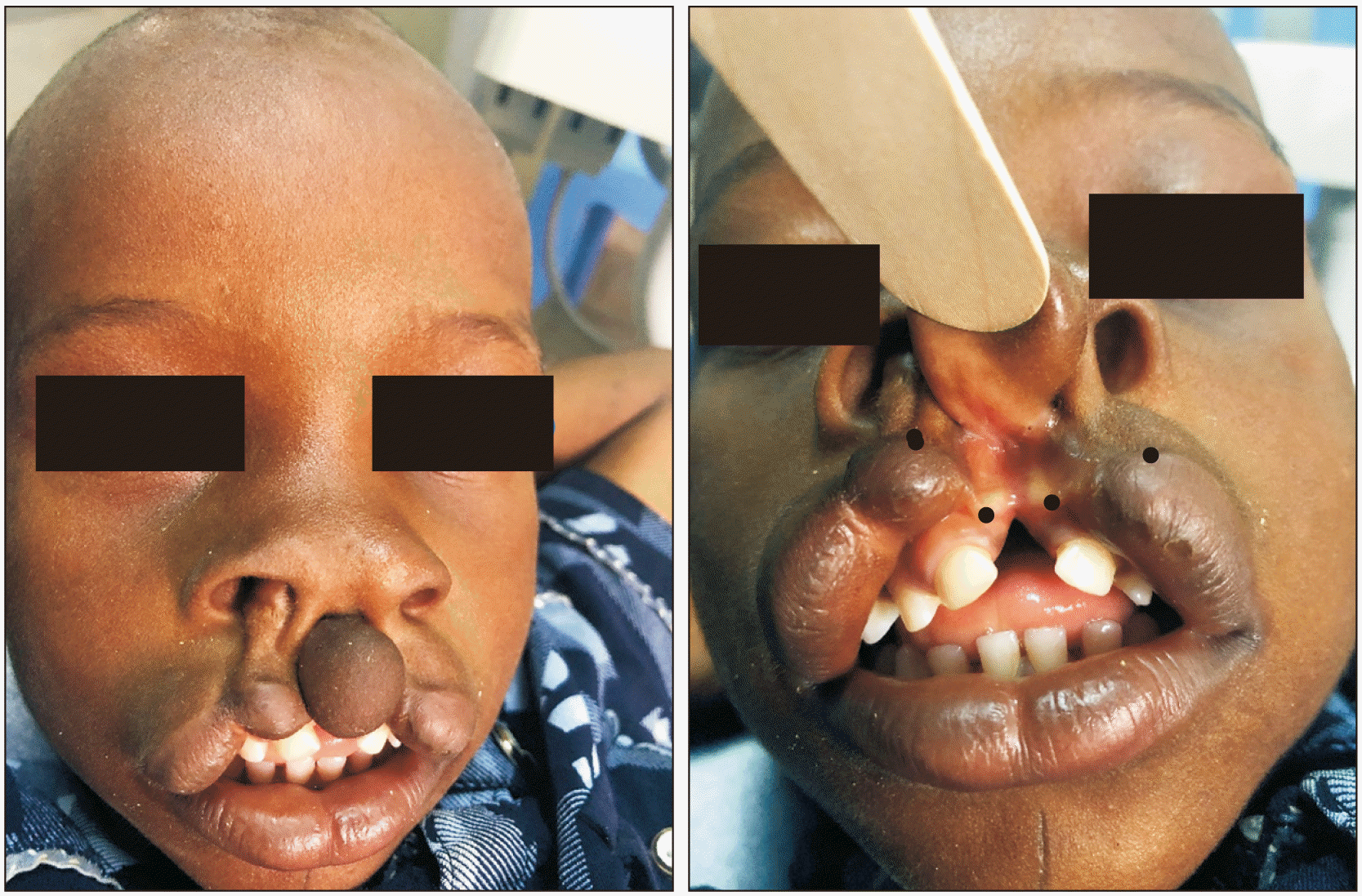
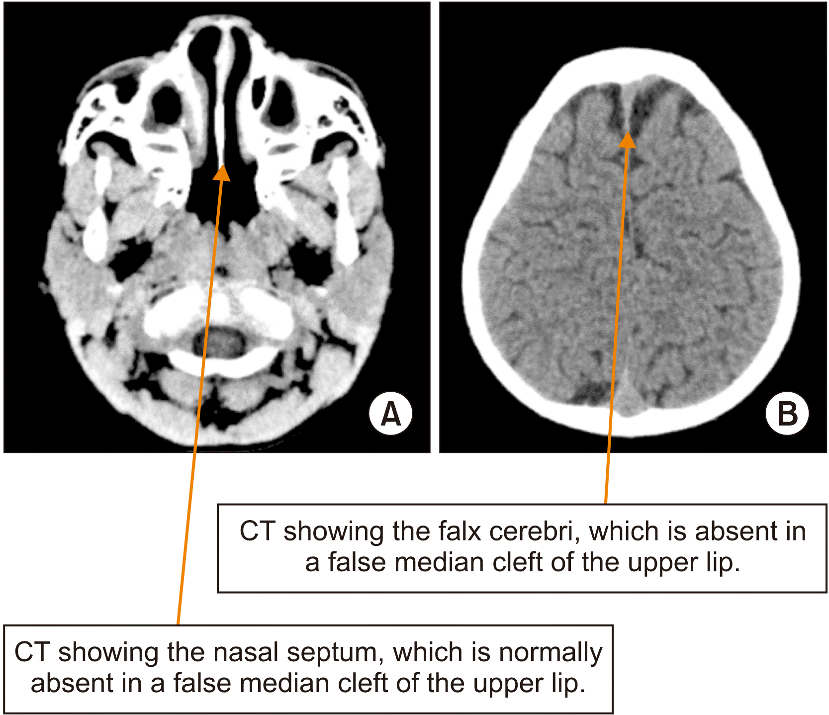
On examination, the patient weighed 20 kg and demonstrated good intellect. Limbs, genitals, abdomen, and chest were clinically normal. There was a complete midline cleft of the upper lip and a right microform cleft of the upper lip. The midline cleft extended into the vermilion, white roll, the philtrum, and up into the columella, splitting them into two halves. Between the two halves of the columella, the base of a balloon-like tissue was attached, the proboscis, composed of skin externally and labial mucosa internally. The width of the cleft was 2.5 cm. The intercanthal distance was normal (35 mm). Intraorally, there were two upper labial frenula followed by an extension of the cleft into the upper alveolus. There was also a coloboma of the right eye with a medial defect of the lower eyelid.(Fig. 1) Computerized tomography of the brain showed that the nasal septum, falx cerebri, frontal bones, cranial vault, corpus callosum, and other intracranial structures were intact.(Fig. 2)
The patient was prepared for primary cheilorrhaphy, but the family was unable to pay for surgery until a charity foundation paid for the treatment.
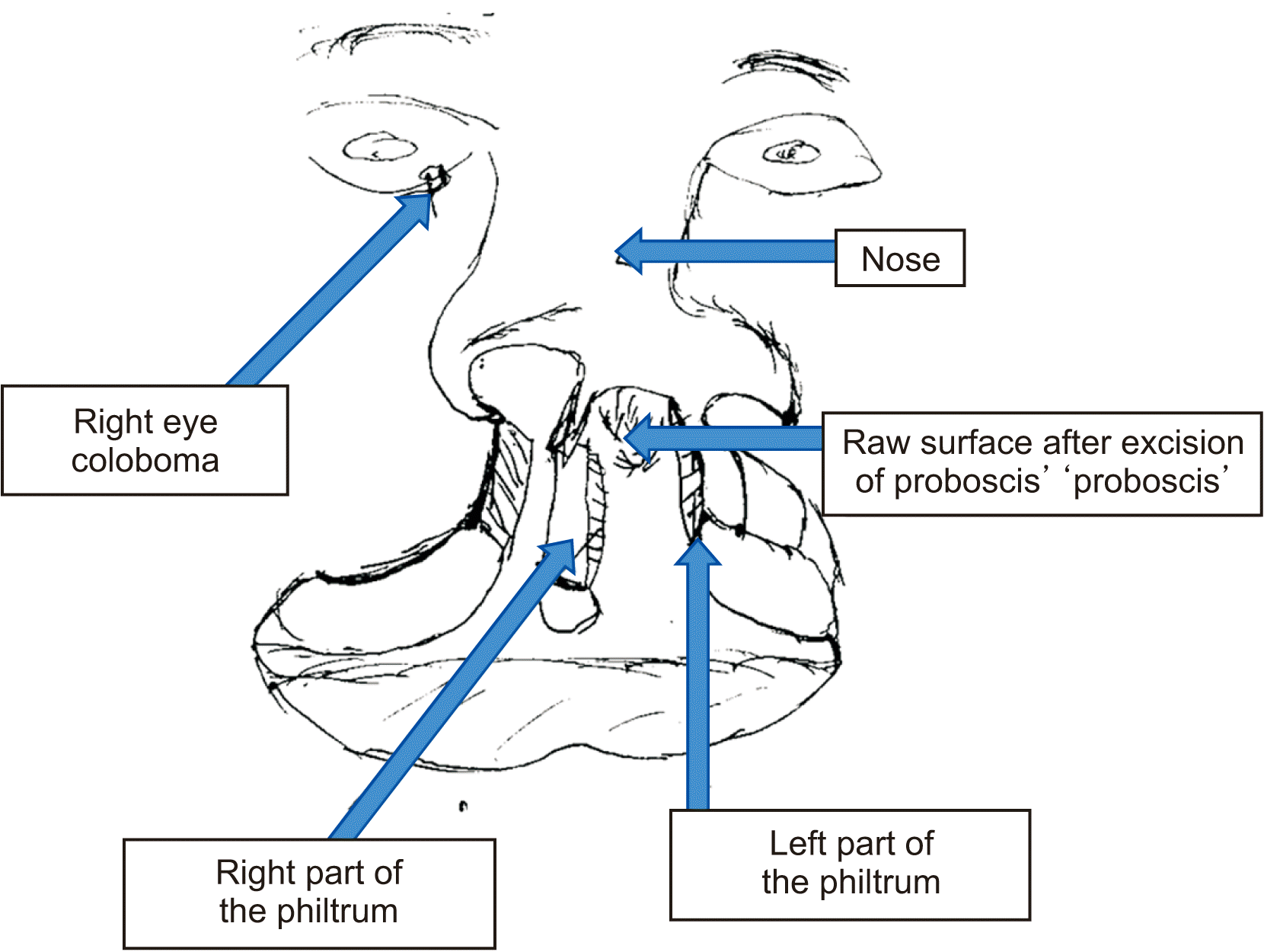
Surgery was done under general anesthesia (endotracheal intubation). The first step was to excise the proboscis at the base of the columella, which resulted in a midline cleft lip and a right microform unilateral cleft lip. A full thickness skin graft was taken from the part of the lip between the midline and right microform clefts and fashioned with a medial curvilinear edge, so that a philtrum could be formed, together with the remnant philtrum on the medial segment.
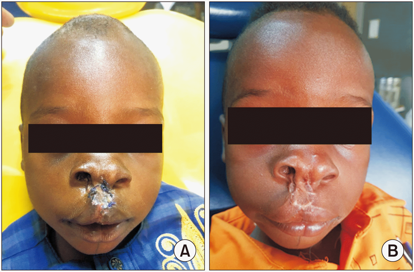
The microform unilateral cleft was converted into a complete one, and the residual tissue from which the full thickness skin had been lifted was excised and discarded. Two opposing C-shaped incisions were made into the two cleft parts of the columella, and a corresponding piece of tissue comprising skin and subcutaneous tissue was removed from the two parts to reduce the width of the columella.(Fig. 3) Markings were then made for a Millard rotation advancement repair. Separation of the orbicularis oris from the skin and oral mucosa at the edges of the cleft was done with a No. 15 scalpel blade. The skin-philtrum flap was sutured, positioning it to form the remaining part of the cupid’s bow, and then the orbicularis oris muscle was repaired to achieve its continuity. Closure was attained through intraoral and extra-oral suturing. An attempt was made to repair the cartilage of the columella by freeing it and bringing it together. Unfortunately, the patient disturbed the healing by repeated traumatic touch, which led to a breakdown, with subsequent migration and re-widening of the nose.(Fig. 4) He is now being worked up for a revisional surgery to reduce the columella width and excise the hypertrophic scar that developed postoperatively.
III. Discussion
A median cleft is defined as a vertical cleft through the center of the upper lip. This condition is rare, and the scientific world is still trying to gather data on the possible etiopathogenesis and clinical features associated with it2,11. The present case of a midline cleft of the upper lip with a right microform cleft of the upper lip is a true median cleft of the upper lip. A duplicated upper labial frenulum, duplicated nasal septum, and wide diastema of the upper incisors are the criteria stated by Ichida et al.2 and re-echoed in Jain12 that must be present in a true median cleft. These structures become duplicated as a result of the midline cleft. The false median cleft is regarded as arhinencephaly by Kundrat13 and as holotelencephaly2 and a component of holoprosencephaly by DeMyer et al.14. This suggests that the etiopathogenesis of this category of cleft is still largely unclear, but it has been traced to the third week of intrauterine life, when failure of fusion or merging of the median nasal prominences caused by reduced mesodermal in-growth results in a median cleft, as observed in rats6,15. The presence of a microform unilateral cleft compounds the possible etiopathogenesis of this case. Mazzola16 stated that the upper lip is formed from the fusion of the medial and lateral nasal prominences and the medial aspect of the maxillary process. Thus, this unilateral cleft might result from a failure of fusion between the maxillary process and lateral nasal prominence or between the lateral and median nasal prominences.
James et al.17 found craniofacial clefts to be commoner on the right sides and in male persons. In line with this, the right-sided microform cleft lip and the presence of a coloboma of the right eye in this case suggest that the cleft on the right is a craniofacial cleft. The implication of the microform unilateral cleft in this case is that the previously described techniques1,2,5 for median cleft lip repair would not (and did not) suffice. The initial challenge in this case was creating a philtrum. However, knowing that in clefts, especially unilateral clefts, most of the tissues are present but displaced18, we looked and found the remaining philtral half attached to the medial edge of the unilateral microform cleft lip. The curvilinear edge of the philtral flap allowed us to make a curvilinear repair, ensuring that the resulting scar simulates the philtral ridge. This technique for median cleft repair is similar to the one advocated by Millard and Williams1 and used by James et al.17. The excess tissue, including the proboscis-like structure7, was excised to allow the cleft edges to approximate a linear or curvilinear shape1,17. A technique involving wide and extensive dissection of nasal cartilage and the incorporation of z-plasty near the vermilion in a median cleft that looked like the one presented in this report was described by Kolker et al.19. The drawback of that technique, as pointed out by Millard and Williams1, is the retardation of nasal growth caused by the extensive nasal dissection. Jian et al.20 described a ‘proportioned’ technique with markings and numbers that incorporated z-plasty to get adequate lip height and width. Unsightly scars have, however, been documented as complications of repair with z-plasty18. Our use of the Millard rotational advancement repair for this case offered flexibility, and the repair produced symmetrical lip height and width in a subjective assessment1. The columella, however, is still relatively large, despite our attempt to narrow it. Therefore, a revisional surgery is being planned to reposition the alar cartilage and correct the patient’s columella widening. Financial challenge remains the main obstacle. While we await additional financial support, the patient is being reviewed every 3 months pending revision surgery.
In summary, a midline cleft of the upper lip is rare, and the clinical features associated with it are still being gathered. The techniques used for surgical repair will obviously depend on the phenotype. Thus, it is important to develop an array of techniques, rather than a one-size-fits-all approach, as seen in the case presented here.
 XML Download