Abstract
Objectives
This case series aims to introduce the nasal floor approach for extracting inverted mesiodens.
Materials and Methods
Through a retrospective chart review between January 2022 and February 2023, we included the mesiodens patients using nasal floor approach, and analysis the location of mesiodens from the anterior nasal spine (ANS), total operation time, and complications.
Dental dysplasia is a condition that affects the eruption of teeth and is responsible for 5.6% to 18.8% of mixed dentition cases in children and adolescents, with a higher occurrence in the maxilla than the mandible1. Among the types of dental dysplasia, supernumerary teeth frequently cause midline displacement, diastema, rotation, cyst formation, delayed eruption of permanent teeth, and adjacent root resorption. To prevent those complications, mesiodens extraction is required, typically at ages 5 to 102.
The most common type of supernumerary tooth is a mesiodens, which is usually located near the maxillary central incisors1. Because a mesiodens mostly develops on the palatal side3, many surgeons prefer a palatal approach for surgical extraction. However, this approach is associated with poor accessibility and a risk of nasopalatine nerve damage. Therefore, some surgeons opt for the labial approach, despite the high risk of injury to nearby permanent teeth2.
In some cases, a mesiodens can be positioned at the base of the nasal cavity, which is known as an inverted position4. In those cases, the traditional approach can injure the adjacent root or nasopalatine nerve and require excessive osteotomy. Therefore, a modified approach, such as the nasal floor approach, should be considered when extracting an inverted mesiodens. This report introduces the nasal floor approach as a viable option for extracting an inverted mesiodens.
The Jeonbuk National University Hospital’s Institutional Review Board (IRB) approved this retrospective study (IRB No. 2023-02-054), which was conducted according to the principles of the Declaration of Helsinki for research on humans.
This study involved a chart review of three patients with deeply inverted mesiodens who underwent extraction through the nasal floor between January 2022 and February 2023.(Table 1)
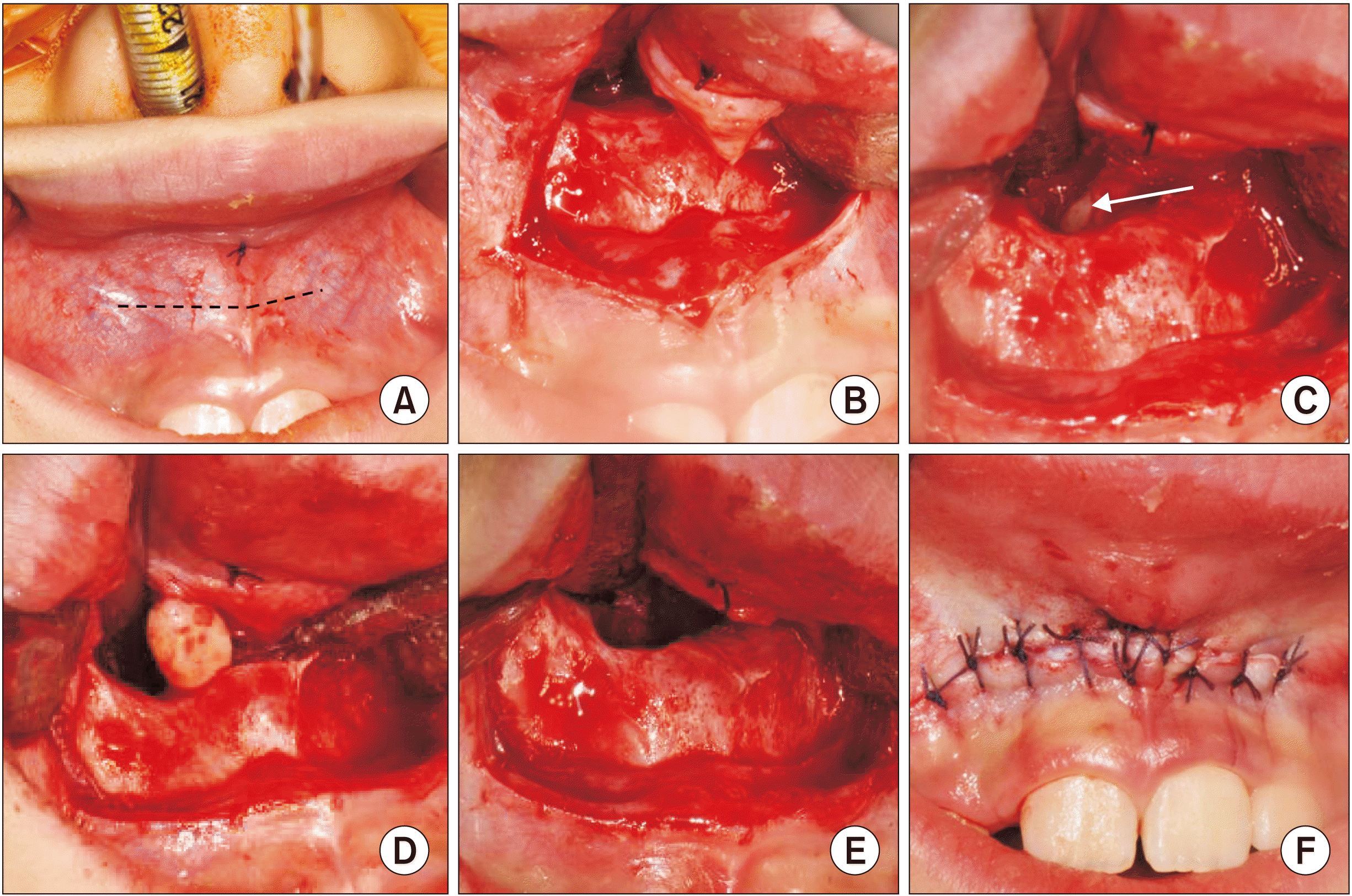
The patient was sent to the operating room for general anesthesia with nasotracheal intubation and routine draping. After placing a marking suture for the midline, a labial vestibular incision was made under local anesthesia.(Fig. 1. A) The mucoperiosteal flap was elevated toward the anterior nasal spine (ANS) and nasal floor. The nasal mucosa was elevated from the base of the piriform aperture and ANS with a periosteal elevator.(Fig. 1. B) Where necessary, the nasal floor was ground using a 2 mm round burr as measured in the preoperative cone-beam computed tomography (CBCT) until the mesiodens was exposed.(Fig. 1. C) The mesiodens was then removed through the nasal floor using extraction elevators in the usual manner.(Fig. 1. D) After profuse saline irrigation, bleeding control was achieved with oxidized regenerated cellulose (Surgigaurd; Samyang Biopharmaceuticals Corp.) and a collagen plug (Hyundai Bioland).(Fig. 1. E) Then, the mucoperiosteal flap was repositioned and sutured with 4-0 Vicryl (Ethicon, Johnson & Johnson International).(Fig. 1. F)
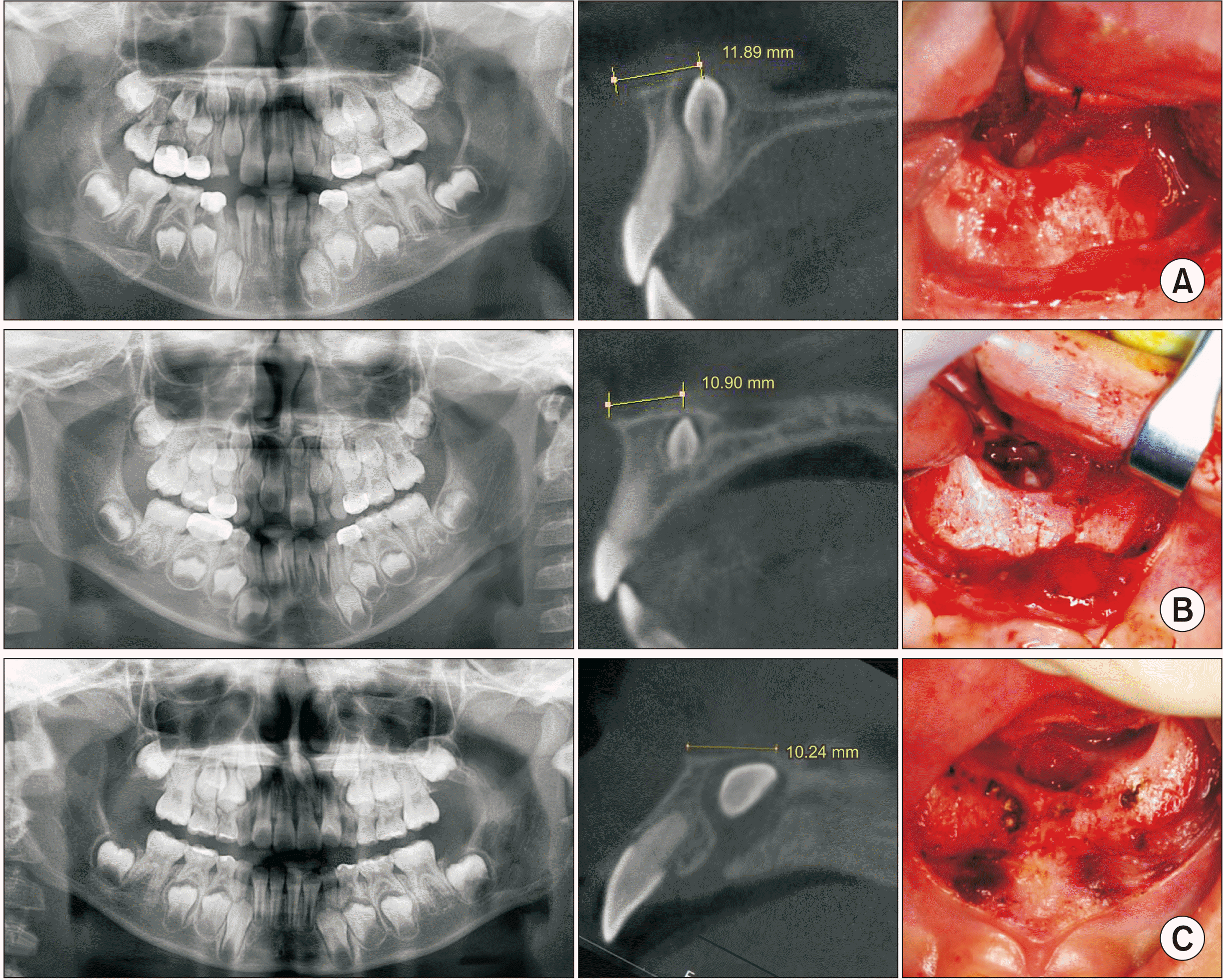
In each case, a panoramic radiograph showed a deeply impacted mesiodens in an inverted position toward the nasal floor, near the roots of adjacent permanent incisors.(Fig. 2) On the sagittal section of the CBCT images, each mesiodens was located 10 to 12 mm from the ANS and was covered with a cortical layer of the nasal floor.(Fig. 2)
Using the nasal floor approach (Fig. 1), all mesiodens were successfully extracted without exposing the adjacent incisors or nasopalatine nerve, and the procedure took approximately 30 minutes from draping to postoperative dressing. Postoperative care consisted of a 5-day course of antibiotics and nonsteroidal anti-inflammatory drugs. Patients were instructed to brush their teeth normally, and they did not require a surgical splint or dietary restrictions.
In this study, three patients with mesiodens underwent surgical extraction using the nasal floor approach, which was performed according to a uniform surgical protocol and caused no anatomical damage. No intraoperative or postoperative complications occurred, and the patients, who were all younger than 10 years, were able to resume normal food intake and tooth brushing immediately after surgery. No patients complained of nasal discomfort, difficulty breathing, bleeding, swelling, or postoperative pain.
Supernumerary teeth are predominantly found in the maxilla, with a frequency in the Asian population of about 3%2. Among people with supernumerary teeth, the prevalence in the inverted position varies from 24% to 67%1,2. Because many mesiodens are found in young children whose maxilla has not yet developed fully, some supernumerary teeth are located closer to the nasal floor than the palatal bone, particularly when they are inverted. In such cases, extraction surgery is recommended because complications can arise from root development of the adjacent permanent incisor, even if the eruption pathway is not directly inhibited by the impacted mesiodens2. In those patients (Fig. 2), however, the traditional approach carries a high risk of damaging the developing permanent incisor, especially when the inverted mesiodens is located very close to the adjacent root5. To prevent tooth injury, the palatal approach has been widely used, but it carries a high risk of neurovascular injury to the nasopalatine nerve, and patients frequently experience difficulty in mastication and oral hygiene due to the incision line and stitches around the teeth. The nasal approach can minimize those risks by approaching from the opposite direction to the incisor and deep into the vestibular fornix. This approach can provide a direct view of the surgical site and prevent excessive removal of palatal or buccal bone to access the mesiodens. In that way, it prevents complications associated with the traditional approach, such as injury to the nasopalatine bundle, paresthesia of the anterior maxilla, and difficulties with food intake and oral hygiene management6.
In 2011, a transoral approach to intranasal teeth was suggested to prevent excessive bone removal and complications related to the traditional intraoral buccal or palatal approaches6. In 2023, Kimura et al.7 classified mesiodens according to vertical and horizontal positioning and suggested the nasal floor approach for highly impacted mesiodens anterior to the nasopalatine duct, where the angle between the nasal floor line and the axis of the mesiodens is less than 90°. Yamamoto and Hanazawa8 reported three mesiodens patients with an angle of 85° to 106° between the palatal plane and the axis of the mesiodens and a distance of 10 to 13 mm from the ANS and suggested using the nasal floor approach when the angle is less than 90°. Previous studies have suggested that the angle and luxation of the inverted mesiodens are important factors in deciding on the nasal floor approach. However, we decided whether to use the nasal floor approach based on the distance from the ANS; we could easily reach the mesiodens if the distance was about 10 mm.
In conclusion, within the limitations of this study, the nasal floor approach appears to be safer and more effective than the traditional palatal approach for extracting deeply impacted, inverted mesiodens with a crown approximately 10 mm from the ANS. However, further studies with larger sample sizes are necessary to compare the clinical outcomes and indications between the nasal floor and traditional approaches.
Notes
Authors’ Contributions
J.K.K. wrote and draft the manuscript. J.K.K. and W.Y.J. participated in data collection. J.K.K., W.Y.J., and J.A.B. participated in the study design. All authors read and approved the final manuscript.
References
1. Syriac G, Joseph E, Rupesh S, Philip J, Cherian SA, Mathew J. 2017; Prevalence, characteristics, and complications of supernumerary teeth in nonsyndromic pediatric population of South India: a clinical and radiographic study. J Pharm Bioallied Sci. 9(Suppl 1):S231–6. https://doi.org/10.4103/jpbs.jpbs_154_17. DOI: 10.4103/jpbs.JPBS_154_17. PMID: 29284970. PMCID: PMC5731020.

2. Shih WY, Hsieh CY, Tsai TP. 2016; Clinical evaluation of the timing of mesiodens removal. J Chin Med Assoc. 79:345–50. https://doi.org/10.1016/j.jcma.2015.10.013. DOI: 10.1016/j.jcma.2015.10.013. PMID: 27090104.

3. Mukhopadhyay S. 2011; Mesiodens: a clinical and radiographic study in children. J Indian Soc Pedod Prev Dent. 29:34–8. https://doi.org/10.4103/0970-4388.79928. DOI: 10.4103/0970-4388.79928. PMID: 21521916.

4. Mathew M, Sasikumar P, Vivek VJ, Kala R. 2020; Surgical management of a rare case of inverted intranasal mesiodens. BMJ Case Rep. 13:e237982. https://doi.org/10.1136/bcr-2020-237982. DOI: 10.1136/bcr-2020-237982. PMID: 33127709. PMCID: PMC7604802.

5. Primosch RE. 1981; Anterior supernumerary teeth--assessment and surgical intervention in children. Pediatr Dent. 3:204–15. PMID: 6945564.
6. Sammartino G, Trosino O, Perillo L, Cioffi A, Marenzi G, Mortellaro C. 2011; Alternative transoral approach for intranasal tooth extraction. J Craniofac Surg. 22:1944–6. https://doi.org/10.1097/scs.0b013e31821151ba. DOI: 10.1097/SCS.0b013e31821151ba. PMID: 21959476.

7. Kimura M, Yasui T, Asoda S, Nagamine H, Soma T, Karube T, et al. 2023; Evaluation of the surgical approach based on impacted position and direction of mesiodens. J Oral Maxillofac Surg Med Pathol. 35:23–9. https://doi.org/10.1016/j.ajoms.2022.07.015. DOI: 10.1016/j.ajoms.2022.07.015.

8. Yamamoto A, Hanazawa Y. 2019; An effective mesiodens extraction method involving an intraoral approach through the nasal floor bone. Oral Sci Int. 16:193–5. https://doi.org/10.1002/osi2.1025. DOI: 10.1002/osi2.1025.

Fig. 1
Intraoperative images from the nasal floor approach. A. A marking suture was placed on the base of the frenulum to identify the midline and incision line (dotted line). B. After mucoperiosteal flap elevation, the nasal floor was exposed. C. Using osteotomy on the nasal floor, the impacted mesiodens was detected (arrow). D. The mesiodens was extracted in the common manner. E. After extraction, minimized osteotomy was verified without exposure of the adjacent root or nasopalatine nerve. F. Primary closure.

Fig. 2
Radiographs of the patients. A. Patient No. 1 (8 years, male) had a mesiodens about 11.89 mm from the anterior nasal spine (ANS) in Fig. 1. B. Patient No. 2 (7 years, female) had a mesiodens about 10.90 mm from the ANS. C. Patient No. 3 (9 years, male) had a mesiodens about 10.24 mm from the ANS.


 PDF
PDF Citation
 Print
Print


 XML Download