This article has been
cited by other articles in ScienceCentral.
Abstract
Objectives
Surgical extraction of maxillary third molars is routine in departments devoted to oral and maxillofacial surgery. Because maxillary third molars are anatomically adjacent to the maxillary sinus, complications such as oroantral fistula and maxillary sinusitis can occur. Here we explore the factors that can cause radiographic postoperative swelling of the maxillary sinus mucosa after surgical extraction.
Materials and Methods
This retrospective study reviewed the clinical records and radiographs of patients who underwent maxillary third-molar extraction. Preoperative panoramas, Waters views, and cone-beam computed tomography were performed for all patients. The patients were divided into two groups; those with and those without swelling of the sinus mucosa swelling or air-fluid level in a postoperative Waters view. We analyzed the age and sex of patients, vertical position, angulation, number of roots, and relation to the maxillary sinus between groups. Statistical analysis used logistic regression and P<0.05 was considered statistically significant.
Results
A total of 91 patients with 153 maxillary third molars were enrolled in the study. Variables significantly related to swelling of the maxillary sinus mucosa after surgical extraction were the age and the distance between the palatal cementoenamel junction (CEJ) and the maxillary sinus floor (P<0.05). Results of the analysis show that the relationship between the CEJ and sinus floor was likely to affect postoperative swelling of the maxillary sinus mucosa.
Conclusion
Maxillary third molars are anatomically adjacent to the maxillary sinus and require careful handling when the maxillary sinus is pneumatized to the CEJ of teeth.
Keywords: Maxilla, Third molar, Maxillary sinus, Cone-beam computed tomography, Panoramic radiography
I. Introduction
Maxillary third molars are routinely extracted in oral and maxillofacial surgery for therapeutic and prophylactic purposes
1. The maxilla is a relatively soft and elastic bone compared with the mandible. Maxillary third molars therefore require less force to extract compared with mandibular third molars, and it is often possible to extract them without sectioning the teeth
2. Because maxillary third molars are anatomically close to the maxillary sinus, complications such as perforation of the maxillary sinus, postoperative maxillary sinusitis, oroantral fistula, and dislocation into the maxillary sinus may occur
3-5. Few studies have addressed the complexity of removing maxillary third molars. Body mass index, tooth morphology, and surgical experience are associated with extraction time for maxillary third molars
6. The proximity of the roots to the maxillary sinus is associated with increased difficulty
7. The purpose of this study was to predict the radiological factors that can cause maxillary sinus complications during maxillary third-molar extraction.
II. Materials and Methods
This study was a retrospective radiographic evaluation of patients who underwent maxillary third-molar extraction. Patients were admitted to the Department of Oral and Maxillofacial Surgery at Seoul National University Dental Hospital between March 1, 2019, and December 31, 2021. All patients underwent preoperative panorama, a Waters view, and cone-beam computed tomography (CBCT). One week after surgical extraction, the patients were followed up with a postoperative panorama and Waters view. This study was conducted according to the guidelines of the Declaration of Helsinki and approved by the Institutional Review Board of Seoul National University (IRB No. S-D20220018).
1. Inclusion criteria
Patients who underwent surgical extraction of a maxillary third molar and received a preoperative panorama, CBCT, postoperative panorama, and Waters views were enrolled in the study.
2. Exclusion criteria
Patients who did not complete follow-ups after extraction, those who had chronic sinusitis and allergic rhinosinusitis, and those younger than 16 years of age were excluded.
3. Methods
The objective was to identify the factors that affect the occurrence of postoperative radiographic swelling of the maxillary mucosa or air-fluid level after third-molar extraction. The following variables in medical records and radiographs were retrospectively reviewed and investigated: patient sex and age; panoramic findings such as depth of impaction (vertical position) and angulation of teeth; CBCT findings including inclination of teeth, number of roots, distance between the cementoenamel junction (CEJ) and maxillary sinus floor, distance between root apex, and maxillary sinus floor.
Extracted maxillary third molars were divided into two groups. Members of Group A had no postoperative abnormalities, either clinically or radiographically, and members of Group B had postoperative swelling of the maxillary mucosa or an air-fluid level in a Waters view. The sinus mucosa is not typically imaged radiographically, but in cases of inflammation the mucosa appears as a non-corticated radiopaque band along the floor and wall of the sinus
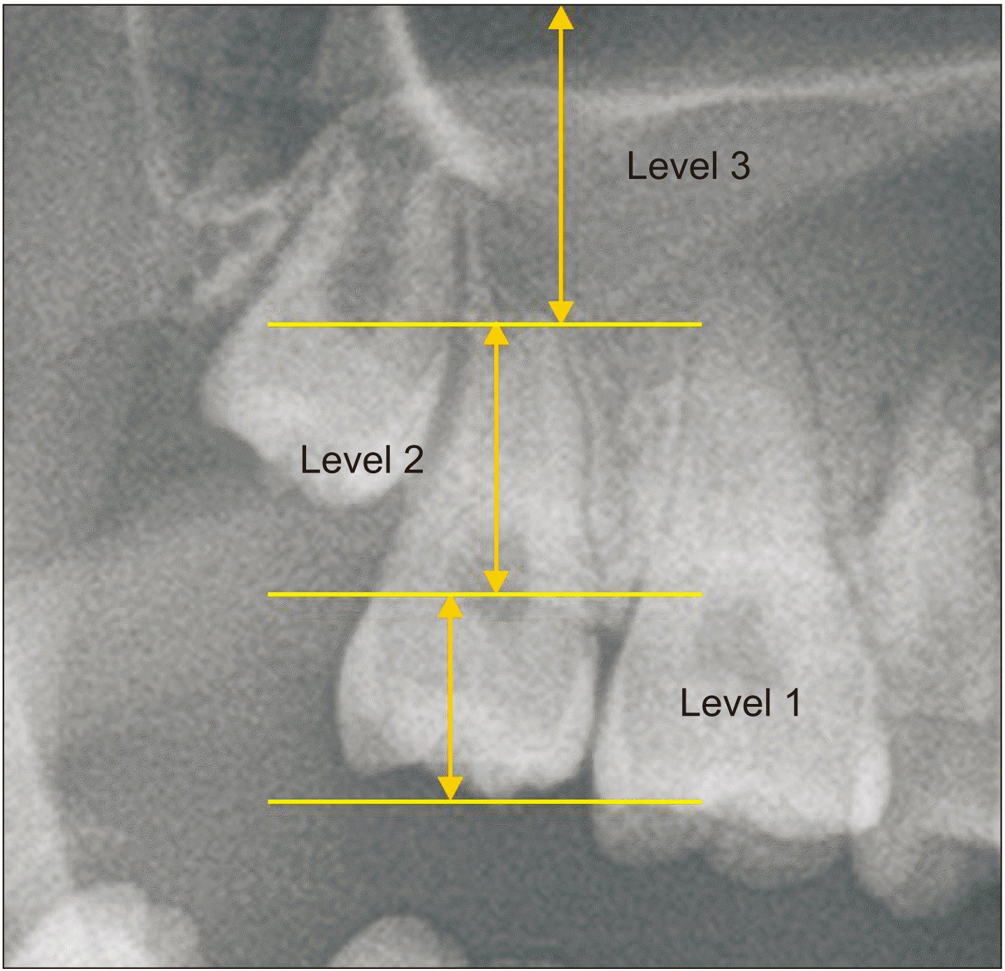
8. Vertical position and tooth angle were evaluated on a preoperative panorama. If the occlusal surface was at the same level or below the CEJ of the maxillary second molar, it was classified as level 1. If the occlusal surface was located between the CEJ and the apex of the maxillary second molar, it was classified as level 2, and if it was located above the apex, it was classified as level 3.(
Fig. 1) The angulations were classified as vertical, mesial, and distal according to the angle between the long axes of the maxillary third and second molars. Those with an angle difference between the two long axes of less than 15° were classified as vertical, those representing negative angles based on the long axes of the second molar were classified as mesial, and those representing positive angles were classified as distal.(
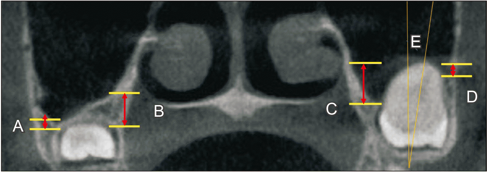
Fig. 2) In CBCT, the number of roots and buccopalatal inclination of the long axis of the tooth in the coronal section were identified. Depending on the angle between the vertical line through the midpalate and the long axis of the tooth, inclination was divided into buccal, vertical, and palatal based on a threshold of 15°. Teeth presenting along the positive angle between the midline and long axis of the tooth were classified as buccal and teeth presenting on the negative angle were classified as palatal. On CBCT, the distance from the tooth to the floor of the maxillary sinus was measured, as was the shortest distance from the buccal and palatal sides of the CEJ and root apex to the floor of the maxillary sinus. When the root apex or CEJ was above the maxillary sinus floor, the distance was expressed as a negative value.(
Fig. 3) Logistic regression was performed using IBM SPSS Statistics (ver. 26.0; IBM) for statistical analysis, for which
P<0.05 was considered statistically significant.
III. Results
A total of 91 patients were included in this study, including 42 males and 49 females. The mean age was 25.47±7.99 years. The age distribution of patients is displayed in
Table 1, with 9 patients aged 16 to 19 years, 69 patients in their 20s, 7 patients in their 30s, 3 patients in their 40s, and 3 patients 50 years or older.(
Table 1) A total of 153 impacted maxillary third molars were included; 120 in Group A and 33 in Group B. In Group B, only 1 patient exhibited clinical postoperative sinusitis. Clinically, the symptoms of maxillary sinusitis were postoperative maxillary pain and swelling, nasal congestion, a runny nose, postnasal drip, and a foul odor. In most cases, postoperative swelling of the maxillary sinus mucosa or air-fluid level was shown on a radiograph without clinical symptoms. The results of radiographic evaluations using panoramas and CBCTs are described in
Table 2. In the vertical position, 67 teeth (43.8%), 85 teeth (55.6%), and 1 tooth (0.7%) were in the level 1, 2, and 3 categories, respectively. For angulations, 57 teeth (37.3%), 62 teeth (40.5%), and 34 teeth (22.2%) exhibited mesial, vertical, and distal angulations, respectively. For the number of roots, 73 teeth (47.7%), 23 teeth (15.0%), 53 teeth (34.6%), and 4 teeth (2.6%), exhibited 1, 2, 3, and 4 roots, respectively in the coronal section, with 88 teeth (57.5%), 38 teeth (24.8%), and 27 teeth (17.6%) exhibiting buccal, vertical, and palatal inclinations, respectively.
Age and distance between the palatal CEJ and maxillary sinus floor were statistically significantly associated with postoperative maxillary sinus mucosa swelling or haziness after surgical extraction.(
Table 3)
IV. Discussion
The maxillary sinus is the largest and first paranasal sinus to develop, and its development is completed with the eruption of the third molar
2. The floor of the maxillary sinus is formed from the alveolar process of the maxilla. The volume of the maxillary sinuses changes with growth, eruption, and extraction of maxillary teeth
9.
In general, maxillary third-molar extraction causes fewer postoperative complications compared with mandibular third-molar extraction
10. However, surgical extraction of maxillary third molars can be a causative factor in odontogenic maxillary sinusitis because of the anatomic relationship. The selection of an appropriate surgical procedure should be based on preoperative evaluation of the morphology and anatomical relationship with adjacent structures
11. Lim et al.
12 reported that the depth of impaction of the maxillary third molar is a possible predictor of oroantral perforation. Rothamel et al.
1, who studied the incidence and predictive factors of perforation of the maxillary sinus in surgical extraction, reported that an intraoperative root fracture, higher degree of impaction, and older patient age were associated with oroantral perforation. The present study produced similar results. As most maxillary third molars are removed by luxation without odontomy, we hypothesized that the factors that increase the difficulty of extraction may also increase the possibility of maxillary sinusitis. Statistically significant differences in sex and depth of impaction were evident in postoperative X-rays of the groups with and without mucosal thickening. Previous studies on the anatomical relationship between maxillary teeth and sinuses have focused on the position of the root apex
13,14.
In this study, we explored whether there was a difference between the two experimental groups in the positional relationships between the root apex and maxillary sinus floor and between the CEJ and maxillary sinus floor. There were statistically significant differences in the positional relationship between the CEJ and maxillary sinus but none in the location of the root apex in relation to the sinus floor. Age was also found to be related to maxillary sinus changes after surgical extraction. As a patient ages, bone resilience decreases, and maxillary tuberosity fracture can be treated by surgical extraction
15. There were no significant relationships with postoperative radiographic maxillary sinus changes in variables such as sex, depth, tooth direction, or number of roots. As a result, we determined that the relationship of the CEJ and the sinus floor was likely to affect the maxillary sinus mucosa. Because the CEJ is close to the sinus floor, caution is required when luxating the tooth with an elevator to avoid iatrogenic maxillary sinusitis.
A limitation of this study was that, due to the small sample size, the proportion of patients who developed clinical sinusitis was small. Additional studies involving a large number of patients with radiographic postoperative maxillary mucosal swelling or air-fluid level are needed.
V. Conclusion
Maxillary third molars are anatomically adjacent to the maxillary sinus and require careful extraction. When the maxillary sinus is pneumatized to the CEJ, swelling of the maxillary sinus mucosa often appears after surgery. If patients are old or if the sinus floor and CEJ of tooth are too close, the possibility of postoperative maxillary sinusitis is high.


 PDF
PDF Citation
 Print
Print


 XML Download