Abstract
Objectives
Understanding the lingual nerve’s precise location is crucial to prevent iatrogenic injury. This systematic review seeks to determine the lingual nerve’s most probable topographical location in the posterior mandible.
Materials and Methods
Two electronic databases were searched, identifying studies reporting the lingual nerve’s position in the posterior mandible. Anatomical data in the vertical and horizontal dimensions at the retromolar and molar regions were collected for meta-analyses.
Results
Of the 2,700 unique records identified, 18 studies were included in this review. In the vertical plane, 8.8% (95% confidence interval [CI], 1.0%-21.7%) and 6.3% (95% CI, 1.9%-12.5%) of the lingual nerves coursed above the alveolar crest at the retromolar and third molar regions. The mean vertical distance between the nerve and the alveolar crest ranged from 12.10 to 4.32 mm at the first to third molar regions. In the horizontal plane, 19.9% (95% CI, 0.0%-62.7%) and 35.2% (95% CI, 13.0%-61.1%) of the lingual nerves were in contact with the lingual plate at the retromolar and third molar regions.
The lingual nerve is a branch of the mandibular division of the trigeminal nerve, containing general somatic and special visceral afferent (gustation) fibers to the anterior two-thirds of the tongue. It also provides parasympathetic innervation of the submandibular and sublingual glands, enabling salivary secretion1. During its course from the infratemporal fossa to the tongue, the lingual nerve tracks between the lateral surface of the medial pterygoid muscle and the lingual surface of the mandible. At this region, the nerve is located superficially and in close proximity to the retromolar pad, and mandibular molars, predisposing it to iatrogenic injury during dentoalveolar surgeries.
While most injuries (~90%) are transient2,3, resolving within 8 weeks, a minority are permanent, interfering with daily functions including mastication, speech, and even sleeping, which adversely impacts the quality of life4. The incidence of iatrogenic lingual nerve injury varies depending on the procedure with the highest incidence of 0.3%-18% during orthognathic surgeries involving sagittal split osteotomy5. Common procedures such as the surgical excision of impacted third molars have also been reported to have an incidence of 0.37%-13% of permanent nerve injury6-10. The lingual nerve may also be damaged during periodontal surgery, implant placement, or ridge augmentation procedures11,12, especially when the lingual flap is advanced during the vertical augmentation of an atrophic posterior mandible13.
To avoid such complications, a precise understanding of the anticipated course of the lingual nerve and anatomical relations in the posterior mandible is fundamental. Prior reviews on this topic have been focused on the clinical risk factors encountered during third molar removal, with less emphasis on the anatomy of the lingual nerve14,15. Furthermore, much of the anatomical literature is based on cadaveric dissection, often involving a small sample size due to the limited availability of cadavers, thus introducing potential sampling and sparse data biases due to an over-representation of older individuals. This resulted in a reduction in the statistical power of comparisons between groups.
Therefore, this systematic review seeks to evaluate, from previous literature, its position with reference to surgically relevant hard and soft tissue landmarks in the posterior mandible, providing a clinically relevant perspective of the anatomy of the lingual nerve.
This systematic review was designed and conducted according to the Cochrane Handbook for Systematic Reviews of Interventions16, and reported according to the Preferred Reporting Items for Systematic Review and Meta-analysis (PRISMA) statement17. The study protocol was registered in the International Prospective Register of Systematic Reviews (CRD42022352971).
The focused question of this review was: “What is the anatomical position of the lingual nerve with reference to the soft tissue, dental and bony landmarks of the posterior mandible?”
An electronic systematic search of MEDLINE (PubMed) and Embase was conducted by two independent reviewers (P.R.S. and J.R.J.C.) for articles published until 12/9/2022. The results were imported into EndNote reference management software (EndNote ver. 20.4; Clarivate Analytics), merging the search results and removing duplicate records. To identify any additional eligible studies, the reference lists of included studies were screened. The detailed search strategy was recorded in Supplementary Table 1.
This review included studies that report on the position of the lingual nerve with reference to the soft tissue, dental and bony landmarks of the posterior mandible. This encompassed both anatomical studies involving cadaveric dissection, as well as clinical studies employing various imaging modalities or surgical exploration to identify the lingual nerve. Studies were excluded based on the following criteria: (1) in vitro studies, (2) in vivo studies involving animals, (3) case reports or case series with a sample size of fewer than 5 patients, (4) narrative reviews, opinion abstracts, and letters to the editor, and (5) publications in languages other than English.
Study selection was performed in stages by two independent and calibrated reviewers (P.R.S. and J.R.J.C.). The title and abstracts of the retrieved records were screened after which full-text reports were retrieved and reviewed for inclusion based on the above eligibility criteria. Studies without or with unclear abstracts were included for full-text analysis to minimize the exclusion of potentially relevant articles. The agreement between the two reviewers for the title and abstract screening was evaluated using Cohen’s kappa. Any disagreements encountered were resolved through discussion with a third author (S.X.Y.L.).
Two independent reviewers (P.R.S. and J.R.J.C.) extracted data from the main text and tables by using standardized pre-tested electronic data collection forms. All data extracted were confirmed by a third reviewer (S.X.Y.L.). In the event of incomplete/missing data, attempts were made to contact the corresponding authors for clarification. The extracted data included study characteristics (author, year of publication, country), methodological details (type of study, study design, methodology for lingual nerve identification, sample size, and reference points for nerve measurements), and subject characteristics (age, sex, ethnicity, presence of pathology).
The primary outcome of this review was the vertical relationship between the lingual nerve and the dental, hard, and soft tissue landmarks of the posterior mandible. This was quantified as either the prevalence at which the lingual nerve is located above the lingual alveolar crest or the vertical distance between these landmarks and the nerve. The secondary outcome is the horizontal relationship defined either by the prevalence of the lingual nerve contacting the lingual plate or the distance between this landmark and the nerve.
The risk of bias in individual studies was assessed using the Anatomical Quality Assessment (AQUA) Tool18, by two independent reviewers (S.X.Y.L. and P.R.S.). In cases of disagreements, the risk of bias assessments was resolved by discussions with a third reviewer (J.R.J.C.). The AQUA Tool18 comprised 5 domains: (1) objectives(s) and subject characteristics, (2) study design, (3) methodology characterization, (4) descriptive anatomy, and (5) reporting of results. Each domain was graded as either high, low, or unclear. Attempts were made to contact the authors of the included studies when clarification was necessary.
The number of lingual nerves presenting above the alveolar crest, and that contacting the lingual plate were recorded as a percentage of the total number of nerves in each study. The reported vertical and horizontal distance between the lingual nerve and the lingual alveolar crest and lingual plate respectively were also recorded as the mean and standard deviation (SD). If the SD was not reported, it was estimated from the standard error.
For both primary and secondary outcomes, individual meta-analyses were performed to estimate the weighted effect sizes with 95% confidence interval (CI). The prevalence was transformed using the arcsine transformation to stabilize the variance. Subgroup analyses were conducted to determine if the weighted effect size were different for the retromolar and third molar regions. The random effects model was used in the meta-analyses to account for heterogeneity among studies. The heterogeneity between studies was assessed using the I2 index and Cochran’s Q test. An I2 value greater than 50% indicated a high heterogeneity whereas a P<0.1 for the Cochran’s Q test indicated evidence of heterogeneity. All analyses were performed using meta and metafor package in R software (2019; R Foundation for Statistical Computing) (https://www.R-project.org/), at a level of significance of 5% (α=0.05).
The systematic review process is summarized in the PRISMA flowchart.(Fig. 1) The electronic search yielded a total of 2,700 unique records. After screening the titles and abstracts, 47 full-text reports were retrieved for eligibility assessment (inter-examiner agreement: κ=0.942). Thirty-one reports were excluded for the reasons listed in Fig. 1. An additional three studies were identified during the hand search. A total of 18 studies fulfilled the eligibility criteria, and 15 studies were included for meta-analyses.
The characteristics of the 18 included studies19-36 are summarized in Table 1. These studies were published between 1984 and 2022, reporting on a total of 1,674 nerves. There was a wide geographical distribution with the majority of studies from Asia19,22,27,29,31,32,36 and North America23,25,30,34,35. Five out of the 18 studies were clinical studies20,21,23,30,34. The lingual nerve was identified using MRI in three studies20,21,34, clinical ultrasonography23, and intra-operative observation during third molar surgery30 in one study each, respectively. Among the five clinical studies, two studies also reported on cadaveric dissection23,30. In total, 15 studies utilized cadaveric dissection to identify the lingual nerve19,22-33,35,36. In addition, the position of the lingual nerve was identified using radiographic imaging in three studies. The nerve was visualized by painting water-soluble barium on the nerve31, or by attaching a wire to the nerve or into its sheath25,29. The reported position of the lingual nerve and landmarks used in each study are summarized in Tables 2-5.
The lingual nerve was localized using both hard and soft tissue landmarks at the retromolar, third, second, and first molar regions, in five, thirteen, four, and three studies respectively. At the first molar region, the lingual nerve was located 25.20±4.42 mm, 14.38±4.35 mm, and 13.0±4.0 mm apical to the occlusal plane36, alveolar crest19, and cementoenamel junction25 respectively. At the second molar region, the lingual nerve was more apically positioned, located 17.90±5.26 mm, 12.34±3.16 mm, 11.46±2.98 mm, and 9.6±3.5 mm apical to the occlusal plane36, distolingual attached gingiva21, alveolar crest19, and cementoenamel junction25 respectively. Notably, there were no differences in nerve’s position on the left and right19,25, and no lingual nerves were found to have a supracrestal location in the first and second molar regions19,21,25,36.
For the third molar and retromolar regions, the majority of studies evaluated the position of the lingual nerve with reference to the alveolar crest. Meta-analyses were performed to estimate the prevalence of lingual nerves located above this landmark and the distance from the alveolar crest to the nerve. The studies included in each meta-analysis were consistently found to have a high pooled heterogenicity (I2=74% and 99% respectively; Cochran Q test, P<0.01).(Fig. 2) In the vertical plane, 6.3% (95% CI, 1.9%-12.5%) and 8.8% (95% CI, 1.0%-21.7%) of the lingual nerves had coursed above the alveolar crest in the third molar region and retromolar pad regions respectively.(Fig. 2. A) Similarly, the vertical distance between the alveolar crest and the lingual nerve was estimated to be 7.58 mm (95% CI, 4.32-10.84 mm) and 7.70 mm (95% CI, 6.27-9.14 mm) at the third molar and retromolar regions respectively.(Fig. 2. B)
In addition, several alternative hard and soft tissue landmarks were also utilized, including the retromolar pad, the attached gingiva, and the occlusal plane. The lingual nerve was located 9.64±2.98 mm, 10.77±2.76 mm, and 9.30±6.15 mm from the retromolar pad21, attached gingiva of the third molar21 and its occlusal plane36 respectively.
The horizontal relationship between the lingual nerve and the lingual plate was examined in 10 studies20,22,26-30,33-35. Meta-analyses were performed using these studies to determine the prevalence at which the nerve contacts the lingual plate, and the distance between the two structures. The included studies for the respective meta-analysis had presented with a high pooled heterogenicity (I2=94% and 99% respectively; Cochran Q test, P<0.01).(Fig. 3) In the horizontal plane, 35.2% (95% CI, 13.0%-61.1%) and 19.9% (95% CI, 0.0%-62.7%) of the lingual nerves would contact the lingual plate in the third molar region and retromolar region respectively.(Fig. 3. A) Similarly, the horizontal distance between the two structures was estimated to be 3.43 mm (95% CI, 1.24-5.62 mm), and 3.30 mm (95% CI, –0.27 to 6.87 mm) for the third molar and retromolar regions respectively.(Fig. 3. B)
Fig. 4 summarises the risk of bias assessment of the included studies. In general, the studies were found to be of a low risk of bias for Domains 2, 4, and 5. For Domain 1, 66.7% of studies had a high risk of bias due to incomplete reporting of the subject ages, ethnicity, and sex19,24,27-33,35,36. For Domain 3, 11 studies were found to have a high risk of bias due to inadequate reporting of methodological details required for replicating the study or the lack of examiner calibration or alignment22-24,27-30,32,33,35,36. For Domain 5, one study was identified to have a high risk of bias for incomplete reporting of the sample size and results36.
Most iatrogenic lingual nerve injuries are known to occur in the posterior mandible, particularly in the retromolar and molar regions7,12,13. It is crucial for clinicians to have a precise understanding of the location of the lingual nerve in these regions to identify high-risk zones that require extra caution during surgeries, mitigating the risks for iatrogenic injury. To provide a clinically relevant perspective to these anatomical findings, the potential course of the lingual nerve was mapped out in this review. In general, the course of the lingual nerve is located apical to the alveolar crest, with a safety margin of approximately 4 to 12 mm as illustrated in Fig. 5. However, the nerve may be located within the lingual soft tissues above the alveolar crest in 8.8% of the retromolar and 6.3% of the partially erupted third molars. Notably, although none of the studies had reported lingual nerves with a supracrestal position at the first and second molar regions, a rare variant was observed where the lingual nerve was present within the retromolar pad of one specimen22. Since clinicians cannot accurately determine the exact location of the lingual nerve peri-operatively, during dentoalveolar surgery, there is a need to adopt conservative precautions to account for these anatomical variants.
Firstly, crestal incisions should be made closer to the center of the alveolar ridge and kept within keratinized tissue away from the retromolar pad to minimize the risk of sectioning the lingual nerve. When considering a distal wedge procedure for pocket reduction or crown lengthening of mandibular second molars in periodontal procedures, the surgical feasibility should be first assessed by evaluating the intraoral distance between the tooth and the retromolar pad, and the radiographic proximity to the ascending ramus. If there is a need to perform an internal bevel incision near the lingual alveolar crest, it is prudent to ensure that the tip of the scalpel is oriented towards and maintains contact with the alveolar crest and not directed towards the lingual tissues. In contrast, at the first and second molar regions, there are substantially lower risks of sectioning the lingual nerve, thus incisions and flap design would be dictated by other surgical considerations.
The position of the lingual nerve within the lingual tissues can also have other implications for implant-related and pre-prosthetic surgeries. The regions analyzed in this review correspond to the first two zones described in the “mylohyoid preservation technique” used for managing an atrophic posterior mandible13,37. The results of this review substantiate the precautions described in the technique, where the sharp dissection of the lingual tissues is discouraged, and the lingual flap is gently elevated off the retromolar pad with a periosteal elevator. The lingual flap is then separated from the underlying mylohyoid muscle with the use of blunt instruments. This technique maintains the integrity of the periosteum, preventing iatrogenic injury to the lingual nerve and other vital structures.
Furthermore, in the horizontal dimension, the position of the lingual nerve is closely related to the mandible, with 19.9% and 35.2% contacting the lingual plate at the retromolar and third molar regions respectively.(Fig. 3) Thus, considering its proximity to the periosteum, additional caution is necessary when elevating and manipulating the lingual flap since excessive pressure and stretching during flap retraction can also result in iatrogenic injury. It is not recommended to routinely place in a periosteal elevator in the lingual tissues for protection. During the surgical division of impacted third molars, to avoid iatrogenic perforation of the thin lingual cortical plate by the bur, clinicians should first perform an incomplete sectioning with the bur before completing it by rotating a hand instrument in the surgically-created cleft38. This is especially critical for deeply and transversely impacted mandibular third molars which may have pre-existing bony fenestrations of the lingual cortical plate at or near the level of the lingual nerve.
Interestingly, this review has also identified several clinical studies that have employed novel non-invasive modalities to image the position of the lingual nerve20,21,23,34. These techniques may help clinicians single out patients with lingual nerves with a supracrestal position during pre-surgical evaluation, enabling greater precision during treatment execution and thus mitigating the risk of iatrogenic injury. Additional research will be required to validate their diagnostic accuracies. However, their clinical benefits may be limited to procedures with higher risks for iatrogenic injury such as sagittal split osteotomy in orthognathic surgeries since the above-mentioned precautions are applicable and sufficient for most routine dentoalveolar surgeries.
Although this review has identified the zones of higher risks, its clinical applicability is limited by the variable results reflected by the heterogeneity of the existing literature. Firstly, this review incorporated studies from different geographical locations, involving subjects of different ethnic groups. Their anatomical variations contributed to the observed heterogenicity. Moreover, the included studies involved subjects with a wide age range, thus encompassing varying extents of edentulism, periodontal disease, and other age-related changes. These confounders can affect the position of the alveolar crest, which is the most commonly used landmark used to locate the lingual nerve. Unfortunately, these subject characteristics were not comprehensively reported in the included studies, precluding subgroup analysis and meta-regression, thus highlighting the need for improved reporting in future studies. In addition, separate analyses for dentate and edentulous mandibles should be performed to provide a more accurate depiction of the anatomical position of the lingual nerve.
The extent of impaction and position of the third molar within the mandible is another source of heterogenicity34. Lingually tilted molars would present with a lower alveolar crest, reducing the vertical distance to the lingual nerve. Moreover, considering the course of the lingual nerve, it is also likely that a greater vertical distance will be measured when the third molars are mesially positioned following the early loss of the second molar or when there is sufficient space for its complete eruption. Unfortunately, these confounders were poorly reported in the included studies. This is further confounded by studies that have attempted to estimate the position of the third molar when it is absent19,26,28,36, and the combined analysis of the third molar and retromolar region as a single entity14. In the present review, the two zones were clearly distinguished, and subgroup analyses were performed to determine if there were any differences in the position of the lingual nerve. Interestingly, similar mean horizontal and vertical distances were observed. However, the retromolar region presented with a higher frequency for lingual nerves with supracrestal positions, whereas the lingual nerve was more likely to contact the lingual plate at the third molar region. While the differences between the retromolar and third molar regions remained inconclusive due to the heterogenous results, it highlighted the need for clearer reporting of the presence and position of the third molar. Alternatively, future studies can consider reporting the position of the nerve at the retromolar region instead since it would be a more reproducible landmark when the third molar is absent, or when analysing atrophic edentulous mandibles.
Another plausible confounder that explains the heterogeneous vertical distances may be the inclusion of the nerves with supracrestal positions. Notably, it was observed that those studies, demonstrating a higher prevalence of the lingual nerve coursing above the alveolar crest, also reported shorter vertical distances. The shortest mean vertical distance of 2.28 mm was reported by the same study that also observed the highest prevalence (17.6%) of the lingual nerves that coursed above the alveolar crest30. Considering the vertical position of these anatomical variants is different from the majority that courses apical to the alveolar crest. Thus, including these nerves with a supracrestal position when measuring the vertical distance between the nerve and the alveolar crest would skew the measurements towards shorter distances. Future studies should report the respective distances for the nerves that course above and below the alveolar crest, accurately conveying the course and position of the lingual nerve. A similar approach should also be adopted for horizontal measurements and when there are unusual anatomical variations, such as the accessory gingival branch32.
This review has mapped out the topographical anatomy of the lingual nerve in the posterior mandible, identifying the anatomical variants at the retromolar and third molar regions, that are predisposed to iatrogenic injuries. Despite the limitations, this review offers a clinically relevant perspective on the anatomy of the lingual nerve, providing a conservative guideline for clinicians.
Acknowledgements
The authors express their gratitude to the authors who have responded to our requests for clarification regarding the individual study data that was included in this review.
Notes
Authors’ Contributions
S.X.Y.L. and P.R.S. participated in the data collection and wrote the manuscript. W.M.C.L. performed the statistical analysis. J.R.J.C. designed the study and coordination helped to draft the manuscript. J.X.L. and R.C.W.W. supervised the study and helped draft the manuscript.
References
1. Fagan SE, Roy W. Aboubakr S, Abu-Ghosh A, Adibi Sedeh P, Aeby TC, Aeddula NR, Agadi S, editors. 2023. Anatomy, head and neck, lingual nerve. StatPearls. StatPearls Publishing.
2. Blackburn CW. 1990; A method of assessment in cases of lingual nerve injury. Br J Oral Maxillofac Surg. 28:238–45. https://doi.org/10.1016/0266-4356(90)90059-t. DOI: 10.1016/0266-4356(90)90059-T. PMID: 2207042.

3. Mason DA. 1988; Lingual nerve damage following lower third molar surgery. Int J Oral Maxillofac Surg. 17:290–4. https://doi.org/10.1016/s0901-5027(88)80005-5. DOI: 10.1016/S0901-5027(88)80005-5. PMID: 3143774.

4. Van der Cruyssen F, Peeters F, Gill T, De Laat A, Jacobs R, Politis C, et al. 2020; Signs and symptoms, quality of life and psychosocial data in 1331 post-traumatic trigeminal neuropathy patients seen in two tertiary referral centres in two countries. J Oral Rehabil. 47:1212–21. https://doi.org/10.1111/joor.13058. DOI: 10.1111/joor.13058. PMID: 32687637. PMCID: PMC7540026.

5. Shawky M, Mosleh M, Jan AM, Jadu FM. 2016; Meta-analysis of the incidence of lingual nerve deficits after mandibular bilateral sagittal split osteotomy. J Craniofac Surg. 27:561–4. https://doi.org/10.1097/scs.0000000000002450. DOI: 10.1097/SCS.0000000000002450. PMID: 26982111.

6. Gülicher D, Gerlach KL. 2001; Sensory impairment of the lingual and inferior alveolar nerves following removal of impacted mandibular third molars. Int J Oral Maxillofac Surg. 30:306–12. https://doi.org/10.1054/ijom.2001.0057. DOI: 10.1054/ijom.2001.0057. PMID: 11518353.

7. Hillerup S, Stoltze K. 2007; Lingual nerve injury in third molar surgery I. Observations on recovery of sensation with spontaneous healing. Int J Oral Maxillofac Surg. 36:884–9. https://doi.org/10.1016/j.ijom.2007.06.004. DOI: 10.1016/j.ijom.2007.06.004. PMID: 17766086.

8. Bataineh AB. 2001; Sensory nerve impairment following mandibular third molar surgery. J Oral Maxillofac Surg. 59:1012–7. discussion 1017. https://doi.org/10.1053/joms.2001.25827. DOI: 10.1053/joms.2001.25827. PMID: 11526568.

9. Gomes AC, Vasconcelos BC, de Oliveira e Silva ED, da Silva LC. 2005; Lingual nerve damage after mandibular third molar surgery: a randomized clinical trial. J Oral Maxillofac Surg. 63:1443–6. https://doi.org/10.1016/j.joms.2005.06.012. DOI: 10.1016/j.joms.2005.06.012. PMID: 16182911.

10. Valmaseda-Castellón E, Berini-Aytés L, Gay-Escoda C. 2000; Lingual nerve damage after third lower molar surgical extraction. Oral Surg Oral Med Oral Pathol Oral Radiol Endod. 90:567–73. https://doi.org/10.1067/moe.2000.110034. DOI: 10.1067/moe.2000.110034. PMID: 11077378.

11. Berberi A, Le Breton G, Mani J, Woimant H, Nasseh I. 1993; Lingual paresthesia following surgical placement of implants: report of a case. Int J Oral Maxillofac Implants. 8:580–2. PMID: 8112800.
12. Tay AB, Zuniga JR. 2007; Clinical characteristics of trigeminal nerve injury referrals to a university centre. Int J Oral Maxillofac Surg. 36:922–7. https://doi.org/10.1016/j.ijom.2007.03.012. DOI: 10.1016/j.ijom.2007.03.012. PMID: 17875382.

13. Urban IA, Monje A, Lozada J, Wang HL. 2017; Principles for vertical ridge augmentation in the atrophic posterior mandible: a technical review. Int J Periodontics Restorative Dent. 37:639–45. https://doi.org/10.11607/prd.3200. DOI: 10.11607/prd.3200. PMID: 28817126.

14. Pippi R, Spota A, Santoro M. 2017; Prevention of lingual nerve injury in third molar surgery: literature review. J Oral Maxillofac Surg. 75:890–900. https://doi.org/10.1016/j.joms.2016.12.040. DOI: 10.1016/j.joms.2016.12.040. PMID: 28142010.

15. Lee J, Feng B, Park JS, Foo M, Kruger E. 2023; Incidence of lingual nerve damage following surgical extraction of mandibular third molars with lingual flap retraction: a systematic review and meta-analysis. PLoS One. 18:e0282185. https://doi.org/10.1371/journal.pone.0282185. DOI: 10.1371/journal.pone.0282185. PMID: 36848347. PMCID: PMC9970109.

16. Higgins JPT, Thomas J, Chandler J, Cumpston M, Li T, Page MJ, et al. 2022. Cochrane handbook for systematic reviews of interventions version 6.3 [Internet]. Cochrane;Available from: https://training.cochrane.org/handbook. cited 2023 Apr 18.
17. Page MJ, McKenzie JE, Bossuyt PM, Boutron I, Hoffmann TC, Mulrow CD, et al. 2021; The PRISMA 2020 statement: an updated guideline for reporting systematic reviews. BMJ. 372:n71. https://doi.org/10.1136/bmj.n71. DOI: 10.1136/bmj.n71. PMID: 33782057. PMCID: PMC8005924.

18. Henry BM, Tomaszewski KA, Ramakrishnan PK, Roy J, Vikse J, Loukas M, et al. 2017; Development of the anatomical quality assessment (AQUA) tool for the quality assessment of anatomical studies included in meta-analyses and systematic reviews. Clin Anat. 30:6–13. https://doi.org/10.1002/ca.22799. DOI: 10.1002/ca.22799. PMID: 27718281.

19. Al-Amery SM, Nambiar P, Naidu M, Ngeow WC. 2016; Variation in lingual nerve course: a human cadaveric study. PLoS One. 11:e0162773. https://doi.org/10.1371/journal.pone.0162773. DOI: 10.1371/journal.pone.0162773. PMID: 27662622. PMCID: PMC5035068.

20. Al-Haj Husain A, Valdec S, Stadlinger B, Rücker M, Piccirelli M, Winklhofer S. 2022; Preoperative visualization of the lingual nerve by 3D double-echo steady-state MRI in surgical third molar extraction treatment. Clin Oral Investig. 26:2043–53. https://doi.org/10.1007/s00784-021-04185-z. DOI: 10.1007/s00784-021-04185-z. PMID: 34586501. PMCID: PMC8816737.

21. Aljamani S, Youngson C, Jarad F, O'Neill F. 2022; Electrical stimulation to clinically identify position of the lingual nerve: results of 50 subjects with reliability and correlation with MRI. Oral Maxillofac Surg. 26:253–60. https://doi.org/10.1007/s10006-021-00985-5. DOI: 10.1007/s10006-021-00985-5. PMID: 34255234. PMCID: PMC9162997.

22. Behnia H, Kheradvar A, Shahrokhi M. 2000; An anatomic study of the lingual nerve in the third molar region. J Oral Maxillofac Surg. 58:649–51. discussion 652–3. https://doi.org/10.1016/s0278-2391(00)90159-9. DOI: 10.1016/S0278-2391(00)90159-9. PMID: 10847287.

23. Benninger B, Kloenne J, Horn JL. 2013; Clinical anatomy of the lingual nerve and identification with ultrasonography. Br J Oral Maxillofac Surg. 51:541–4. https://doi.org/10.1016/j.bjoms.2012.10.014. DOI: 10.1016/j.bjoms.2012.10.014. PMID: 23182453.

24. Bokindo IK, Butt F, Hassanali J. 2015; Morphology and morphometry of the lingual nerve in relation to the mandibular third molar. Open J Stomatol. 5:6–11. https://doi.org/10.4236/ojst.2015.51002. DOI: 10.4236/ojst.2015.51002.

25. Chan HL, Leong DJ, Fu JH, Yeh CY, Tatarakis N, Wang HL. 2010; The significance of the lingual nerve during periodontal/implant surgery. J Periodontol. 81:372–7. https://doi.org/10.1902/jop.2009.090506. DOI: 10.1902/jop.2009.090506. PMID: 20192863.

26. Dias GJ, de Silva RK, Shah T, Sim E, Song N, Colombage S, et al. 2015; Multivariate assessment of site of lingual nerve. Br J Oral Maxillofac Surg. 53:347–51. https://doi.org/10.1016/j.bjoms.2015.01.011. DOI: 10.1016/j.bjoms.2015.01.011. PMID: 25662169.

27. Erdogmus S, Govsa F, Celik S. 2008; Anatomic position of the lingual nerve in the mandibular third molar region as potential risk factors for nerve palsy. J Craniofac Surg. 19:264–70. https://doi.org/10.1097/scs.0b013e31815c9411. DOI: 10.1097/scs.0b013e31815c9411. PMID: 18216699.

28. Hölzle FW, Wolff KD. 2001; Anatomic position of the lingual nerve in the mandibular third molar region with special consideration of an atrophied mandibular crest: an anatomical study. Int J Oral Maxillofac Surg. 30:333–8. https://doi.org/10.1054/ijom.2001.0064. DOI: 10.1054/ijom.2001.0064. PMID: 11518358.

29. Karakas P, Uzel M, Koebke J. 2007; The relationship of the lingual nerve to the third molar region using radiographic imaging. Br Dent J. 203:29–31. https://doi.org/10.1038/bdj.2007.584. DOI: 10.1038/bdj.2007.584. PMID: 17632483.

30. Kiesselbach JE, Chamberlain JG. 1984; Clinical and anatomic observations on the relationship of the lingual nerve to the mandibular third molar region. J Oral Maxillofac Surg. 42:565–7. https://doi.org/10.1016/0278-2391(84)90085-5. DOI: 10.1016/0278-2391(84)90085-5. PMID: 6590806.

31. Kim SY, Hu KS, Chung IH, Lee EW, Kim HJ. 2004; Topographic anatomy of the lingual nerve and variations in communication pattern of the mandibular nerve branches. Surg Radiol Anat. 26:128–35. https://doi.org/10.1007/s00276-003-0179-x. DOI: 10.1007/s00276-003-0179-x. PMID: 14586562.

32. Kocabiyik N, Varol A, Sencimen M, Ozan H. 2009; An unnamed branch of the lingual nerve: gingival branch. Br J Oral Maxillofac Surg. 47:214–7. https://doi.org/10.1016/j.bjoms.2008.07.197. DOI: 10.1016/j.bjoms.2008.07.197. PMID: 18778880.

33. Mendes MB, de Carvalho Leite Leal Nunes CM, de Almeida Lopes MC. 2014; Anatomical relationship of lingual nerve to the region of mandibular third molar. J Oral Maxillofac Res. 4:e2. https://doi.org/10.5037/jomr.2013.4402. DOI: 10.5037/jomr.2013.4402. PMID: 24478912. PMCID: PMC3904728.

34. Miloro M, Halkias LE, Slone HW, Chakeres DW. 1997; Assessment of the lingual nerve in the third molar region using magnetic resonance imaging. J Oral Maxillofac Surg. 55:134–7. https://doi.org/10.1016/s0278-2391(97)90228-7. DOI: 10.1016/S0278-2391(97)90228-7. PMID: 9024349.

35. Pogrel MA, Renaut A, Schmidt B, Ammar A. 1995; The relationship of the lingual nerve to the mandibular third molar region: an anatomic study. J Oral Maxillofac Surg. 53:1178–81. https://doi.org/10.1016/0278-2391(95)90630-4. DOI: 10.1016/0278-2391(95)90630-4. PMID: 7562172.

36. Shimoo Y, Yamamoto M, Suzuki M, Yamauchi M, Kaketa A, Kasahara M, et al. 2017; Anatomic and histological study of lingual nerve and its clinical implications. Bull Tokyo Dent Coll. 58:95–101. https://doi.org/10.2209/tdcpublication.2016-0010. DOI: 10.2209/tdcpublication.2016-0010. PMID: 28724864.

37. Urban I, Traxler H, Romero-Bustillos M, Farkasdi S, Bartee B, Baksa G, et al. 2018; Effectiveness of two different lingual flap advancing techniques for vertical bone augmentation in the posterior mandible: a comparative, split-mouth cadaver study. Int J Periodontics Restorative Dent. 38:35–40. https://doi.org/10.11607/prd.3227. DOI: 10.11607/prd.3227. PMID: 29240202.

38. Appiah-Anane S, Appiah-Anane MG. 1997; Protection of the lingual nerve during operations on the mandibular third molar: a simple method. Br J Oral Maxillofac Surg. 35:170–2. https://doi.org/10.1016/s0266-4356(97)90557-x. DOI: 10.1016/S0266-4356(97)90557-X. PMID: 9212292.

Fig. 1
Prisma flow chart illustrating the selection process of the included studies and the number of excluded studies at each stage.
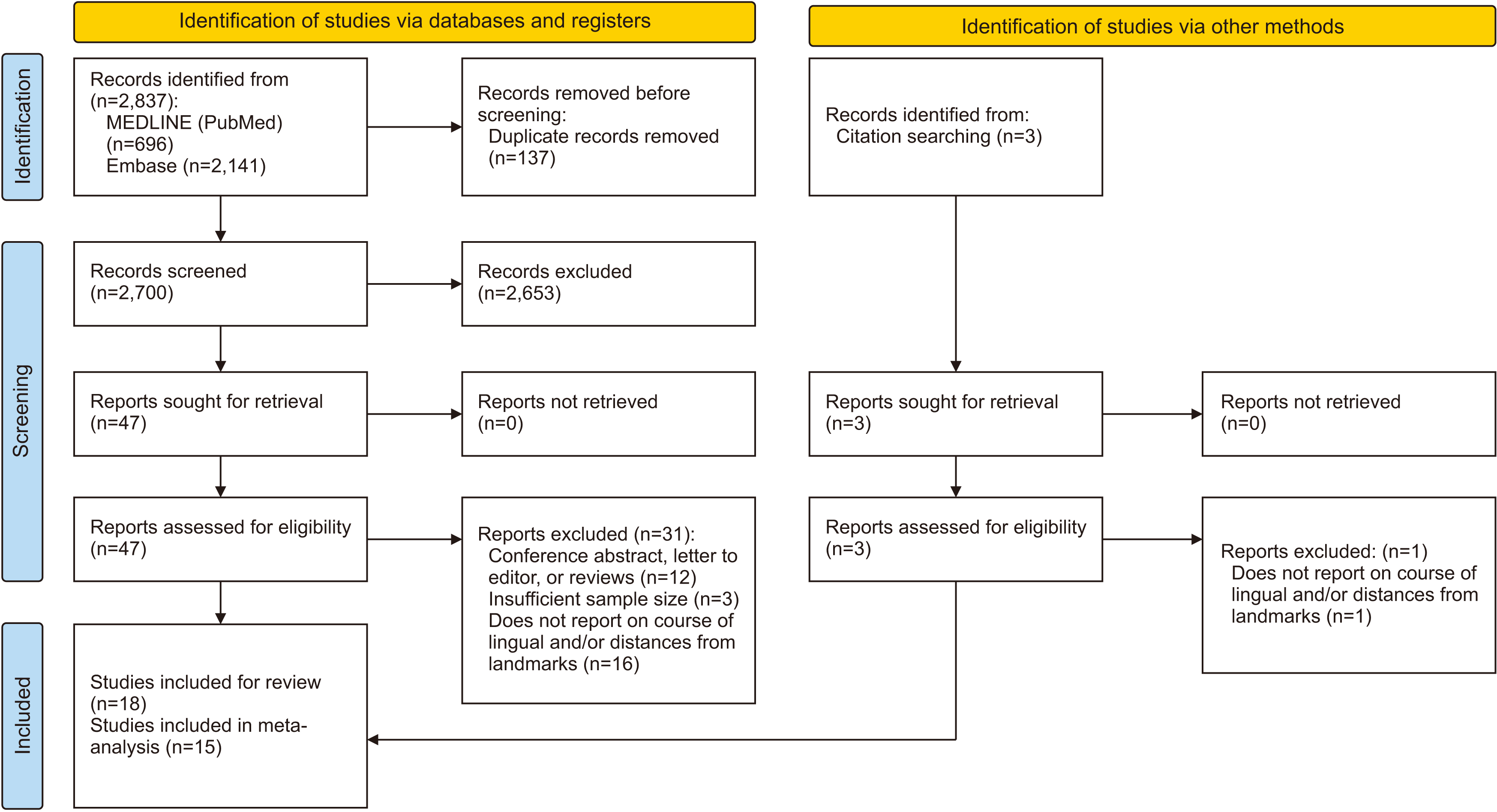
Fig. 2
Forest plots of the vertical position of the lingual nerve. A. Prevalence of the lingual nerve coursing above the alveolar crest at the retromolar and third molar regions. B. Vertical distance between the lingual nerve and the alveolar crest.
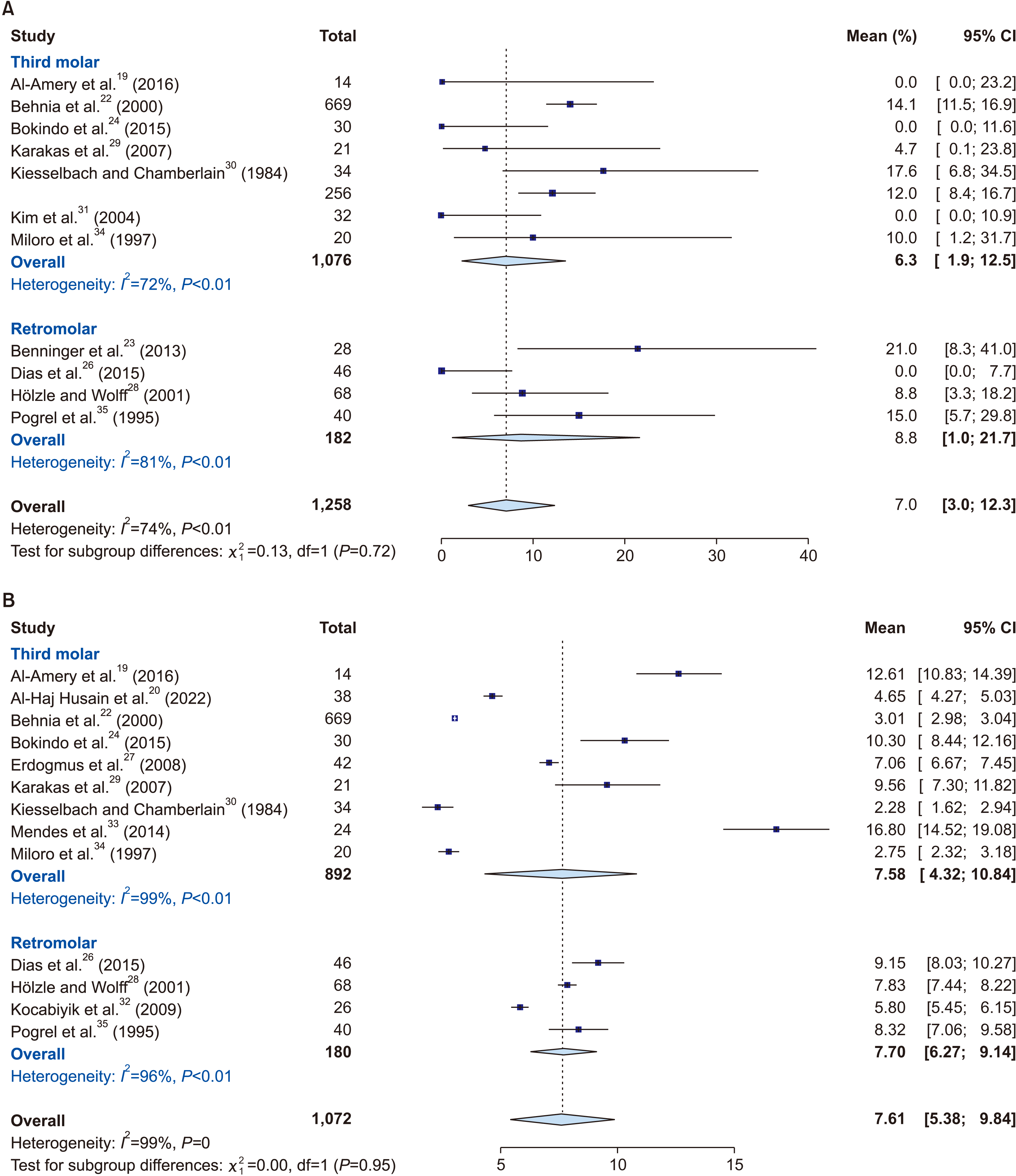
Fig. 3
Forest plots of the horizontal position of the lingual nerve. A. Prevalence of the lingual nerve contacting the lingual plate at the retromolar and third molar regions. B. Horizontal distance between the lingual nerve and the lingual plate.
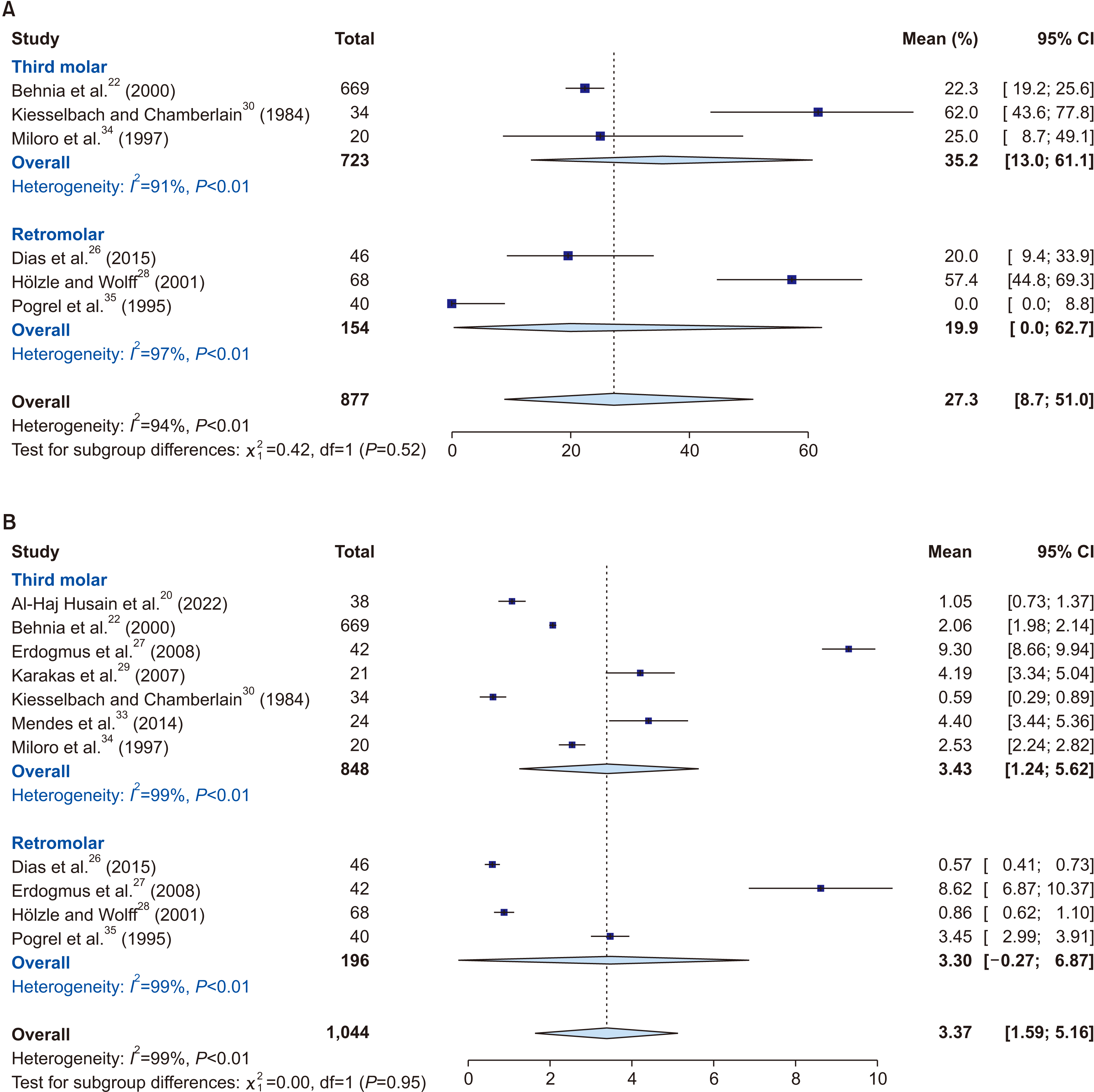
Fig. 4
Risk of bias (ROB) evaluated using the AQUA (Anatomical Quality Assessment) ROB tool, reported as the percentage per criterion.
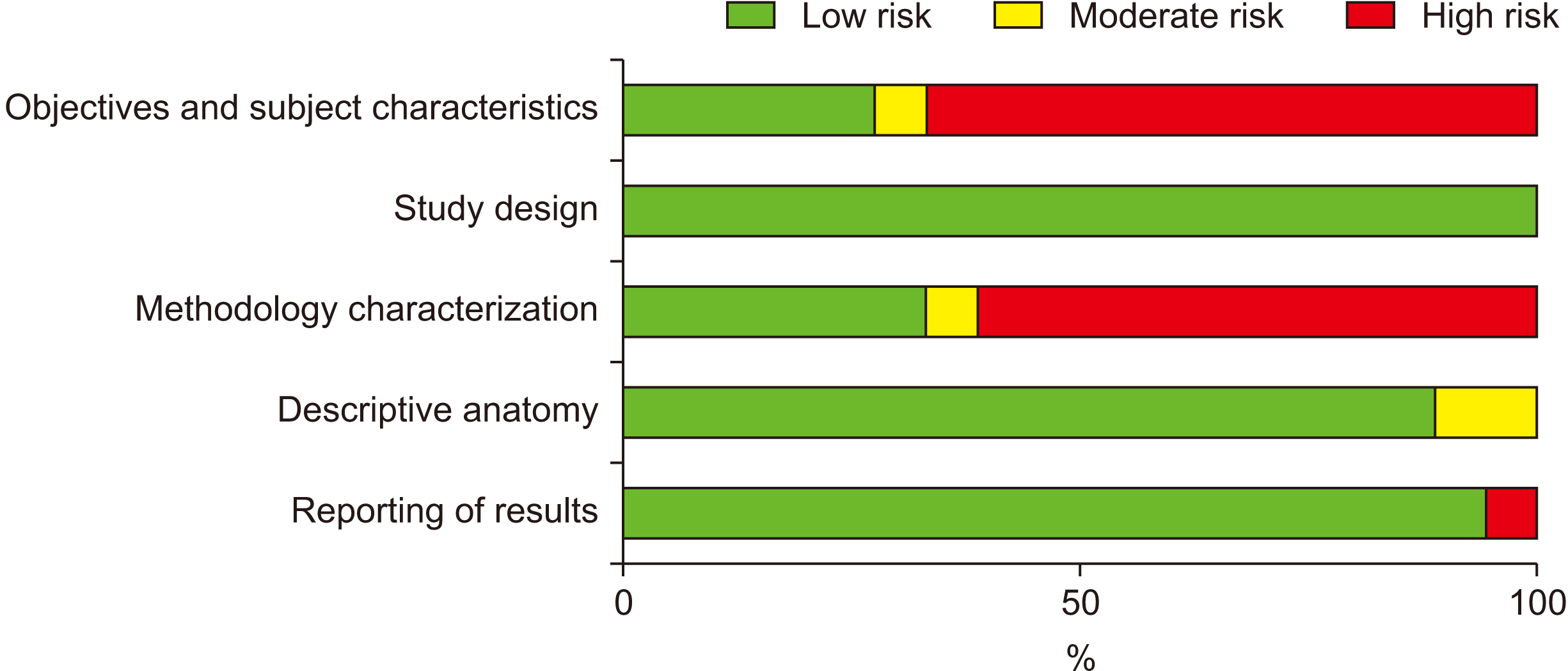
Fig. 5
Illustration of the lingual nerve’s course in relation to the alveolar crest at the retromolar and molar regions of the lingual surface of the mandible. Majority of the nerves is located inferior to the alveolar crest during its course. The red line (①) represents the lingual nerve’s mean vertical location with reference to the alveolar crest, while the red zone represents the 95% confidence intervals. The distance between this zone and the alveolar crest is represented by the green arrows. This distance is the safety margin to the possible location of the lingual nerve at each landmark. A minority of the lingual nerves would course above the alveolar crest at the retromolar (8.8%) and a partially erupted third molar (6.3%) regions as represented by the orange line (②).
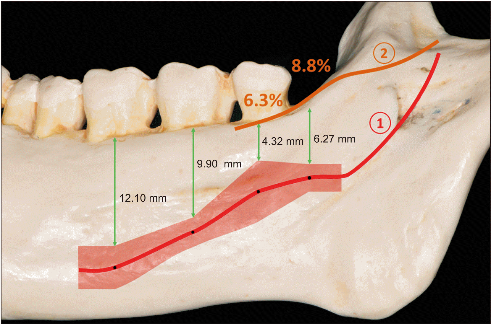
Table 1
Characteristics of the included studies
| Study | Methodology | Country | No. of subjects/cadavers | No. of nerves | Age (yr) | Sex (male/female), ethnicity | Presence of pathology |
|---|---|---|---|---|---|---|---|
| Al-Amery et al.19 (2016) | Cadaveric dissection | Malaysia | 7 | 14 | NR | 7/0, NR | No pathology, absence of previous surgery |
| Al-Haj Husain et al.20 (2022) | Clinical MRI | Switzerland | 19 | 38 | 30.5±13 | 6/13, NR | No pathology except partially erupted and impacted third molars |
| Aljamani et al.21 (2022) | Clinical MRI, electrical stimulation | United Kingdom | 50 | 96 | 24.1 (18-38) | 22/28, White British (28), Asian (17), Arabic (2), African (3) | No pathology (no neuropathy/pain) except partially erupted third molars |
| Behnia et al.22 (2000) | Cadaveric dissection | Iran | 430 | 669 | 25.2 (21-32) | 277/153, NR | No pathology, absence of dental surgery near third molar site |
| Benninger et al.23 (2013) | Cadaveric dissection | United States | 28 | 28 | 76 (44-89) | 14/14, NR | NR |
| Clinical ultrasonography | United States | 140 | 140 | 25 (22-41) | 74/66, NR | NR | |
| Bokindo et al.24 (2015) | Cadaveric dissection | Kenya | 30 | 30 | NR | NR, NR | No pathology |
| Chan et al.25 (2010) | Cadaveric dissection, CBCT with wire | United States | 18 | 30 | 70.2 (33-97) | 10/8, NR | NR |
| Dias et al.26 (2015) | Cadaveric dissection | New Zealand | 30 | 46 | 79 (52-100) | 23/23, White | No pathology |
| Erdogmus et al.27 (2008) | Cadaveric dissection | Turkey | 21 | 42 | NR | 21/0, Aegean | No pathology (no macroscopic pathology of the head) |
| Hölzle and Wolff28 (2001) | Cadaveric dissection | Germany | 34 | 68 | 78.82±7.63 | 15/19, NR | NR |
| Karakas et al.29 (2007) | Cadaveric dissection and radiographic imaging with metal wire on nerve | Turkey | 11 | 21 | NR, 52-98 | 5/6, NR | NR |
| Kiesselbach and Chamberlain30 (1984) | Cadaveric dissection | United States | 34 | 34 | NR | NR, NR | NR except impacted third molars |
| Intra-operative observation | NR | 256 | NR | NR, NR | NR | ||
| Kim et al.31 (2004) | Cadaveric dissection and radiographic imaging | Korea | 32 | 32 | NR, 20-94 | 23/9, Korean | NR |
| Kocabiyik et al.32 (2009) | Cadaveric dissection | Turkey | 13 | 26 | 65, NR | NR, NR | NR |
| Mendes et al.33 (2014) | Cadaveric dissection | Brazil | 24 | 24 | NR | NR, NR | NR |
| Miloro et al.34 (1997) | Clinical MRI | United States | 10 | 20 | 24.7 (22-35) | NR, NR | Absence of history of dental surgery |
| Pogrel et al.35 (1995) | Cadaveric dissection | United States | 20 | 40 | NR | NR, NR | NR |
| Shimoo et al.36 (2017) | Cadaveric dissection | Japan | 10 | 20 | NR | NR, NR | NR |
Table 2
Prevalence of the lingual nerve above the alveolar crest
| Study | No. of nerves | Reference point | Prevalence of lingual nerve above alveolar crest (%) |
|---|---|---|---|
| Retromolar region | |||
| Benninger et al.23 (2013) | 28 | Alveolar crest of lingual plate | 21.0 |
| Dias et al.26 (2015) | 46 | Alveolar crest of lingual plate | 0.0 |
| Hölzle and Wolff28 (2001) | 68 | Alveolar crest of lingual plate | 8.8 |
| Pogrel et al.35 (1995) | 40 | Lingual plate at retromolar pad region | 15.0 |
| Third molar region | |||
| Al-Amery et al.19 (2016) | 14 | Alveolar crest at mandibular third molar region | 0.0 |
| Behnia et al.22 (2000) | 669 | Lingual crest at mandibular third molar region | 14.1 |
| Bokindo et al.24 (2015) | 30 | Posterior point of alveolar crest at mandibular third molar region | 0.0 |
| Karakas et al.29 (2007) | 21 | Lingual crest of mandible at mandibular third molar region | 4.7 |
| Kiesselbach and Chamberlain30 (1984) | 34 | Alveolar crest at mandibular third molar region | 17.6 |
| 2561 | 12.0 | ||
| Kim et al.31 (2004) | 32 | Mandibular lingual plate | 0.0 |
| Miloro et al.34 (1997) | 202 | Lingual crest at mandibular third molar region | 10.0 |
Table 3
Prevalence of the lingual nerve contacting the lingual plate
| Study | No. of nerves | Reference point | Prevalence of lingual nerve contacting lingual plate (%) |
|---|---|---|---|
| Retromolar region | |||
| Dias et al.26 (2015) | 46 | Lingual plate at retromolar region | 20.0 |
| Hölzle and Wolff28 (2001) | 68 | Lingual plate at retromolar region | 57.4 |
| Pogrel et al.35 (1995) | 40 | Lingual plate at retromolar pad region | 0.0 |
| Third molar region | |||
| Behnia et al.22 (2000) | 669 | Lingual plate at alveolar crest of mandibular third molar region | 22.3 |
| Kiesselbach and Chamberlain30 (1984) | 34 | Lingual plate at mandibular third molar region | 62.0 |
| Miloro et al.34 (1997) | 201 | Lingual plate at mandibular third molar region | 25.0 |
Table 4
Vertical distance between the lingual nerve and the respective landmarks
| Study | No. of nerves | Landmark for measurements | Vertical distance (mm) |
|---|---|---|---|
| Retromolar region | |||
| Aljamani et al.21 (2021) | 96 | Retromolar pad | 9.64±2.98 |
| Dias et al.26 (2015) | 46 | Alveolar crest of lingual plate | 9.15±3.87 |
| Hölzle et al.28 (2001) | 68 | Alveolar crest at retromolar region | 7.83±1.65 |
| Kocabiyik et al.32 (2009) | 26 | Alveolar crest at retromolar region | 5.80±0.90 |
| Pogrel et al.35 (1995) | 40 | Alveolar crest of lingual plate | 8.32±4.05 |
| Third molar region | |||
| Al-Amery et al.19 (2016) | 14 | Alveolar ridge | 12.61±3.40 |
| Al-Haj Husain et al.20 (2022) | 381 | Alveolar crest of lingual cortical plate |
Right mandible: 4.87±1.20 Left mandible: 4.42±1.30 Overall: 4.65±1.20 |
| Aljamani et al.21 (2021) | 96 | Mid-point of attached gingiva of third molar | 10.77±2.76 |
| Behnia et al.22 (2000) | 669 | Lingual crest | 3.01±0.42 |
| Benninger et al.23 (2013) | 28 | Superior edge of alveolar bone at the posterior aspect of the third molar or at its extraction site | 7.30 (2.90-13.20) |
| Bokindo et al.24 (2015) | 30 | Most posterior point of the alveolar crest (representing the most distal portion of the third molar) | 10.30±5.20 |
| Erdogmus et al.27 (2008) | 42 | Medial edge of alveolar crest of third molar | 7.06±1.30 |
| Karakas et al.29 (2007) | 21 | Lingual crest of mandible | 9.56±5.28 |
| Kiesselbach and Chamberlain30 (1984) | 34 | Lingual plate at third molar region | 2.28±1.96 (7.00-2.00; below crest to above crest) |
| Kim et al.31 (2004) | 32 | Mesial and distal position of third molar |
Mesial position of third molar: 9.50 (5.10-16.10) Distal position of third molar: 15.00 (8.70-19.90) |
| Mendes et al.33 (2014) | 24 | Alveolar crest at third molar | 16.80±5.70 |
| Miloro et al.34 (1997) | 201 | Lingual crest | 2.75±0.97 |
| Shimoo et al.36 (2017) | 20 | Occlusal planes of mandibular third molar | 9.30±6.15 |
| Second molar region | |||
| Al-Amery et al.19 (2016) | 13 | Alveolar ridge | 11.46±2.98 |
| Aljamani et al.21 (2021) | 96 | Distolingual of attached gingiva of second molar | 12.34±3.16 |
| Chan et al.25 (2010) | 30 | Cementoenamel junctions at mid-lingual sites of second molar |
Right second molar: 9.50±3.90 Left second molar: 9.70±2.90 |
| Shimoo et al.36 (2017) | 20 | Occlusal planes of mandibular second molar | 17.90±5.26 |
| First molar region | |||
| Al-Amery et al.19 (2016) | 4 | Alveolar ridge | 14.38±4.35 |
| Chan et al.25 (2010) | 30 | Cementoenamel junctions at mid-lingual sites of first molar |
Right first molar: 12.70±3.70 Left first molar: 13.20±4.30 |
| Shimoo et al.36 (2017) | 20 | Occlusal planes of mandibular first molar | 25.20±4.42 |
Table 5
Horizontal distance between the lingual nerve and the respective landmarks
| Study | No. of nerves | Landmark for measurements | Horizontal distance (mm) |
|---|---|---|---|
| Retromolar region | |||
| Dias et al.26 (2015) | 46 | Alveolar crest of lingual plate | 0.57±0.56 |
| Erdogmus et al.27 (2008) | 42 | Rear medial edge of retromolar trigone | 8.62±5.80 |
| Hölzle et al.28 (2001) | 68 | Alveolar crest of lingual plate | 0.86±1.00 |
| Pogrel et al.35 (1995) | 40 | Alveolar crest of lingual plate | 3.45±1.48 |
| Third molar region | |||
| Al-Haj Husain et al.20 (2022) | 381 | Alveolar crest of lingual cortical plate |
Right mandible: 0.91±1.00 Left mandible: 1.18±1.10 Overall: 1.05±1.00 |
| Behnia et al.22 (2000) | 669 | Lingual plate at third molar | 2.06±1.10 |
| Erdogmus et al.27 (2008) | 42 | Medial edge of alveolar crest of third molar | 9.30±2.10 |
| Karakas et al.29 (2007) | 21 | Lingual crest of mandible | 4.19±1.99 |
| Kiesselbach and Chamberlain30 (1984) | 34 | Lingual plate at third molar region | 0.59±0.90 |
| Mendes et al.33 (2014) | 24 | Third molar socket | 4.40±2.40 |
| Miloro et al.34 (1997) | 201 | Lingual plate | 2.53±0.67 |


 PDF
PDF Citation
 Print
Print


 XML Download