Journal List > J Korean Med Sci > v.38(32) > 1516083528

Yang, Seo, Lee, Huh, Kim, Park, Kim, Shin, Shin, Park, Lee, and the Research Management Committee, Korean College of Helicobacter and Upper Gastrointestinal Research: Sample Collection Methods in Upper Gastrointestinal Research

Abstract

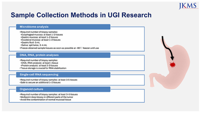
In recent years, significant translational research advances have been made in the upper gastrointestinal (GI) research field. Endoscopic evaluation is a reasonable option for acquiring upper GI tissue for research purposes because it has minimal risk and can be applied to unresectable gastric cancer. The optimal number of biopsy samples and sample storage is crucial and might influence results. Furthermore, the methods for sample acquisition can be applied differently according to the research purpose; however, there have been few reports on methods for sample collection from endoscopic biopsies. In this review, we suggested a protocol for collecting study samples for upper GI research, including microbiome, DNA, RNA, protein, single-cell RNA sequencing, and organoid culture, through a comprehensive literature review. For microbiome analysis, one or two pieces of biopsied material obtained using standard endoscopic forceps may be sufficient. Additionally, 5 mL of gastric fluid and 3–4 mL of saliva is recommended for microbiome analyses. At least one gastric biopsy tissue is necessary for most DNA or RNA analyses, while proteomics analysis may require at least 2–3 biopsy tissues. Single cell-RNA sequencing requires at least 3–5 tissues and additional 1–2 tissues, if possible. For successful organoid culture, multiple sampling is necessary to improve the quality of specimens.

Graphical Abstract

jkms-38-e255-abf001.jpg

INTRODUCTION

In the past few years, vast progress in basic science and technology has dramatically changed scientific perspectives by opening a new era of translational research.1 Translational research refers to the “bench-to-bedside” enterprise of harnessing knowledge from basic sciences to produce new drugs, devices, and treatment options for patients.23 In the last two decades, gastroenterology has become an area of dynamic translational research.1 Basic research requires an adequate number of clinical samples to verify the scientific hypothesis. To date, most tissue samples used for translational research are obtained from surgically resected specimens. However, most patients are not candidates for surgery in clinical practice; therefore, tissue samples from endoscopic biopsies can be applied more widely for gastrointestinal diseases. The quality, amount, and storage of the study sample are crucial for basic research and can differ according to the research purpose. Endoscopic biopsy is necessary to confirm a histopathologic diagnosis, and the optimal number of endoscopic biopsies is crucial for the diagnosis of gastrointestinal diseases, including gastric cancer.4 Previous reports have also shown that the number of biopsy fragments is significantly correlated with disease-specific biomarker status owing to intratumoral heterogeneity.56 However, there have been few reports on the optimal number of endoscopic biopsies or methods for sample storage in upper gastrointestinal research. In this review, we suggest protocols for collecting study samples for upper gastrointestinal research, including microbiome, DNA, RNA, protein, single-cell RNA sequencing (scRNA-seq), and organoid culture.

PROTOCOLS FOR COLLECTING STUDY SAMPLES

Upper gastrointestinal microbiome analysis

Fluid aspiration and mucosal biopsy

1. Fluid aspiration by endoscope7

  • • Clean and disinfect the upper gastrointestinal endoscope.

    • - Follow the standard sterilization protocol.

  • • Keep the participants fasting overnight.

  • • Ask the participants to rinse their mouths with bottled water to remove food debris.

  • • Simethicone prior to gastrointestinal endoscopy is not recommended.

  • • Insert the upper gastrointestinal endoscopy.

    • - Do not aspirate any fluid by endoscope prior to sample collection to avoid contaminating the endoscope channel with gastric or oropharyngeal secretion.

    • - Insert a disposable catheter through the working channel of the endoscope.

    • - Aspirate gastric or duodenal fluid through the catheter using a disposable syringe.

    • - Remove the syringe from the catheter and collect the fluid into a disposable sample container.

      • → No buffer fluid is required in a sample container.

    • - Transfer and store samples at −80°C freezer until use.

      • → Freeze samples as soon as possible.

    • - If unavoidable, exposure at room temperature for 1 hour or in a refrigerator for 4 hours may be allowed.

To collect microbiome samples from the upper gastrointestinal tract through the endoscope, it is important to collect samples as cleanly as possible, because the upper gastrointestinal tract has a relatively low biomass environment compared to the colorectum (see Chapter 2. Low biomass environment and 3. Contamination issues in the low biomass environment).8 Upper gastrointestinal endoscopes were disinfected according to standard sterilization protocols. The participants were instructed to fast overnight before an endoscopic examination. Before initiating the endoscopic examination, the participants were instructed to rinse their mouths with bottled water to remove food debris. In clinical practice, simethicone may be used before an endoscopic examination to improve gastric cleanliness at the clinician’s discretion. However, simethicone administration is not recommended when collecting gastric or duodenal fluid because it may dilute or even contaminate the gastric or duodenal fluid.
When all preparations were complete, an endoscope was inserted into the participant’s upper gastrointestinal tract according to the standard endoscopic examination method. However, no dirty fluid, including oral secretions and sputum, was aspirated before gastric or duodenal fluid aspiration to avoid contamination of the endoscope channel. After the endoscope was placed near the sample, a disposable catheter was inserted through the working channel. The gastric or duodenal fluid was then aspirated through the catheter using a disposable syringe under negative pressure. The syringe was then removed from the catheter, and the fluid was collected in a disposable sample container. Finally, samples were stored at −80°C as soon as possible until use.

2. Endoscopic mucosal biopsy7

  • • Clean and disinfect the upper gastrointestinal endoscope.

    • - Follow the standard sterilization protocol.

  • • Keep the participants fasting overnight.

  • • Ask the participants to rinse their mouths with bottled water to remove food debris.

  • • Simethicone prior to gastrointestinal endoscopy is not recommended.

  • • Insert the upper gastrointestinal endoscopy.

  • • Do not aspirate any fluid by endoscope prior to sample collection to avoid contaminating the endoscope channel with gastric or oropharyngeal secretion.

    • - Prepare blunt 18 Gauge syringe needle to prepare for procurement and a tissue container.

    • - Insert a disposable biopsy forceps into the working channel of the endoscope.

      • → Aseptic biopsy device (encased biopsy forceps) may be used.

    • - Take a biopsy of mucosa using a forceps.

    • - Use a blunt 18 Gauge syringe needle to extract biopsy sample from the forceps.

    • - Place the biopsy sample into a disposable sample container.

      • → No buffer fluid is required in a sample container.

    • - Transfer and store samples at −80°C freezer until use.

      • → Freeze samples as soon as possible.

      • → If unavoidable, exposure at room temperature for 1 hour or in a refrigerator for 4 hours may be allowed.

The protocol for collecting mucosal samples via biopsy was similar to that used for fluid collection. First, an upper gastrointestinal endoscope was prepared and disinfected according to a standard sterilization protocol. The participants were instructed to fast overnight before the endoscopic examination. To remove food debris from the oral cavity, participants were instructed to rinse their mouths with bottled water.
When inserting an endoscope into a participant’s upper gastrointestinal tract, aspiration of dirty fluid, including oral secretions and sputum, was avoided before the endoscopic biopsy. To collect the biopsied sample, the assistant prepared a needle (such as a blunt 18 Gauge syringe needle) before the biopsy. After placing the endoscope near the sample, the disposable biopsy forceps were inserted into the working channel. To reduce contamination, aseptic biopsy devices, such as encased biopsy forceps, may be used if available.9 After taking a biopsy of the mucosa, a sample was extracted from the forceps using the prepared needle. The biopsy samples were placed in disposable sample containers. Finally, samples were stored at −80°C as soon as possible until use.
Researchers may wash the gastric mucosa with water before mucosal biopsy to remove contaminants or colonizing microorganisms. However, the effect of washing before a biopsy has not been fully elucidated. A previous study on washed biopsies demonstrated that the number of cultured non-Helicobacter bacteria decreased in washed biopsies; however, there was no significant difference in bacterial diversity between unwashed and washed biopsies.3

3. Saliva1011

  • • Keep the participants fasting at least an hour prior to sample collection

  • • Ask the participants to rinse their mouths with bottled water to remove food debris (30–60 minutes before the sample collection)

  • • Collect saliva (spit twice, 3–4 mL) into a disposable sample container.

    • - A stabilization kit (e.g., OMNIgene collection kit) may be used.

  • • Transfer and store samples at −80°C freezer until use.

  • • After swabbing the mucosa, break the head of the swab off into a disposable sample container

    • - No buffer fluid is required in a sample container.

  • • Transfer and store samples at −80°C freezer until use.

    • - Freeze samples as soon as possible.

    • - If unavoidable, exposure at room temperature for 1 hour or in a refrigerator for 4 hours may be allowed.

The collection of saliva samples is relatively easy. First, participants were recommended to fast for at least 1 hour before sample collection to minimize contamination from food or beverages. Half an hour before sample collection, we recommended that participants rinse their mouths with bottled water to remove food debris. The sample was collected by making the participants spit twice into a sample container. Researchers may use a stabilization kit (e.g., OMNIgene collection kit) if available. 12 Then, samples were stored at −80°C as soon as possible until use.

4. Oropharyngeal swab13

  • • Keep the participants fasting at least an hour prior to sample collection

  • • Ask the participants to rinse their mouths with bottled water to remove food debris (30–60 minutes before the sample collection)

  • • Swab the mucosa at the posterior pharyngeal wall using a swab kit

  • • After swabbing the mucosa, break the head of the swab off into a disposable sample container

    • - No buffer fluid is required in a sample container.

  • • Transfer and store samples at −80°C freezer until use.

    • - Freeze samples as soon as possible.

    • - If unavoidable, exposure at room temperature for 1 hour or in a refrigerator for 4 hours may be allowed.

The swab method can also be used to collect oropharyngeal microbiome samples. Similar to saliva sample collection, participants were recommended to fast for at least 1 hour before sample collection. Additionally, the participants were advised to rinse their mouths with bottled water to remove food debris 30–60 minutes before sample collection. The mucosa was swabbed up to the posterior pharyngeal wall using a swab kit. After collecting the mucosal swab, the head was broken off into a disposable sample container. Then, samples were stored at −80°C as soon as possible until use.

Required number of samples for microbiome analysis

  • • Esophageal mucosal biopsy: at least 1–2 tissues

  • • Gastric mucosal biopsy: at least 1–2 tissues

  • • Duodenal mucosal biopsy: at least 1–2 tissues

  • • Gastric fluid: 5 mL

  • • Saliva: spit twice, 3–4 mL

For microbiome analysis, only a minimal amount of DNA is required, because amplification of the 16S (or 18S) rRNA gene through polymerase chain reaction (PCR) is performed before sequencing. In our experience, 100 ng of genomic DNA is sufficient for analyzing the upper gastrointestinal microbiome. Only one piece of biopsied material obtained using standard endoscopic forceps can satisfy the required amount of DNA. In case further experiments are needed, an additional piece can be collected if possible. Additionally, 5 mL of gastric fluid and 3–4 mL of saliva is recommended for microbiome analyses.

Other considerations

1. Performing experiments following sample collection

To maintain the sample quality, dissolving the frozen sample and freezing it again is not recommended. If many study samples and several experiments are needed, the same DNA extraction and amplification techniques across the experiments should be used to minimize the batch effect. In addition, it is necessary to perform 16S (or 18S) rRNA gene sequencing in a single run, if possible. If there were too many samples to run sequencing simultaneously, a positive control sample was used.

2. Low biomass environment

A low biomass environment indicates an environment in which a mass of living organisms is present. The low biomass environments include the blood,1415 placenta,16 meconium,17 airways,18 and milk.19 Although the stomach and duodenum have relatively higher biomass than the environments mentioned above, their biomass is significantly lower than that in the colon. The bacterial density is 101–103 colony-forming units (CFU)/mL in the stomach or duodenum, 104–107 CFU/mL in the jejunum or ileum, and 1011–1012 CFU/mL in the colon.8 The stomach and duodenum may be regarded as relatively low biomass environments.

3. Contamination issues in the low biomass environment

During microbiome analysis, contamination may occur from many sources, including participants’ or researchers’ bodies,2021 laboratory surfaces and air,222324 sample collection instruments, DNA extraction kits, and PCR reagents.142526 Such contamination is inevitable and more significant in samples from low-biomass environments. Various methods for eliminating microbial DNA contamination, including ultraviolet irradiation,27 restriction endonuclease digestion,272829 DNase treatment,2730 ultrafiltration to remove high-molecular-weight DNA,31 and treatment with DNA-intercalating products,1432 have been attempted. However, there is no practical way to physically remove contaminants in microbiome analysis.
Therefore, it is important to prepare negative controls. The preparation of at least one no-template control (NTC) for each batch (e.g., DNA extraction and amplification) is required. This NTC can be used for in silico removal of contaminants. Currently, the following in silico contaminant removal methods are available: 1) filtration based on the relative abundance in study samples,23333435 2) filtration based on the blacklist method, and 3) filtration based on the relative abundance of NTCs.2336 However, there is a risk of cross-contamination from study samples to NTCs (i.e., well-to-well effects).25373839 Therefore, the removal of taxa appearing in NTCs should be avoided. In addition, randomization of the study samples and NTCs across the plate was required during the experiments, including DNA extraction, amplification, and sequencing. Computational algorithms (e.g., the decontam package in R) may also be used to adjust for contamination.4041

DNA, RNA, and protein analyses

Required number of endoscopy biopsy samples

1. DNA sample requirement

  • • PCR and quantitative PCR analysis: at least one biopsy tissue

  • • Whole genome sequencing analysis: at least one biopsy tissue

One gastric biopsy tissue is usually sufficient for PCR and quantitative PCR analysis.42 In previous studies, 0.5–1 µg genomic DNA was converted into 40 µL bisulfite modified DNA, and 1–2 µL was used for one reaction of methylation-specific PCR or quantitative methylation-specific PCR.434445 This means that 1 µg genomic DNA is sufficient for 20–40 methylation-specific PCR reactions. In other studies with quantitative methylation-specific PCR, the starting amount of genomic DNA was 1 µg.424647484950 From one gastric biopsy tissue, approximately 20–30 µg of DNA is usually extracted. Therefore, if four reactions were required in duplicate wells for each unknown sample of methylated and unmethylated sequences, one gastric biopsy tissue sample would ideally be sufficient to measure more than 25 target genes.
For whole genome sequencing analysis, genomic DNA of > 1.0–1.5 µg with a concentration of > 20 ng/µL is usually recommended and a minimum of > 100–500 ng of DNA is required. In a previous study of whole genome sequencing analysis, 0.2 µg of blood DNA and 1 µg of saliva DNA showed little difference compared with 3 µg of blood DNA on the quality of sequencing and variant calling.51 Therefore, at least one gastric biopsy tissue is required for whole genome sequencing analysis. While one biopsy tissue can often fulfill the requirements for most DNA analyses, the collection of additional tissues becomes valuable in situations where the size or quality of the initial tissue is inadequate or when supplementary experiments need to be conducted.

2. RNA sample requirement

  • • Reverse transcriptase (RT)-PCR and quantitative RT-PCR analysis: at least one biopsy tissue

  • • RNA sequencing analysis: at least one biopsy tissue

One gastric biopsy tissue is also usually sufficient for RT-PCR and quantitative RT-PCR analysis when RNA is extracted from samples collected in the RNA-stabilizing reagent using the TRIzol method. The median amount of RNA extracted from one esophageal, gastroduodenal, and colonic biopsy tissue was 3.1 µg, 4.5 µg, and 4.0–9.5 µg, respectively.52535455 Total RNA of 1 µg is used for the synthesis of 50 µL of cDNA. Because 2 µL of cDNA (0.04 µg total RNA) per well is required for quantitative PCR, a total of 8 µL of cDNA (2 × 2 µL for the target gene and 2 × 2 µL for the housekeeping gene as an internal control) is required to measure in duplicate wells for each unknown sample. Thus, one gastric biopsy tissue sample was sufficient for the quantification of up to 25 templates.
For RNA sequencing analysis, the input amounts of total RNA recommended by the manufacturers range from 100 ng–1 µg.56 However, in a previous study, the tested protocols performed well for low-quantity inputs down to 5 ng.57 Therefore, at least one gastric biopsy tissue is required for RNA sequencing analysis. Collecting multiple biopsy tissue samples can be beneficial in many RNA analyses, similar to DNA analyses. In particular, RNA sequencing can be influenced by the quality of RNA. Hence, it is advisable to monitor the yield and quality of extracted RNA during the collection process to ensure an adequate number of samples are obtained.

3. Protein sample requirement

  • • Proteomics analysis: at least 2–3 biopsy tissues may be needed

At least 2–3 gastric biopsy tissue samples may be required for proteomic analysis. In a previous study, 30–50 µg of protein was extracted from an average of two colonic tissues obtained from colonoscopic or sigmoidoscopic biopsy, which was sufficient for proteomics analysis.58 In another study, an average of 70 µg of protein was extracted from two or more (average of three) colonic tissue biopsies for proteomic analysis.59 One study reported that at least 100 µg of protein could be obtained from one gastric biopsy tissue. A comprehensive phosphoproteomic analysis could be conducted from a minimum of 320 µg protein (three endoscopic biopsy tissues) by identifying more than 10,000 class 1 phosphosites.60 Therefore, further validation is required to determine the required number of endoscopic biopsy samples for proteomics analysis.

Tissue collection and storage (especially for RNA)

1. Tissue collection and RNA stabilization5361

  • • One of the following methods are recommended

    • - Process immediately after harvest

    • - Snap freeze in liquid nitrogen

    • - Stabilize in RNA stabilizing reagents (e.g., RNAlater) to inhibit RNase activity

It is practically impossible to extract RNA immediately after sample collection using an endoscopic biopsy. Immediate processing is typically performed for RNA extraction from cultured cells. In many studies, endoscopic biopsy samples are snap-frozen in liquid nitrogen until storage in the vapor phase of liquid nitrogen (VPLN) or a mechanical freezer. In situations where snap-freezing is not possible immediately after endoscopic biopsy, tissue samples can be stabilized by immersion in RNA-stabilizing reagents (e.g., RNAlater) to inhibit RNase activity. Sample storage in RNAlater has a minimal impact on the quality of protein quantification compared with fresh frozen tissue.62

2. Long-term storage

  • • One of the following storage options can be chosen

    • - VPLN, −150°C

    • - Mechanical freezer at −80°C

  • • Storage duration: Storage at −80°C or in VPLN may not affect RNA and DNA quality up to 10 years

Storage of samples in VPLN is an ideal option. The theoretical background for storage in VPLN (at −150°C) is that aqueous-based chemical reactions are thought to cease at the glass transition temperature (−108°C to −137°C).63 At −80°C, enzymatic reactions may continue though cells do not remain viable. In previous studies, there was no time-dependent decrease in the RNA quality in the fresh-frozen tissue samples stored in VPLN for 2–11 years64 or in the fresh-frozen gastric cancer tissue samples stored at −80°C for 2–12 years.65 In a comparative study of 49 specimens stored for 5–12 years, there was no decrease in the RNA quality stored at −80°C compared with those stored in VPLN.63 However, in another study of 16 pairs of parallel samples stored for more than 10 years (10.0–12.2 years), the RNA quality was significantly lower in the samples stored at −80°C than those stored in VPLN although there was no difference in the DNA quality.66 Therefore, the long-term storage of endoscopic biopsy tissues at −80°C or in VPLN may not affect RNA and DNA quality for up to 10 years. However, further validation studies are required to evaluate the RNA quality in samples stored for more than 10 years at −80°C because of conflicting study results.

scRNA-seq

Required number of endoscopy biopsy samples

  • • scRNA-seq analysis: at least 3–5 biopsy tissues

  • • It is safe to secure an additional 1–2 tissues

Sample requirement

Preparing high-quality single-cell suspensions is essential for successful single-cell studies. All specimens were obtained from one or two sites using conventional upper gastrointestinal endoscopy with routine forceps. In most published studies,67686970 freshly biopsied tumor samples and adjacent normal tissue samples were obtained. To preserve single-cell transcriptomes before scRNA-seq, it is recommended to proceed immediately from single-cell isolation to cell lysis and mRNA capture. 717273 Although the required number of cells increases with the complexity of the sample under investigation, scRNA-seq generally requires at least 50,000 cells. There is no standard number of biopsy tissues for scRNA-seq method. Since the sample requirements of esophagus and stomach are different, and it is affected by the dissociation method or process of the laboratory or analysis room, it is recommended to collect at least 3-5 biopsy tissues. During the evaluation of tissue quality, such as tumor content, necrosis, fat, and hemorrhage, some tissues may be unsuitable for examination; therefore, it is safe to secure an additional 1–2 pieces. Because scRNA-seq requires the use of fresh live cells, special care is required. Therefore, it is desirable to start with sufficient tissues, especially when unfamiliar with scRNA-seq, because the final number of cells obtained may be reduced depending on the number of endoscopic biopsies and the tissue processing. Considering various limitations, although it is recommended to collect a total of 5-7 or more tissues at one time, it is advisable to monitor the yield and quality of extracted scRNA during the collection process to ensure an appropriate number of samples.

Tissue collection and preparation

  • • One of the following methods is recommended:

    • - Fresh viable single cells

    • - Preserved samples

    • - Nuclear RNA from frozen tissue

All common scRNA-seq methods were designed to use fresh viable cells, and this method has been established. Collection of fresh tumor tissue should be performed to reduce the time elapsed between the removal of fresh tissue from the body, placement of the specimen in media, and processing for scRNA-seq. However, immediate sample processing can be difficult in real research and clinical practice because of the lack of essential infrastructure or specialized equipment, such as fluorescence-activated cell sorting devices. Although most methods use fresh viable cells, preserved samples,747576 and nuclear RNA from frozen tissue77787980 can be used as an alternative. Among the preserved samples, cryopreservation is relatively well-established for scRNA-seq.

1. Fresh samples67686970

  • 1) Fresh tissues are collected in plain Roswell Park Memorial Institute (RPMI) medium on ice immediately after resection.

  • 2) Tissues are dissected with iris scissors into small pieces. Approximately 0.5–1.5 cm3 of tissue from each resection is used.

  • 3) Tissues are digested for 30 minutes at 37°C with a digestion solution containing phosphate-buffered saline (PBS) and collagenase II and IV.

  • 4) The cell suspension is further filtered using a 45 µm or 70 µm filter to remove cell aggregates and resuspended in PBS with 10% fetal bovine serum (FBS).

2. Preserved samples (cryopreservation)747576

  • 1) The sample is minced on ice, placed into a freezing solution (10% dimethyl sulfoxide [DMSO], 90% non-inactivated FBS), and frozen by gradually decreasing the temperature (1°C/min) to −80°C (cryopreserved).

  • 2) After storage for one week at −80°C, the sample is rapidly thawed in a water bath in continuous agitation and placed into 25 mL of cold 1× HBSS.

  • 3) For single-cell separation the fresh and conserved samples are minced on ice and enzymatically digested in 5 mL 1× HBSS and 83 µL collagenase IV (10,000 U/mL) for 10 minutes at 37°C.

3. Nuclear RNA from frozen tissue77787980

  • 1) Whole cells are dissociated.

  • 2) The sample is excised and immediately placed into a nuclei isolation medium.

  • 3) Tissue is Dounce homogenized, allowing for the mechanical separation of nuclei from cells.

  • 4) Samples are washed, resuspended in nuclei storage buffer, and filtered. Solutions and samples are kept cold.

Gastric organoid culture

Required number of endoscopy biopsy samples

  • • Required number of biopsy samples: at least 3–5 tissues

  • • Multipoint deep biopsy in different parts of the tumor

  • • Avoid the contamination of normal mucosal tissue

To date, almost all gastric cancer organoid studies have been conducted using surgical specimens. There are few studies on gastric cancer organoids with endoscopic biopsy specimens.818283 Compared with surgical specimens, the size of the tumor tissue obtained by endoscopic biopsy is small, and the inflammatory reaction may affect the tissue; thus, it determines successful organoid culture. There is no standard number of biopsies for organoid culture in patients with suspected gastric cancer undergoing endoscopy. Multiple sampling and larger-diameter biopsy forceps are more conducive to improving the quality of specimens. Additionally, it should be combined with high-definition or magnifying endoscopy for an intensive examination to improve the accuracy of tumor detection. Tumor biopsy should avoid normal mucosal tissue, preferring the center of the tumor and increasing the number of biopsies. For successful organoid culture, tissue samples require high activity, stem cells, sufficient tissue, and proper tumor cell quality. Organoid construction faces great challenges if the number of biopsy samples, living cells, or positive cells is low.

Primary organoid culture and maintenance 8485868788

1. Primary organoid culture

  • 1) Biopsied tissues were washed three times with advanced Dulbecco’s Modified Eagle Medium (DMEM)/F12 supplemented with antibiotics/antifungal (Primocin®; InvivoGen, San Diego, CA, USA) and chopped with sterile blades.

  • 2) The specimen is chopped and minced and incubated in digestion buffer (PBS containing 2.0 mg/mL collagenase type I, 1× penicillin/streptomycin, and 100 ug/mL Primocin®) at 37°C for 1 hour. The digestion time can be extended overnight, depending on the tissue stiffness and amount.

  • 3) The digestion is stopped with advanced DMEM/F12. The suspension was strained over a 100 µm filter to remove any debris.

  • 4) After centrifugation at 500 relative centrifugal force (rcf), the supernatants are discarded. Isolated cells are resuspended in ice-cold 500 µL matrigel and 20 µL of matrigel-cell mixture are sowed in a pre-warmed 48-well culture plate.

  • 5) The matrigel is solidified for 10 minutes at 37°C and overlaid with 250 µL organoid culture medium. Y-27632 (10 µM) is added for the first 2 days to prevent cell death (anoikis). The culture is kept at 37°C, 5% CO2 in a humidified incubator.

2. Organoid subculture

  • 1) Culture medium is exchanged every 2–3 days, and organoids were passaged every 1–2 weeks at a ratio of 1:4–1:8 according to the growth rate and density of the organoids.

  • 2) For passaging, organoids in matrigel were harvested with culture medium and then transferred to a new 1.7 mL tube.

  • 3) Organoids are mechanically dissociated by vigorous pipetting or 1 mL syringe flushing.

  • 4) The dissociated organoids are centrifuged at 500 rcf for 5 minutes.

  • 5) The supernatant is discarded, the pellet resuspended in ice-cold Matrigel, and re-seeded as described above.

CONCLUSION

In summary, we reviewed the methods for collecting study samples for upper GI research, including microbiome, DNA, RNA, protein, scRNA-seq, and organoid culture. For microbiome analysis, one or two pieces of biopsied material obtained using standard endoscopic forceps may be sufficient. Additionally, 5 mL of gastric fluid and 3–4 mL of saliva is recommended for microbiome analyses. The low-biomass environment in the stomach and duodenum is another consideration for microbiome analysis. At least one gastric biopsy tissue is necessary for most DNA or RNA analyses, while proteomics analysis may require at least 2–3 biopsy tissues. Tissue storage is crucial for RNA stabilization. In the scRNA-seq, it is recommended to collect at least 3–5 biopsy tissues, and safe to secure additional 1–2 tissues. For successful organoid culture, tissue samples require high activity, stem cells, sufficient tissue, and proper tumor cell quality. Further validated studies on sample acquisition may be required in the future.

Notes

Disclosure: The authors have no potential conflicts of interest to disclose.

Author Contributions:

  • Conceptualization: Yang HJ, Seo SI, Park CH, Lee SK.

  • Investigation: Lee SK.

  • Methodology: Yang HJ, Seo SI.

  • Project administration: Lee SK.

  • Supervision: Park CH, Lee SK.

  • Validation: Kim H, Shin H, Shin CM.

  • Writing - original draft: Yang HJ, Seo SI, Lee J, Huh CW, Kim JS, Park JC, Park CH, Lee SK.

  • Writing - review & editing: Kim H, Shin H, Shin CM, Park CH, Lee SK.

References

1. Di Sabatino A, Moschetta A, Conte D, Tiribelli C, Caprioli FA, et al. Translational Committee of the Italian Society of Gastroenterology. The impact of translational research on gastroenterology. Dig Liver Dis. 2014; 46(4):293–294. PMID: 24508099.
2. Zerhouni EA. Translational and clinical science--time for a new vision. N Engl J Med. 2005; 353(15):1621–1623. PMID: 16221788.
3. Woolf SH. The meaning of translational research and why it matters. JAMA. 2008; 299(2):211–213. PMID: 18182604.
4. Choi Y, Choi HS, Jeon WK, Kim BI, Park DI, Cho YK, et al. Optimal number of endoscopic biopsies in diagnosis of advanced gastric and colorectal cancer. J Korean Med Sci. 2012; 27(1):36–39. PMID: 22219611.
5. Ahn S, Ahn S, Van Vrancken M, Lee M, Ha SY, Lee H, et al. Ideal number of biopsy tumor fragments for predicting HER2 status in gastric carcinoma resection specimens. Oncotarget. 2015; 6(35):38372–38380. PMID: 26460823.
6. Xu C, Liu Y, Ge X, Jiang D, Zhang Y, Ji Y, et al. Tumor containing fragment number influences immunohistochemistry positive rate of HER2 in biopsy specimens of gastric cancer. Diagn Pathol. 2017; 12(1):41. PMID: 28549444.
7. Dreskin BW, Luu K, Dong TS, Benhammou J, Lagishetty V, Vu J, et al. Specimen collection and analysis of the duodenal microbiome. J Vis Exp. 2021; 167(167):61900.
8. O’Hara AM, Shanahan F. The gut flora as a forgotten organ. EMBO Rep. 2006; 7(7):688–693. PMID: 16819463.
9. Shanahan ER, Zhong L, Talley NJ, Morrison M, Holtmann G. Characterisation of the gastrointestinal mucosa-associated microbiota: a novel technique to prevent cross-contamination during endoscopic procedures. Aliment Pharmacol Ther. 2016; 43(11):1186–1196. PMID: 27086880.
10. Lim Y, Totsika M, Morrison M, Punyadeera C. The saliva microbiome profiles are minimally affected by collection method or DNA extraction protocols. Sci Rep. 2017; 7(1):8523. PMID: 28819242.
11. Ji Y, Liang X, Lu H. Analysis of by high-throughput sequencing: Helicobacter pylori infection and salivary microbiome. BMC Oral Health. 2020; 20(1):84. PMID: 32197614.
12. Luo T, Srinivasan U, Ramadugu K, Shedden KA, Neiswanger K, Trumble E, et al. Effects of specimen collection methodologies and storage conditions on the short-term stability of oral microbiome taxonomy. Appl Environ Microbiol. 2016; 82(18):5519–5529. PMID: 27371581.
13. Liu AQ, Vogtmann E, Shao DT, Abnet CC, Dou HY, Qin Y, et al. A comparison of biopsy and mucosal swab specimens for examining the microbiota of upper gastrointestinal carcinoma. Cancer Epidemiol Biomarkers Prev. 2019; 28(12):2030–2037. PMID: 31519703.
14. Glassing A, Dowd SE, Galandiuk S, Davis B, Chiodini RJ. Inherent bacterial DNA contamination of extraction and sequencing reagents may affect interpretation of microbiota in low bacterial biomass samples. Gut Pathog. 2016; 8(1):24. PMID: 27239228.
15. Peters RP, Mohammadi T, Vandenbroucke-Grauls CM, Danner SA, van Agtmael MA, Savelkoul PH. Detection of bacterial DNA in blood samples from febrile patients: underestimated infection or emerging contamination? FEMS Immunol Med Microbiol. 2004; 42(2):249–253. PMID: 15364111.
16. Lauder AP, Roche AM, Sherrill-Mix S, Bailey A, Laughlin AL, Bittinger K, et al. Comparison of placenta samples with contamination controls does not provide evidence for a distinct placenta microbiota. Microbiome. 2016; 4(1):29. PMID: 27338728.
17. Stinson LF, Keelan JA, Payne MS. Comparison of meconium DNA extraction methods for use in microbiome studies. Front Microbiol. 2018; 9:270. PMID: 29515550.
18. Drengenes C, Wiker HG, Kalananthan T, Nordeide E, Eagan TM, Nielsen R. Laboratory contamination in airway microbiome studies. BMC Microbiol. 2019; 19(1):187. PMID: 31412780.
19. Dahlberg J, Sun L, Persson Waller K, Östensson K, McGuire M, Agenäs S, et al. Microbiota data from low biomass milk samples is markedly affected by laboratory and reagent contamination. PLoS One. 2019; 14(6):e0218257. PMID: 31194836.
20. Kitchin PA, Szotyori Z, Fromholc C, Almond N. Avoidance of PCR false positives. Nature. 1990; 344(6263):201. PMID: 2156164.
21. Meadow JF, Altrichter AE, Bateman AC, Stenson J, Brown GZ, Green JL, et al. Humans differ in their personal microbial cloud. PeerJ. 2015; 3:e1258. PMID: 26417541.
22. Adams RI, Bateman AC, Bik HM, Meadow JF. Microbiota of the indoor environment: a meta-analysis. Microbiome. 2015; 3(1):49. PMID: 26459172.
23. Bittinger K, Charlson ES, Loy E, Shirley DJ, Haas AR, Laughlin A, et al. Improved characterization of medically relevant fungi in the human respiratory tract using next-generation sequencing. Genome Biol. 2014; 15(10):487. PMID: 25344286.
24. Knights D, Kuczynski J, Charlson ES, Zaneveld J, Mozer MC, Collman RG, et al. Bayesian community-wide culture-independent microbial source tracking. Nat Methods. 2011; 8(9):761–763. PMID: 21765408.
25. Jousselin E, Clamens AL, Galan M, Bernard M, Maman S, Gschloessl B, et al. Assessment of a 16S rRNA amplicon Illumina sequencing procedure for studying the microbiome of a symbiont-rich aphid genus. Mol Ecol Resour. 2016; 16(3):628–640. PMID: 26458227.
26. Salter SJ, Cox MJ, Turek EM, Calus ST, Cookson WO, Moffatt MF, et al. Reagent and laboratory contamination can critically impact sequence-based microbiome analyses. BMC Biol. 2014; 12(1):87. PMID: 25387460.
27. Corless CE, Guiver M, Borrow R, Edwards-Jones V, Kaczmarski EB, Fox AJ. Contamination and sensitivity issues with a real-time universal 16S rRNA PCR. J Clin Microbiol. 2000; 38(5):1747–1752. PMID: 10790092.
28. Sharma JK, Gopalkrishna V, Das BC. A simple method for elimination of unspecific amplifications in polymerase chain reaction. Nucleic Acids Res. 1992; 20(22):6117–6118. PMID: 1334263.
29. Carroll NM, Adamson P, Okhravi N. Elimination of bacterial DNA from Taq DNA polymerases by restriction endonuclease digestion. J Clin Microbiol. 1999; 37(10):3402–3404. PMID: 10488219.
30. Hilali F, Saulnier P, Chachaty E, Andremont A. Decontamination of polymerase chain reaction reagents for detection of low concentrations of 16S rRNA genes. Mol Biotechnol. 1997; 7(3):207–216. PMID: 9219235.
31. Wages JM Jr, Cai D, Fowler AK. Removal of contaminating DNA from PCR reagents by ultrafiltration. Biotechniques. 1994; 16(6):1014–1017. PMID: 8074862.
32. Hein I, Schneeweiss W, Stanek C, Wagner M. Ethidium monoazide and propidium monoazide for elimination of unspecific DNA background in quantitative universal real-time PCR. J Microbiol Methods. 2007; 71(3):336–339. PMID: 17936386.
33. Flores GE, Henley JB, Fierer N. A direct PCR approach to accelerate analyses of human-associated microbial communities. PLoS One. 2012; 7(9):e44563. PMID: 22962617.
34. Willner D, Daly J, Whiley D, Grimwood K, Wainwright CE, Hugenholtz P. Comparison of DNA extraction methods for microbial community profiling with an application to pediatric bronchoalveolar lavage samples. PLoS One. 2012; 7(4):e34605. PMID: 22514642.
35. Lazarevic V, Gaïa N, Girard M, Schrenzel J. Decontamination of 16S rRNA gene amplicon sequence datasets based on bacterial load assessment by qPCR. BMC Microbiol. 2016; 16(1):73. PMID: 27107811.
36. Nejman D, Livyatan I, Fuks G, Gavert N, Zwang Y, Geller LT, et al. The human tumor microbiome is composed of tumor type-specific intracellular bacteria. Science. 2020; 368(6494):973–980. PMID: 32467386.
37. Callahan BJ, DiGiulio DB, Goltsman DS, Sun CL, Costello EK, Jeganathan P, et al. Replication and refinement of a vaginal microbial signature of preterm birth in two racially distinct cohorts of US women. Proc Natl Acad Sci U S A. 2017; 114(37):9966–9971. PMID: 28847941.
38. Larsson AJ, Stanley G, Sinha R, Weissman IL, Sandberg R. Computational correction of index switching in multiplexed sequencing libraries. Nat Methods. 2018; 15(5):305–307. PMID: 29702636.
39. Minich JJ, Sanders JG, Amir A, Humphrey G, Gilbert JA, Knight R. Quantifying and understanding well-to-well contamination in microbiome research. mSystems. 2019; 4(4):e00186-19.
40. Davis NM, Proctor DM, Holmes SP, Relman DA, Callahan BJ. Simple statistical identification and removal of contaminant sequences in marker-gene and metagenomics data. Microbiome. 2018; 6(1):226. PMID: 30558668.
41. Claassen-Weitz S, Gardner-Lubbe S, Mwaikono KS, du Toit E, Zar HJ, Nicol MP. Optimizing 16S rRNA gene profile analysis from low biomass nasopharyngeal and induced sputum specimens. BMC Microbiol. 2020; 20(1):113. PMID: 32397992.
42. Yang HJ, Kim SG, Lim JH, Choi JM, Kim WH, Jung HC. Helicobacter pylori-induced modulation of the promoter methylation of Wnt antagonist genes in gastric carcinogenesis. Gastric Cancer. 2018; 21(2):237–248. PMID: 28643146.
43. Maekita T, Nakazawa K, Mihara M, Nakajima T, Yanaoka K, Iguchi M, et al. High levels of aberrant DNA methylation in Helicobacter pylori-infected gastric mucosae and its possible association with gastric cancer risk. Clin Cancer Res. 2006; 12(3 Pt 1):989–995. PMID: 16467114.
44. Nanjo S, Asada K, Yamashita S, Nakajima T, Nakazawa K, Maekita T, et al. Identification of gastric cancer risk markers that are informative in individuals with past H. pylori infection. Gastric Cancer. 2012; 15(4):382–388. PMID: 22237657.
45. Ando T, Yoshida T, Enomoto S, Asada K, Tatematsu M, Ichinose M, et al. DNA methylation of microRNA genes in gastric mucosae of gastric cancer patients: its possible involvement in the formation of epigenetic field defect. Int J Cancer. 2009; 124(10):2367–2374. PMID: 19165869.
46. Kim HJ, Kim N, Kim HW, Park JH, Shin CM, Lee DH. Promising aberrant DNA methylation marker to predict gastric cancer development in individuals with family history and long-term effects of H. pylori eradication on DNA methylation. Gastric Cancer. 2021; 24(2):302–313. PMID: 32915372.
47. Asada K, Nakajima T, Shimazu T, Yamamichi N, Maekita T, Yokoi C, et al. Demonstration of the usefulness of epigenetic cancer risk prediction by a multicentre prospective cohort study. Gut. 2015; 64(3):388–396. PMID: 25379950.
48. Shin CM, Kim N, Lee HS, Park JH, Ahn S, Kang GH, et al. Changes in aberrant DNA methylation after Helicobacter pylori eradication: a long-term follow-up study. Int J Cancer. 2013; 133(9):2034–2042. PMID: 23595635.
49. Shin CM, Kim N, Jung Y, Park JH, Kang GH, Park WY, et al. Genome-wide DNA methylation profiles in noncancerous gastric mucosae with regard to Helicobacter pylori infection and the presence of gastric cancer. Helicobacter. 2011; 16(3):179–188. PMID: 21585603.
50. Shin CM, Kim N, Jung Y, Park JH, Kang GH, Kim JS, et al. Role of Helicobacter pylori infection in aberrant DNA methylation along multistep gastric carcinogenesis. Cancer Sci. 2010; 101(6):1337–1346. PMID: 20345486.
51. Zhu Q, Hu Q, Shepherd L, Wang J, Wei L, Morrison CD, et al. The impact of DNA input amount and DNA source on the performance of whole-exome sequencing in cancer epidemiology. Cancer Epidemiol Biomarkers Prev. 2015; 24(8):1207–1213. PMID: 25990554.
52. Wex T, Treiber G, Lendeckel U, Malfertheiner P. A two-step method for the extraction of high-quality RNA from endoscopic biopsies. Clin Chem Lab Med. 2003; 41(8):1033–1037. PMID: 12964810.
53. Cui G, Olsen T, Christiansen I, Vonen B, Florholmen J, Goll R. Improvement of real-time polymerase chain reaction for quantifying TNF-alpha mRNA expression in inflamed colorectal mucosa: an approach to optimize procedures for clinical use. Scand J Clin Lab Invest. 2006; 66(3):249–259. PMID: 16714253.
54. Moen AE, Tannæs TM, Vatn S, Ricanek P, Vatn MH, Jahnsen J, et al. Simultaneous purification of DNA and RNA from microbiota in a single colonic mucosal biopsy. BMC Res Notes. 2016; 9(1):328. PMID: 27352784.
55. Stiekema J, Cats A, Boot H, Langers AM, Balague Ponz O, van Velthuysen ML, et al. Biobanking of fresh-frozen endoscopic biopsy specimens from esophageal adenocarcinoma. Dis Esophagus. 2016; 29(8):1100–1106. PMID: 26541751.
56. Chao HP, Chen Y, Takata Y, Tomida MW, Lin K, Kirk JS, et al. Systematic evaluation of RNA-Seq preparation protocol performance. BMC Genomics. 2019; 20(1):571. PMID: 31296163.
57. Schuierer S, Carbone W, Knehr J, Petitjean V, Fernandez A, Sultan M, et al. A comprehensive assessment of RNA-seq protocols for degraded and low-quantity samples. BMC Genomics. 2017; 18(1):442. PMID: 28583074.
58. Liu X, Xu Y, Meng Q, Zheng Q, Wu J, Wang C, et al. Proteomic analysis of minute amount of colonic biopsies by enteroscopy sampling. Biochem Biophys Res Commun. 2016; 476(4):286–292. PMID: 27230957.
59. Saleem S, Tariq S, Aleem I, Sadr-Ul Shaheed , Tahseen M, Atiq A, et al. Proteomics analysis of colon cancer progression. Clin Proteomics. 2019; 16(1):44. PMID: 31889941.
60. Abe Y, Hirano H, Shoji H, Tada A, Isoyama J, Kakudo A, et al. Comprehensive characterization of the phosphoproteome of gastric cancer from endoscopic biopsy specimens. Theranostics. 2020; 10(5):2115–2129. PMID: 32089736.
61. Wang M, Ji X, Wang B, Li Q, Zhou J. Simultaneous evaluation of the preservative effect of RNAlater on different tissues by biomolecular and histological analysis. Biopreserv Biobank. 2018; 16(6):426–433. PMID: 30484701.
62. Bennike TB, Kastaniegaard K, Padurariu S, Gaihede M, Birkelund S, Andersen V, et al. Comparing the proteome of snap frozen, RNAlater preserved, and formalin-fixed paraffin-embedded human tissue samples. EuPA Open Proteom. 2015; 10:9–18. PMID: 29900094.
63. Auer H, Mobley JA, Ayers LW, Bowen J, Chuaqui RF, Johnson LA, et al. The effects of frozen tissue storage conditions on the integrity of RNA and protein. Biotech Histochem. 2014; 89(7):518–528. PMID: 24799092.
64. Kelly R, Albert M, de Ladurantaye M, Moore M, Dokun O, Bartlett JM. RNA and DNA Integrity remain stable in frozen tissue after long-term storage at cryogenic temperatures: a report from the Ontario Tumour Bank. Biopreserv Biobank. 2019; 17(4):282–287. PMID: 30762427.
65. Zhang X, Han QY, Zhao ZS, Zhang JG, Zhou WJ, Lin A. Biobanking of fresh-frozen gastric cancer tissues: impact of long-term storage and clinicopathological variables on RNA quality. Biopreserv Biobank. 2019; 17(1):58–63. PMID: 30457887.
66. Babel M, Mamilos A, Seitz S, Niedermair T, Weber F, Anzeneder T, et al. Compared DNA and RNA quality of breast cancer biobanking samples after long-term storage protocols in - 80 °C and liquid nitrogen. Sci Rep. 2020; 10(1):14404. PMID: 32873858.
67. Sathe A, Grimes SM, Lau BT, Chen J, Suarez C, Huang RJ, et al. Single-cell genomic characterization reveals the cellular reprogramming of the gastric tumor microenvironment. Clin Cancer Res. 2020; 26(11):2640–2653. PMID: 32060101.
68. Zhang P, Yang M, Zhang Y, Xiao S, Lai X, Tan A, et al. Dissecting the single-cell transcriptome network underlying gastric premalignant lesions and early gastric cancer. Cell Reports. 2019; 27(6):1934–1947.e5. PMID: 31067475.
69. Zhang M, Hu S, Min M, Ni Y, Lu Z, Sun X, et al. Dissecting transcriptional heterogeneity in primary gastric adenocarcinoma by single cell RNA sequencing. Gut. 2021; 70(3):464–475. PMID: 32532891.
70. Kim J, Park C, Kim KH, Kim EH, Kim H, Woo JK, et al. Single-cell analysis of gastric pre-cancerous and cancer lesions reveals cell lineage diversity and intratumoral heterogeneity. NPJ Precis Oncol. 2022; 6(1):9. PMID: 35087207.
71. Haque A, Engel J, Teichmann SA, Lönnberg T. A practical guide to single-cell RNA-sequencing for biomedical research and clinical applications. Genome Med. 2017; 9(1):75. PMID: 28821273.
72. Lafzi A, Moutinho C, Picelli S, Heyn H. Tutorial: guidelines for the experimental design of single-cell RNA sequencing studies. Nat Protoc. 2018; 13(12):2742–2757. PMID: 30446749.
73. Slyper M, Porter CB, Ashenberg O, Waldman J, Drokhlyansky E, Wakiro I, et al. A single-cell and single-nucleus RNA-Seq toolbox for fresh and frozen human tumors. Nat Med. 2020; 26(5):792–802. PMID: 32405060.
74. Guillaumet-Adkins A, Rodríguez-Esteban G, Mereu E, Mendez-Lago M, Jaitin DA, Villanueva A, et al. Single-cell transcriptome conservation in cryopreserved cells and tissues. Genome Biol. 2017; 18(1):45. PMID: 28249587.
75. Alles J, Karaiskos N, Praktiknjo SD, Grosswendt S, Wahle P, Ruffault PL, et al. Cell fixation and preservation for droplet-based single-cell transcriptomics. BMC Biol. 2017; 15(1):44. PMID: 28526029.
76. Wang W, Penland L, Gokce O, Croote D, Quake SR. High fidelity hypothermic preservation of primary tissues in organ transplant preservative for single cell transcriptome analysis. BMC Genomics. 2018; 19(1):140. PMID: 29439658.
77. Kim J, Kwon J, Kim M, Do J, Lee D, Han H. Low-dielectric-constant polyimide aerogel composite films with low water uptake. Polym J. 2016; 48(7):829–834.
78. Krishnaswami SR, Grindberg RV, Novotny M, Venepally P, Lacar B, Bhutani K, et al. Using single nuclei for RNA-seq to capture the transcriptome of postmortem neurons. Nat Protoc. 2016; 11(3):499–524. PMID: 26890679.
79. Habib N, Li Y, Heidenreich M, Swiech L, Avraham-Davidi I, Trombetta JJ, et al. Div-Seq: single-nucleus RNA-Seq reveals dynamics of rare adult newborn neurons. Science. 2016; 353(6302):925–928. PMID: 27471252.
80. Habib N, Avraham-Davidi I, Basu A, Burks T, Shekhar K, Hofree M, et al. Massively parallel single-nucleus RNA-seq with DroNc-seq. Nat Methods. 2017; 14(10):955–958. PMID: 28846088.
81. Gao M, Lin M, Rao M, Thompson H, Hirai K, Choi M, et al. Development of patient-derived gastric cancer organoids from endoscopic biopsies and surgical tissues. Ann Surg Oncol. 2018; 25(9):2767–2775. PMID: 30003451.
82. Li J, Chen Y, Zhang Y, Peng X, Wu M, Chen L, et al. Clinical value and influencing factors of establishing stomach cancer organoids by endoscopic biopsy. J Cancer Res Clin Oncol. 2023; 149(7):3803–3810. PMID: 35987927.
83. Song H, Park JY, Kim JH, Shin TS, Hong SA, Huda MN, et al. Establishment of patient-derived gastric cancer organoid model from tissue obtained by endoscopic biopsies. J Korean Med Sci. 2022; 37(28):e220. PMID: 35851862.
84. Bartfeld S, Bayram T, van de Wetering M, Huch M, Begthel H, Kujala P, et al. In vitro expansion of human gastric epithelial stem cells and their responses to bacterial infection. Gastroenterology. 2015; 148(1):126–136.e6. PMID: 25307862.
85. Lau HC, Kranenburg O, Xiao H, Yu J. Organoid models of gastrointestinal cancers in basic and translational research. Nat Rev Gastroenterol Hepatol. 2020; 17(4):203–222. PMID: 32099092.
86. Nanki K, Toshimitsu K, Takano A, Fujii M, Shimokawa M, Ohta Y, et al. Divergent routes toward Wnt and R-spondin niche independency during human gastric carcinogenesis. Cell. 2018; 174(4):856–869.e17. PMID: 30096312.
87. Seidlitz T, Merker SR, Rothe A, Zakrzewski F, von Neubeck C, Grützmann K, et al. Human gastric cancer modelling using organoids. Gut. 2019; 68(2):207–217. PMID: 29703791.
88. Zhang M, Liu Y, Chen YG. Generation of 3D human gastrointestinal organoids: principle and applications. Cell Regen (Lond). 2020; 9(1):6.
TOOLS
Similar articles