Journal List > J Rhinol > v.30(2) > 1516083491

An and Kim: Two Cases of Frontal Sinus Inverted Papilloma Treated With a Combined Bifrontal Craniotomy and Endonasal Endoscopic Approach

Abstract

An inverted papilloma of the frontal sinus is a challenging lesion for surgeons, for both anatomical and pathological reasons. Despite the trend away from an external approach and towards an endonasal endoscopic approach, indications for an external approach remain. The options for an external approach include endoscopic frontal trephination, transpalpebral orbital craniotomy, a supraorbital transeyebrow approach, an osteoplastic flap, and bifrontal craniotomy with cranialization. Each approach has advantages and disadvantages. Deciding on the appropriate approach is important for the patient’s prognosis and risk of complications. We report two cases of frontal sinus inverted papilloma treated with a combined bifrontal craniotomy with cranialization and endonasal endoscopic approach. We also present a general review of the external approaches mentioned above.

INTRODUCTION

Although inverted papilloma (IP) is the most common benign epithelial tumor of the sinonasal tract, it is a challenging lesion for the surgeon to treat [1]. Various characteristics make it difficult to manage, such as its recurrence after incomplete resection, association with malignancy, bony alterations of the surrounding bone, and tendency towards multicentricity [1,2]. Approximately 16% of IPs originate from the frontal sinus, where surgery is more difficult due to the complex anatomy of the frontoethmoidal cells, the upward-angled location of the frontal sinus, the tendency toward osteoneogenesis and mucocele formation, and the nearby location of critical structures [2-4].
With advances in endoscopic instruments and the accumulation of surgical experience, the indications for endoscopic sinus surgery have expanded. There has been a paradigm shift away from external approaches towards endonasal endoscopic approaches for resecting IPs of the frontal sinus [4,5]. However, for frontal sinus pathology with bony alterations of the skull base or the orbital roof, or for lateral extension towards the lamina papyracea or supraorbital recess, an endoscopic approach alone is not suitable for complete resection [4]. External approaches including bifrontal craniotomy are also necessary.
We report two cases of IP of the frontal sinus treated with bifrontal craniotomy with cranialization combined with an endoscopic technique.

CASE REPORTS

Case 1

A 55-year-old man presented to our hospital with a 1-year history of nasal stuffiness. At his local hospital, polyps had been found in both nasal cavities, and the patient was referred to our center. His underlying diseases included hypertension, dyslipidemia, and lumbar spinal stenosis. He had bilateral endoscopic polypectomies 10 years ago but had no history of trauma or dental procedures. Sinus fiberscopy showed both nasal cavities filled with polyps. Nonenhanced paranasal sinus computed tomography (CT) was done, showing soft tissue attenuation occupying both frontal, ethmoid, and maxillary sinuses and middle meatus. The left frontal sinus was expanded due to possible ossification or bony erosion.
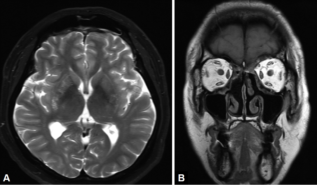
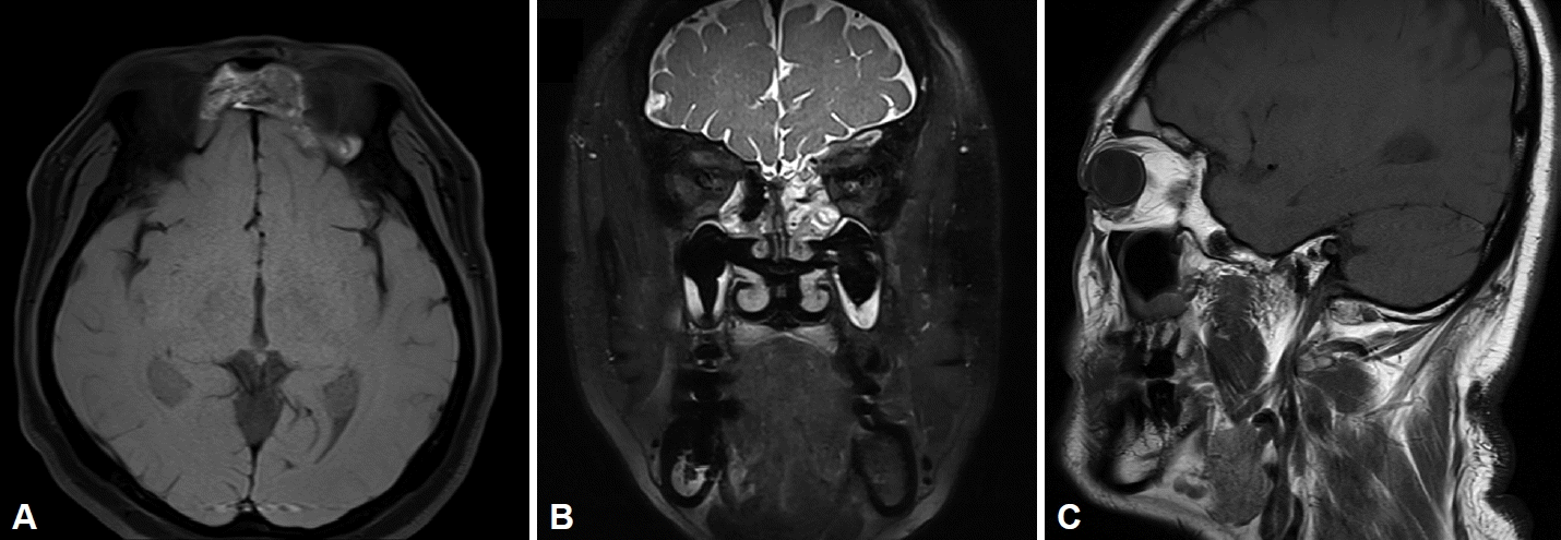
Endoscopic sinus surgery was done to explore the frontal, ethmoid, and maxillary sinuses. The explored sinuses were filled with polypoid tissue that was removed with a debrider and forceps. During surgery, frozen biopsies were done for the polyps in both nasal cavities and the pathologist suggested the possibility of IP. This was confirmed by pathology results for both nasal cavity and frontal sinus tissues, showing inverted sinonasal papilloma. Enhanced magnetic resonance imaging (MRI) of the paranasal sinus was done, and a heterogeneously enhanced, mildly convoluted, cerebriform 3.7-cm mass was seen on a T2-weighted image of the frontal sinus (Fig. 1). A large bony spur attached to the anterior wall of the left frontal sinus was also found and was suggested as the IP attachment site. Since the IP was suspected to extensively involve the far lateral frontal sinus, the supraorbital cell, and the frontal bullar cell (Fig. 2), and to be attached to the anterior wall of the left frontal sinus, an external approach was considered. For wide exposure of the lateral frontal sinus, multiple surgical axes, and access to the supraorbital cell, a bifrontal craniotomy with cranialization and a modified Lothrop operation was planned in consultation with a neurosurgeon.
In the second surgery, the frontal process, the floor of the frontal sinus, and the interfrontal sinus septum were removed with a drill, preserving the olfactory fascicle after endoscopically making an anterior septum window. The neurosurgeon exposed the frontal cranial bone with a bicoronal incision and a pericranial flap harvest. Frontal craniotomy was done and the exposed tumor was dissected and excised. The tumor attached to the detached frontal cranial bone was also removed, and the suspicious invasion site was drilled. With the posterior wall of the frontal sinus removed, the remaining tumor at the posterior and lateral aspects was removed using an endoscopic approach. After frozen biopsy of the mucosa at the frontal sinus border confirmed that no tumor remained, the pericranial flap was repositioned between the frontal lobe and the frontal sinus for obliteration, and the frontal cranial bone was reattached using a plate and screws. After checking for cerebrospinal fluid leakage, the nasal cavity was packed using packing materials and the surgery ended (Supplementary Video 1 in the online-only Data Supplement).
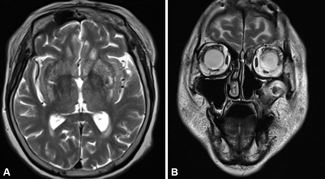
The pathology results were consistent with an inverted sinonasal papilloma. After 7 days of hospitalization with intravenous antibiotics (ceftriaxone, 2 g daily), the patient took additional per-oral antibiotics for 7 days. On postoperative day 13, at the outpatient department follow-up, the intranasal and scalp operation sites were intact, and the patient had no complaints. The 6-month follow-up MRI showed that the heterogeneous mass in both frontal sinuses was removed, and the only postoperative change seen was frontal dura thickening (Fig. 3). The patient had a normal sense of smell before the initial surgery and did not complain of a decreased sense of smell at the last follow-up. Annual follow-up examinations with MRI and endoscopy are planned to check for recurrence.

Case 2

The second patient was a 69-year-old woman who had endoscopic sinus surgery 6 years ago for a left nasal cavity mass at another hospital with a pathological result of squamous cell carcinoma. After the patient was referred to our center, the head and neck department did an additional biopsy and radiologic exam. The pathological result was squamous cell carcinoma in situ and IP. Positron emission tomography-CT and enhanced MRI of the paranasal sinus showed a hypermetabolic malignant tumor in the left posterior ethmoid sinus (T2N0M0, stage II). Two months after the initial surgery (left partial maxillectomy), reconstruction was done with left anterolateral thigh myofasciocutaneous free flap surgery. The pathological result was IP.
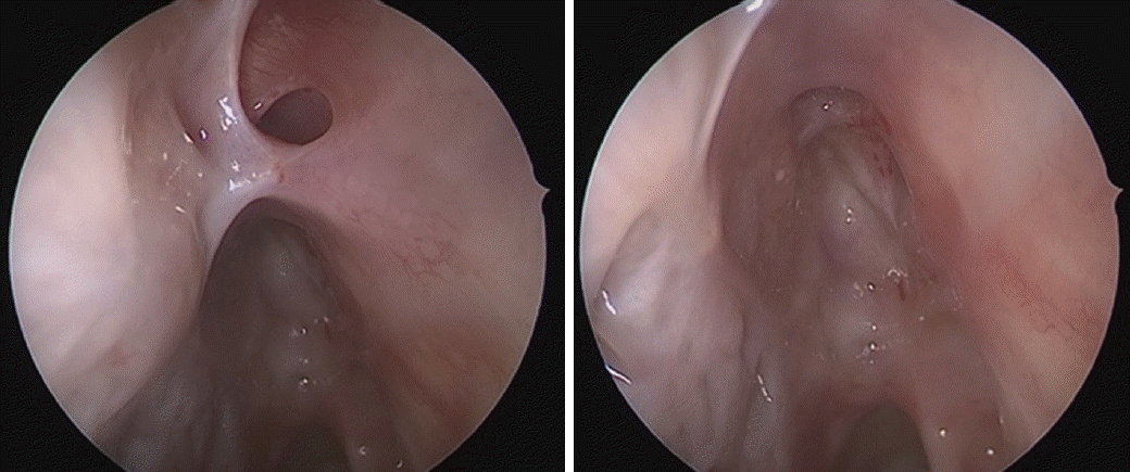
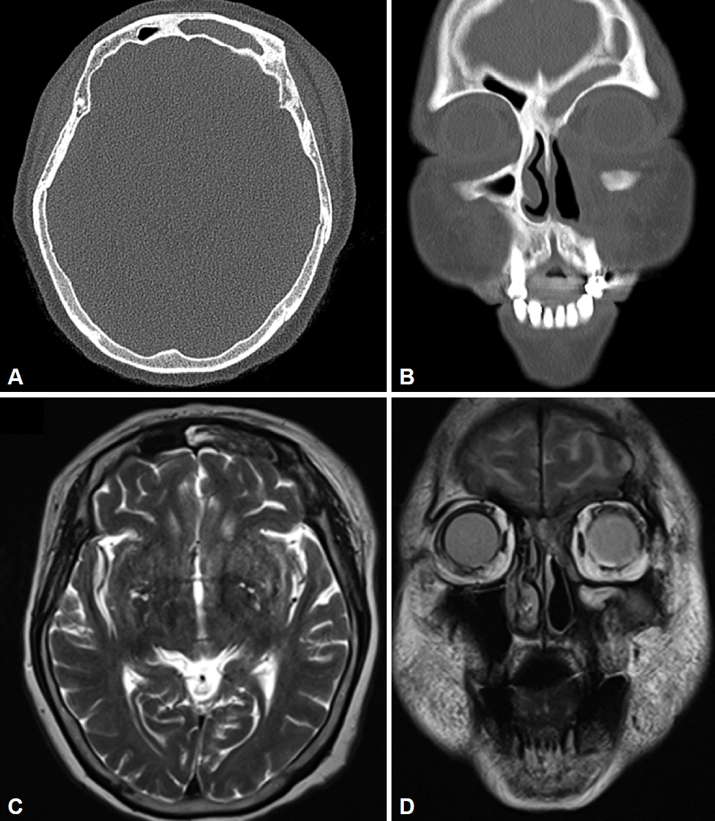
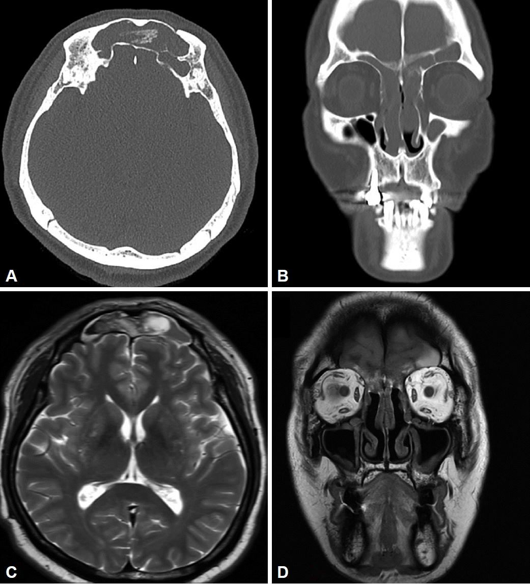
During the follow-up period, the patient underwent repeated endoscopic sinus surgery three times for recurrent IP. The chronological sites of recurrence were the left posterior ethmoid and frontal recess, the left posterior ethmoid, and the left frontoethmoidal sinus. In a 6-year follow-up at the rhinology outpatient department after the last surgery, the patient had rhinorrhea and headache, which had started 1 month earlier. She had no underlying disease. Sinus fiberscopy showed fibrosis of the left frontostomy site (Fig. 4). A nonenhanced paranasal sinus CT was done and a soft tissue density filling the left frontal, ethmoid, and maxillary sinuses was seen. The patient was referred to a neurosurgeon for suspected left frontal sinus IP. Enhanced MRI of the paranasal sinus was performed and a suspicious enhancing mass in the left frontal sinus was seen (Fig. 5). Since an IP was suspected to involve the far lateral frontal sinus and the supraorbital cell, and because a postoperative scar from the previous surgery was seen on endoscopic exam, an external approach was considered. For wide exposure of the lateral frontal sinus, multiple surgical axes, and access to the supraorbital cell, bifrontal craniotomy with cranialization and endoscopic sinus surgery were planned in consultation with the neurosurgeon.
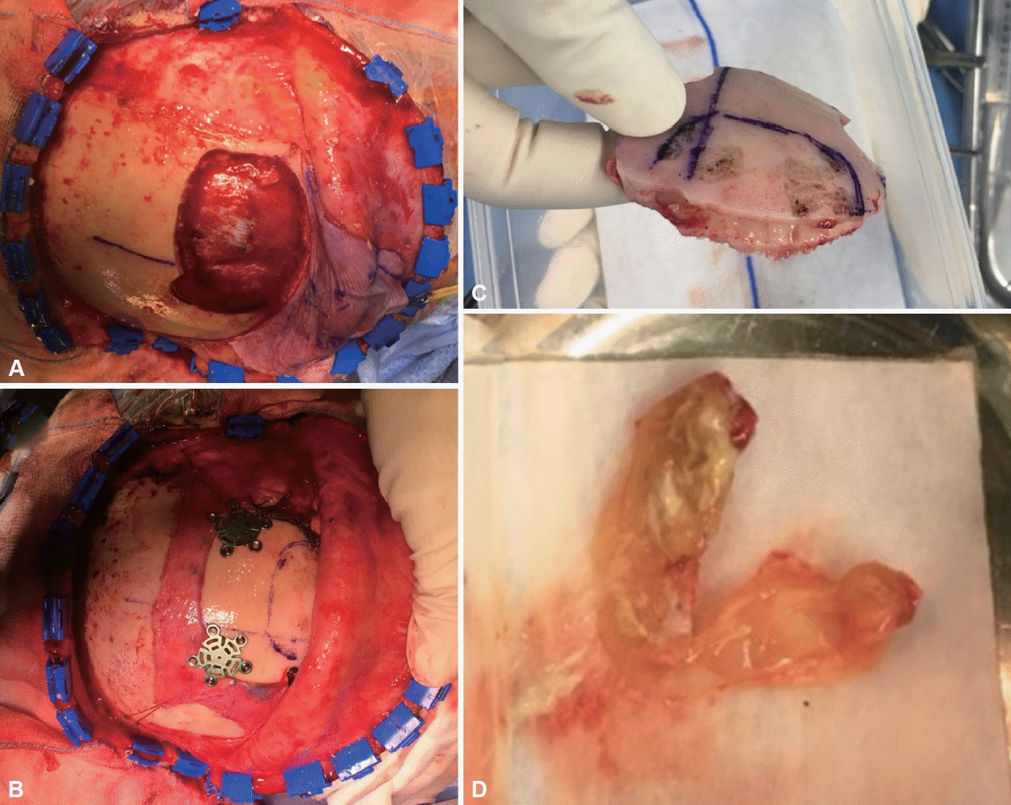
The neurosurgeon first made a bicoronal incision and approached the frontal cranium, harvesting a galeal flap. Frontal craniotomy was done, and a greenish, colloidal mass in the frontal sinus was found. The ear, nose, and throat surgeon then approached the frontal sinus by endoscopy, widening the frontal recess and frontal sinus opening, where the left frontal sinus mass was. The frontal sinus posterior wall and frontal recess were removed and biopsied. The neurosurgeon then removed the remnant frontal sinus tumor using endoscopy through the craniotomy site. A suspicious invasion site in the detached frontal cranium was drilled. After placement of the galeal flap on the sinus wall defect, the detached frontal cranium was reattached to the skull by miniplate (Fig. 6). After placement of a silastic sheet over both the frontal sinus and the scalp suture, surgery ended. On postoperative day 1, follow-up enhanced paranasal sinus MRI was done, and no evidence of a remnant or recurrent sinus lesion was seen. After treatment with intravenous dexamethasone for 6 days and intravenous antibiotics for 8 days, the patient was discharged.
At the 1-month follow-up, the left frontal sinus silastic sheet was removed. The operation field was intact with no abnormality. The pathological results of the left frontal sinus, frontal recess, and frontal sinus posterior wall were all sinonasal papilloma of the oncocytic type. At a 2-month follow-up, the patient expressed an improved sense of smell. Three serial enhanced paranasal sinus MRI examinations and one enhanced paranasal sinus CT examination were performed as of June 2021, and no newly developed abnormality or mass was seen (Fig. 7). Follow-up at 3 years is planned.

DISCUSSION

IP is the second most frequent benign tumor of the frontal sinus [6]. As local extension and bony remodeling with expansion into adjacent tissue are typical characteristics of IP, accurate excision of the IP and mucoperiosteal dissection of the attachment site are important principles for successful surgery [4,7]. For microscopic digitation extending into underlying bone, the bone underneath the attachment site should be removed or drilled to avoid recurrence [1,2,6].
The importance of eradication at the attachment site and complete excision emphasizes the need for preoperative radiologic and endoscopic evaluation to analyze the origin and extent of the IP [1]. With high-resolution CT, the site of attachment can be identified by focal bony thickening [5]. MRI has even greater value in assessing tumor extent and can distinguish the border between mucus retention and the tumor [5].
To date, no definitive guideline exists for the best technique to remove an IP of the frontal sinus, but Pietrobon et al. [1] suggested a practical algorithm for the surgical management of frontal sinus IP based on a retrospective review of 47 patients. Generally, the authors preferred an endonasal endoscopic approach, if possible, for lower morbidity and a shorter hospitalization time. However, in cases where an exclusive endonasal endoscopic approach was contraindicated because incomplete resection could result, an external approach such as osteoplastic flap or endoscopic frontal trephination was considered [1]. The contraindications included: 1) a small anteroposterior diameter of the frontal sinus (<1 cm) and a short interorbital distance, 2) erosion of the posterior wall of the frontal sinus with intracranial extension, 3) extensive lateral supraorbital attachment of the lesion in the laterally-pneumatized frontal sinus, 4) attachment of the tumor to the anterior wall or to the upper half of the posterior wall of the frontal sinus, 5) extensive involvement of the mucosa of the frontal sinus and/or of the supraorbital cell, 6) histological evidence of squamous cell carcinoma in the IP in preoperative biopsies or intraoperative frozen sections, and 7) the presence of abundant scar tissue from previous surgeries or relevant posttraumatic anatomic changes of the frontal bone [1,4]. The full extent of tumor invasion could only be assessed intraoperatively, so informed consent regarding the possibility of an external approach should be obtained [1].
For an exclusive endonasal approach, Gotlib et al. [5] suggested Draf IIA or IIB when the lesion was limited to the frontal recess, and frontal sinus opacification was due to mucus retention. When the opening made by an initial Draf IIA procedure is insufficient to prevent postoperative stenosis and additional access is needed, Draf IIB is indicated [8]. When a lateralized middle turbinate has resulted from a previous Draf I or IIA procedure, Draf IIB is indicated for complete resection of the remnant middle turbinate, thus preventing narrowing of the frontal recess [8]. When the tumor originates within the sinus and involves the contralateral side, a Draf III procedure is preferred [5]. Additional indications for performing Draf III as the primary surgery include severe polyposis, as seen in diseases such as cystic fibrosis, ciliary dyskinesia, and aspirin-exacerbated respiratory disease [9]. A frontal sinus opening <4 mm on preoperative CT and trauma to the frontal sinus outflow tract are also indications for Draf III [9]. However, the Draf III procedure is contraindicated when the patient has active airway inflammatory disease and is using a systemic steroid, since increased topical application of steroids with the Draf III procedure does not benefit a patient who is already receiving adequate systemic treatment [9]. A distance from the nasofrontal beak skin to the posterior table of <5 mm is also a contraindication for Draf III, as it makes it impossible to perform surgery without violating the skin [8].
For an external approach, endoscopic frontal trephination, transpalpebral orbital craniotomy, a supraorbital trans-eyebrow approach, an osteoplastic flap, and bifrontal craniotomy with cranialization are usually considered (Table 1). Endoscopic frontal trephination is an operative technique in which a skin incision and osteotomy are performed at the frontal table of the frontal sinus. The location and size of the osteotomy are determined by the exposure needed. The osteotomy enables visualization and instrumentation of a far lateral and superior lesion in the frontal sinus, with no significant risk of cosmetic deformity [10]. Although morbidity is minimal and the physiological function of the sinus is preserved without damaging the outflow tract of the frontal sinus, complications such as facial cellulitis and cerebrospinal fluid leakage can occur [10]. Transient forehead paresthesia can also occur, so effort should be made not to damage the supraorbital neurovascular pedicle and supratrochlear nerve bundle [10].
Transpalpebral orbital craniotomy is a surgical technique that involves making a supratarsal crease incision and dissecting the periorbita of the medial and superior orbital wall for craniotomy. Combined with an endonasal endoscopic approach, this could be an alternative to traditional external approaches, with lower morbidity and better cosmetic outcomes [4]. Direct visualization of the frontal bone, orbital rim, orbital roof, root of the nasal bone, and even the bilateral frontal sinus can be achieved, and the outflow tract of the frontal sinus can be preserved to facilitate endoscopic monitoring [4]. However, disadvantages include 1) orbital complications such as an eyelid scar with retraction, 2) ocular disease can be a relative contraindication of the technique, and 3) the titanium plate used to fix the bone flap could be palpable after surgery [4].
The supraorbital trans-eyebrow approach is a technique in which a skin incision is made in the eyebrow, extending from the supraorbital notch medially to the lateral aspect and dissecting the frontal subgaleal flap superiorly and the superior portion of the temporalis muscle laterally for exposure of the craniotomy site [11]. The small incision, hidden by the eyebrow, has a better cosmetic result and, with less temporalis muscle dissection, a lower incidence of facial nerve frontalis branch damage, scalp pain, temporalis muscle atrophy, and mastication disorders [11,12]. Resection of the upper supraorbital rim enables wider access to the anterior cranial fossa and the suprasellar region, and the need for brain retraction is decreased, minimizing neurologic complications [11,12]. However, the disadvantages include 1) a narrow surgical corridor in the vertical and horizontal directions that could limit manipulation [11,12]; 2) the possibility that adjacent structures could hinder visualization, such as the orbital roof limiting view of the anterior cranial fossa and the lesser sphenoid wing limiting view of the middle cranial fossa and cavernous sinus [12]; 3) the risk that injury of the facial nerve frontal branch could lead to paralysis of the frontalis muscle, which raises the eyebrow [12]; and 4) the fact that depending on individual skin characteristics, some patients experience cosmetic problems, including eyebrow alopecia, visible scarring, and bone defects [11].
In the osteoplastic flap procedure, a bicoronal incision is made and the pericranium is entirely elevated [6]. The frontal sinus boundary is delineated and a bone flap is elevated with the anterior wall of the frontal sinus hinged on the flap of periosteum inferiorly [6]. This technique offers wide exposure, especially in cases of multifocal involvement or concomitant malignancy [7]. It is easier to approach the sinus cavity, and operation time becomes shortened [13]. Lee et al. [3] divided the complications of osteoplastic flaps into operative, perioperative, and postoperative categories. Operative complications included dural injury due to the use of an inaccurate template or wrong placement of the template [3]. A bone flap fracture could occur with inadequate osteotomies along the supraorbital rim, thinning related to mucocele, or attachment of the osteoma to the anterior wall [3]. An orbital injury could result from an orbital roof fracture during bone flap elevation and from drilling underlying bone to remove mucosal tissue [3]. Perioperative complications include an operation site infection causing an abscess or fistula [3]. If abdominal fat is used for obliteration, a seroma or hematoma could arise with continuous bleeding or infection [3]. Postoperatively, some patients develop a type of regional neuralgia called chronic frontal pain syndrome [3]. Cosmetically, a frontal contour irregularity could arise from the loss of bone or periosteum and hypertrophy of the bone flap [3]. Although the distal coronal incisions can be hidden in the hairline, some scarring may be visible [3]. A major complication is mucocele formation that can cause skin necrosis, ptosis, and facial nerve injury [3].
In bifrontal craniotomy with the cranialization technique, the craniotomy is done after a bicoronal incision and pericranial flap elevation, without a hinge in the osteoplastic flap [3]. The posterior table of the frontal sinus is removed and later covered with a pericranial flap or autologous fat to fill dead space and prevent infectious sequelae [14]. This technique provides wide exposure with a large medial-lateral dimension and multiple axes, enough for skull base reconstruction [15]. As an alternative to traditional craniofacial resection, it prevents osteomyelitis caused by facial osteotomies [15]. Cranialization enables access to the supraorbital cell by pushing the dura with an endoscopic device, which is made possible by removal of the posterior wall of the frontal sinus. It also lowers the risk of forming a secondary mucocele [14]. When the technique is used to approach an intracranial or anterior skull base lesion, frontal lobe retraction is necessitated, which can lead to brain parenchymal injury and neurocognitive sequelae [15]. However, when the technique is used to approach the frontal sinus alone, frontal lobe retraction rarely occurs. The frontal recess to the entire frontal sinus lesion can be completely resected without damage to the anterior table of the frontal sinus, which can occur in an osteoplastic flap procedure. In case 1 in this report, with the frontal bullar cell pneumatized to the space posterior to the orbit, an osteoplastic flap procedure with the posterior table of the frontal sinus limiting the free movement of the surgical instruments (e.g., drill) would have made complete resection of the lesion impossible.
Although the endonasal endoscopic approach is currently the paradigm for the treatment of frontal sinus IP, the bifrontal craniotomy with cranialization is still indicated for inaccessible, refractory, and malignancy-associated lesions. Because the combined bifrontal craniotomy with cranialization and endonasal endoscopic approach provided wide exposure and multiple axes for surgery, our two cases of frontal sinus IP were successfully treated. Careful selection of the surgical procedure is necessary and bifrontal craniotomy with cranialization still has its place as a viable option.
Supplementary Video Legend

Video 1. Endoscopic and external surgical procedures in case 1.

Supplementary Materials

The online-only Data Supplement is available with this article at https://doi.org/10.18787/jr.2023.00024.
Supplementary Video 1.
jr-2023-00024-Supplementary-Video-1.mp4

Notes

Ethics Statement

The case studies were approved by the Institutional Review Board (KC17RESI0354) of Seoul St. Mary’s Hospital, The Catholic Medical University of Korea. We obtained informed consent in writing from the study subjects.

Availability of Data and Material

All data generated or analyzed during the study are included in this published article and its supplementary information files.

Conflicts of Interest

Do Hyun Kim who is on the editorial board of the Journal of Rhinology was not involved in the editorial evaluation or decision to publish this article. The remaining author has declared no conflicts of interest.

Author Contributions

Conceptualization: Do Hyun Kim. Data curation: Jae Seong An, Do Hyun Kim. Formal analysis: Jae Seong An, Do Hyun Kim. Methodology: Jae Seong An, Do Hyun Kim. Project administration: Do Hyun Kim. Supervision: Do Hyun Kim. Visualization: Jae Seong An. Writing—original draft: Jae Seong An. Writing—review & editing: Jae Seong An, Do Hyun Kim.

Funding Statement

None

References

1. Pietrobon G, Karligkiotis A, Turri-Zanoni M, Fazio E, Battaglia P, Bignami M, et al. Surgical management of inverted papilloma involving the frontal sinus: a practical algorithm for treatment planning. Acta Otorhinolaryngol Ital. 2019; 39(1):28–39.
2. Kamel RH, Abdel Fattah AF, Awad AG. Origin oriented management of inverted papilloma of the frontal sinus. Rhinology. 2012; 50(3):262–8.
3. Lee YH, Lee JY, Lawson W. Indications and outcomes of the osteoplastic flap procedure with or without obliteration. J Craniofac Surg. 2020; 31(8):2243–9.
4. Albathi M, Ramanathan M Jr, Lane AP, Boahene KDO. Combined endonasal and eyelid approach for management of extensive frontal sinus inverting papilloma. Laryngoscope. 2018; 128(1):3–9.
5. Gotlib T, Krzeski A, Held-Ziółkowska M, Niemczyk K. Endoscopic transnasal management of inverted papilloma involving frontal sinuses. Wideochir Inne Tech Maloinwazyjne. 2012; 7(4):299–303.
6. Fokkens W, Harvey R. Management of the frontal sinuses. In : Flint PW, Haughey BH, Lund VJ, Robbins KT, Thomas JR, Lesperance MM, editors. otolaryngology: head and neck surgery. 7th ed. Philadelphia (PA): Elsevier;2021. p. 719–32.
7. Walgama E, Ahn C, Batra PS. Surgical management of frontal sinus inverted papilloma: a systematic review. Laryngoscope. 2012; 122(6):1205–9.
8. Noller M, Fischer JL, Gudis DA, Riley CA. The Draf III procedure: a review of indications and techniques. World J Otorhinolaryngol Head Neck Surg. 2022; 8(1):1–7.
9. Turner JH, Vaezeafshar R, Hwang PH. Indications and outcomes for Draf IIB frontal sinus surgery. Am J Rhinol Allergy. 2016; 30(1):70–3.
10. Patel AB, Cain RB, Lal D. Contemporary applications of frontal sinus trephination: a systematic review of the literature. Laryngoscope. 2015; 125(9):2046–53.
11. Adawi MM, Abdelbaky AM. Validity of the lateral supraorbital approach as a minimally invasive corridor for orbital lesions. World Neurosurg. 2015; 84(3):766–71.
12. Pitskhelauri DI, Sanikidze AZ, Abramov IT, Moshchev DA, Anan’ev EP, Eliseeva NM, et al. [The trans-eyebrow supraorbital approach for removal of anterior cranial fossa and suprasellar meningiomas]. Zh Vopr Neirokhir Im N N Burdenko. 2017; 81(6):89–98. Russian.
13. Yu P, Mao X, Li X, Hu X, Li J, Sun G. Endoscopic frontal trephination verse the osteoplastic flap in patients with frontal sinus disease after bifrontal craniotomy. Br J Neurosurg. 2021; 35(1):65–7.
14. Morse JC, Chandra RK. Is there still a role for cranialization in modern sinus surgery? Curr Opin Otolaryngol Head Neck Surg. 2021; 29(1):53–8.
15. Ozlen F, Abuzayed B, Dashti R, Isler C, Tanriover N, Sanus GZ. Lowprofile 1-piece bifrontal craniotomy for anterior skull base approach and reconstruction. J Craniofac Surg. 2010; 21(1):233–8.

Fig. 1.
Preoperative radiologic exam of case 1. Paranasal nonenhanced CT axial image (A) and coronal image (B); paranasal enhanced MRI axial T2 image (C) and coronal T1 image (D). CT, computed tomography; MRI, magnetic resonance imaging.
jr-2023-00024f1.tif
Fig. 2.
Inverted papilloma involving the left frontal bullar cell in the preoperative paranasal enhanced MRI of case 1: axial T1 image (A), coronal T1 image (B), and sagittal T1 image (C). MRI, magnetic resonance imaging.
jr-2023-00024f2.tif
Fig. 3.
Six-month postoperative follow-up paranasal enhanced MRI of case 1: axial T2 images (A) and coronal T1 images (B). MRI, magnetic resonance imaging.
jr-2023-00024f3.tif
Fig. 4.
Preoperative sinus fiberscopy of case 2. Sinus fiberscopy image of the left frontostomy site.
jr-2023-00024f4.tif
Fig. 5.
Preoperative radiologic exam of case 2. Paranasal nonenhanced CT axial image (A) and coronal image (B), and a paranasal enhanced MRI axial T2 image (C) and coronal T2 image (D). CT, computed tomography; MRI, magnetic resonance imaging.
jr-2023-00024f5.tif
Fig. 6.
Intraoperative pictures of case 2. A: Frontal craniotomy state after bicoronal incision and galeal flap harvest. B: Reattached frontal cranium using miniplates and screws (Jeil Medical, Leforte Neuro system 14-NL-004, NL-BR-040, Seoul, South Korea). C: Detached frontal bone. D: Suspicious invasion site removed from detached frontal cranium.
jr-2023-00024f6.tif
Fig. 7.
Two-year postoperative follow-up paranasal enhanced MRI of case 2: axial T2 image (A) and coronal T2 image (B). MRI, magnetic resonance imaging.
jr-2023-00024f7.tif
Table 1.
The advantages and disadvantages of external approaches in the surgical resection of inverted papilloma
Approaches Advantages Disadvantages
Endoscopic frontal trephination - Visualization and instrumentation of far lateral, superior lesions in the frontal sinus
- No significant risk of cosmetic deformity
- Morbidity is minimal
- Physiological function of the sinus is preserved without damaging the outflow tract of the frontal sinus
- Possibility of facial cellulitis and leakage of cerebrospinal fluid
- Transient forehead paresthesia can occur
Transpalpebral orbital craniotomy - Lower morbidity
- Better cosmetic outcomes
- Direct visualization of the frontal bone, orbital rim, orbital roof, root of the nasal bone, and bilateral frontal sinuses
- The outflow tract of the frontal sinus can be preserved, facilitating endoscopic monitoring
- Orbital complications can occur (e.g., eyelid scar, eyelid retraction)
- Ocular disease is a relative contraindication
- The titanium plate used to fix the bone flap can be palpable at the eyelid
Supraorbital trans-eyebrow approach - The incision is hidden by the eyebrow, providing a better cosmetic result
- Less temporalis muscle dissection decreases the risk of facial nerve frontalis branch damage, scalp pain, temporalis muscle atrophy, and mastication disorders
- Wider access to the anterior cranial fossa and the suprasellar region
- Decreased need for brain retraction, minimizing neurologic complications
- The surgical corridor is narrow in both vertical and horizontal directions, limiting manipulation
- The orbital roof limits the view of the anterior cranial fossa
- The lesser sphenoid wing limits the view of the middle cranial fossa and cavernous sinus
- Injury to the facial nerve frontal branch can lead to paralysis of the frontalis muscle, which raises the eyebrow
- Eyebrow alopecia, visible scar, and bone defect
Osteoplastic flap - Wide exposure to multifocal involvement and concomitant malignancy
- Easier approach to the sinus cavity
- Operation time shortened
- Dural injury if the template is inaccurate or wrongly placed
- Bone flap fracture due to inadequate osteotomy or osteoma attached to the anterior wall
- Orbital injury from an orbital roof fracture or from drilling underlying bone
- Operation site abscess, fistula
- Seroma or hematoma after abdominal fat obliteration
- Chronic frontal pain syndrome
- Frontal contour irregularity
- Forehead scar
- Mucocele formation, causing skin necrosis, ptosis, and facial nerve injury
- In a frontal bullar cell lesion posterior to the orbit, the surgical access of instruments is limited by the posterior table
Bifrontal craniotomy with cranialization - Wide exposure with large medial-lateral dimensions and multiple axes
- Prevention of facial osteomyelitis by facial osteotomy
- Prevention of secondary mucocele formation
- Easier access to the supraorbital cell
- No frontal lobe retraction is required if only a frontal sinus lesion is involved
- The entire frontal recess is widened and the frontal sinus lesion is completely resectable without anterior table damage
- For an intracranial lesion, frontal lobe retraction is needed and can cause brain parenchymal injury
TOOLS
Similar articles