

 PDF
PDF Citation
 Print
Print


INTRODUCTION
Endoscopic sinus surgery (ESS) is indicated for chronic rhinosinusitis refractory to medical treatment [1]. Iatrogenic skull base injuries after ESS are rare (overall complication rate, 0.5%), but they can result in significant morbidity and mortality [2,3]. The skull base defect tends to be larger in these cases than in trauma cases [4]. The most common site of skull base injury following ESS is the anterior ethmoid roof adjacent to the cribriform plate [4]. Conventionally, skull base injuries were repaired using an external approach. However, in recent years, most skull base injuries after ESS have been repaired with an endoscopic transnasal approach due to the advantages of decreased morbidity, fewer postoperative complications, and shorter hospital stays [4,5].
Here, we report two cases of iatrogenic skull base injury following ESS and describe the skull base repair techniques employed in each case. This case report adds value to previously published literature in that these skull base defects with intracranial complications were repaired successfully, and both patients recovered without any neurologic deficits.
Go to : 
CASE REPORTS
Case presentation 1
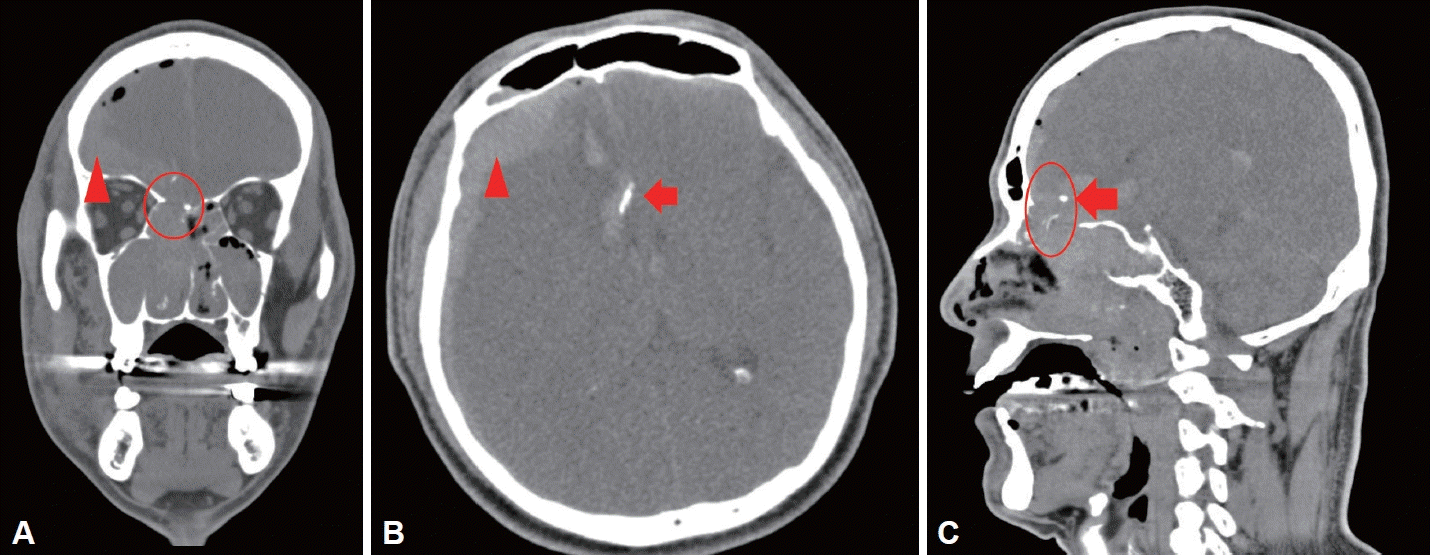
A 55-year-old man underwent bilateral ESS at a local clinic and developed headache and right orbital swelling and ecchymosis immediately afterward. Hypertension was his only underlying health condition. On postoperative day 1, he was transferred to a tertiary care hospital. Endoscopic examination of the right nasal cavity revealed an ethmoid roof defect at the level of the cribriform plate. Because of the patient’s stuporous mental status, brain computed tomography (CT) was done, which demonstrated pneumocephalus, intracranial hemorrhage, subdural hemorrhage, a high-density material suspected to be bony fragments from surgery, and a defect in the right anterior skull base (Fig. 1). Because of epistaxis in both nasal cavities, gauze packing was inserted and conservative care was performed.
On postoperative day 5, the patient was transferred to our facility for skull base repair. He presented with fever and a positive Kernig’s sign and was hospitalized for the treatment of a possible infectious condition and surgical reconstruction of a skull base defect.
Transnasal endoscopic examination showed a bony defect (1×1 cm) in the right anterior skull base (Fig. 2A). To reduce the elevated intracranial pressure, right frontal burr hole evacuation and lumbar drain insertion were performed. Since bony fragments were seen on preoperative CT, intracranial exploration was performed by the neurosurgery team. Craniotomy was performed via a bicoronal scalp incision, followed by elevation of the pericranial flap. When a frontotemporal dura window was created and the frontal lobes were retracted, black hematoma gushed out. Every effort was made to find the bony fragments near the anterior end of the corpus callosum. Following the craniotomy procedure, intracranial exploration was conducted, and a pericranial flap based on the anterior region of the pericranium was harvested. The mucosa within the frontal sinus was entirely removed, and reconstruction of the skull base, including the frontal sinus roof, was carried out using the harvested anterior-based pericranial flap (Fig. 2B). TachoSil® (Takeda Pharmaceutical Co., Zurich, Switzerland) was placed on the reconstructed pericranial flap. Using a transnasal approach, the intranasal defect was covered with a layer of fascia lata and a nasoseptal flap with adequate overlap. To harvest the nasoseptal flap, a mucosal topical solution of 1:100,000 lidocaine-epinephrine was injected around the margin of the nasoseptal flap. Under visualization with a 30° rigid endoscope, a needle-tip extended monopolar electrocautery device was used to make the mucosal incision along the posterior margin of the septum toward the nasal floor. The nasoseptal flap was elevated in a subperichondrial and subperiosteal plane and rotated to cover the skull base defect. The right nasal cavity was packed with Merocel® (Medtronic Inc., Minneapolis, MN, USA) to maintain upward pressure on the skull base repair site.
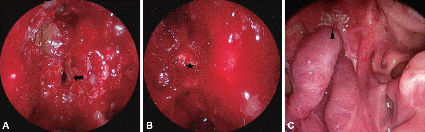
The patient was transferred to the neurosurgery intensive care unit for postoperative care. He remained intubated for the first two weeks and subsequently underwent tracheostomy for approximately 1 month because of persistent stupor. The nasal cavity packings were removed on postoperative day 15, and endoscopic endonasal examination revealed no signs of cerebrospinal fluid (CSF) leakage. On postoperative day 40, the patient was able to obey one-step commands. CT on postoperative day 25 demonstrated reduced volumes of brain hemorrhage and pneumocephalus. The tracheostomy cannula was removed on postoperative day 55, and the patient was able to ambulate independently on postoperative day 60. Endonasal endoscopic examination revealed a well-healing reconstruction site at the 10-month outpatient follow-up appointment. The surgical site had completely healed by the 23-month follow-up appointment (Fig. 2C).
Case presentation 2
An 83-year-old man underwent left-side ESS at a local facility and showed continuous left intranasal bleeding postoperatively. His past medical history included hypertension, cerebrovascular accident, and benign prostate hypertrophy. Head CT was performed after the ESS and revealed a large skull base defect on the left side (8×29 mm) with brain hemorrhage and pneumocephalus (Fig. 3). The patient was transferred to our facility for skull base repair. Upon arrival, he was alert and showed no neurologic deficits other than blurred vision, left orbital swelling, and ecchymosis.
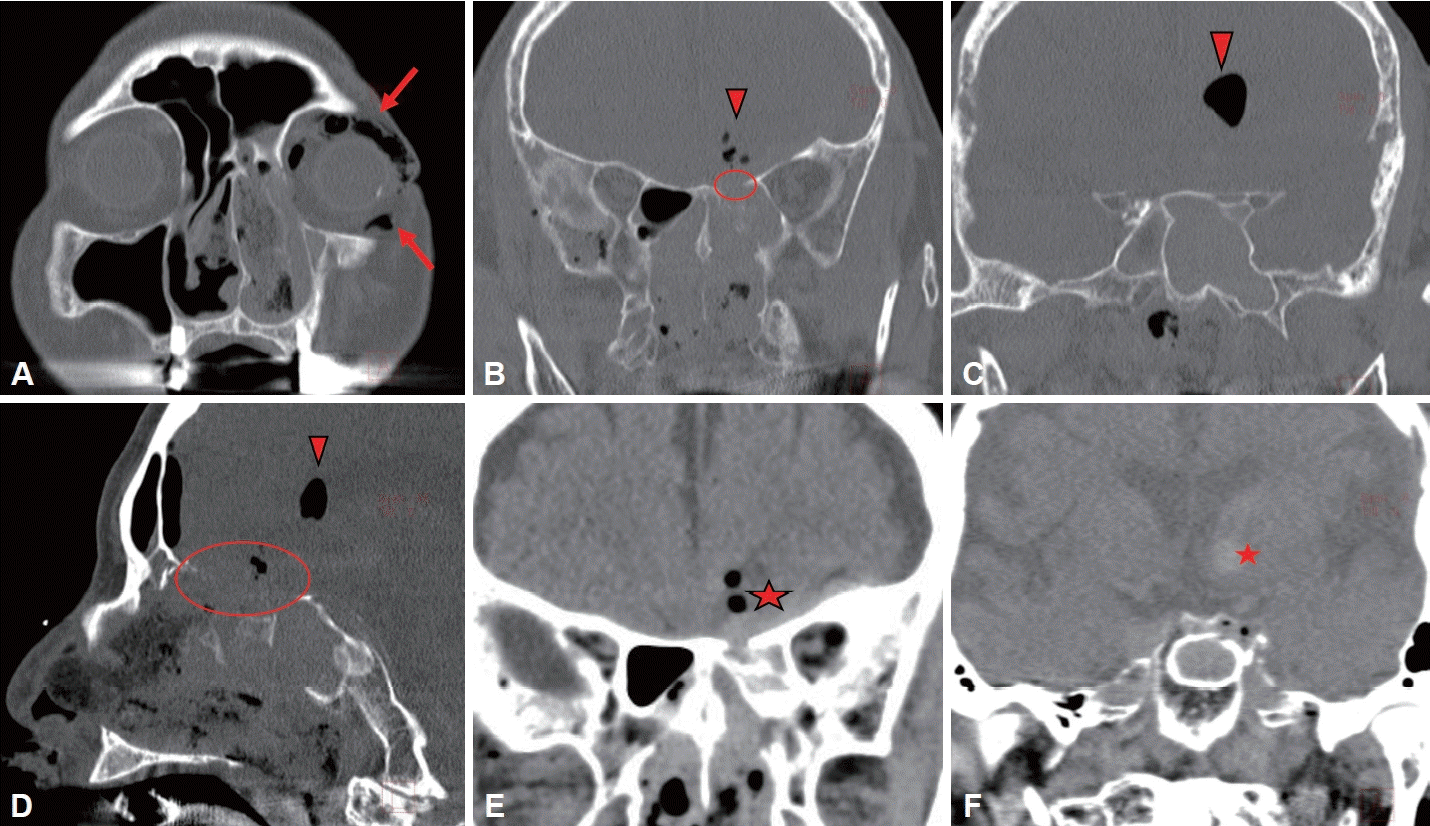 | Fig. 3.Preoperative computed tomography findings. Intraorbital emphysema in the left orbit (arrows) (A). Multifocal intracranial pneumocephalus (arrowhead) (B-D). Intracranial hemorrhage in the left inferomedial frontal lobe and basal ganglia (★) (E, F). A 4×1.5 cm right anterior skull base defect (circle) (B, D).
|
Endonasal exploration revealed an approximately 4.0×1.5 cm defect in the left anterior ethmoid roof (Fig. 4A). The left lateral lamella was intact. Endoscopic endonasal skull base repair was carried out using synthetic materials and a nasoseptal flap. First, a biological, absorbable dura substitute made from bovine pericardium (Lyoplant®, B. Braun, Melsungen AG, Germany) was inserted using the inlay and overlay technique. Over the Lyoplant® graft, TachoSil®, Surgicel® (Ethicon, J&J Surgical Technologies, Arlington, TX, USA), and Tisseel® (Baxter Healthcare Ltd., Norfolk, UK) fibrin sealants were sequentially applied. Finally, the left nasoseptal flap was harvested and positioned over the reconstruction site (Fig. 4B). The graft was packed with Merocel® for support. A lumbar drain was inserted postoperatively.
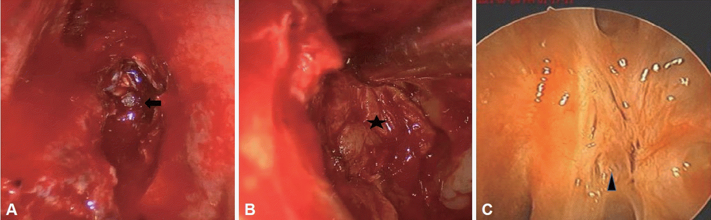
On postoperative day 4, the lumbar drain was removed. On postoperative day 5, the Merocel® packings were removed, and no signs of CSF leakage were found. The patient remained on absolute bed rest for 6 days, with the head of the bed set at 20°. On postoperative day 6, head CT revealed improved pneumocephalus and intracranial hemorrhage. Upon consultation with the infectious disease department of our institution, intravenous antibiotics were administered for approximately 4 weeks postoperatively to prevent ascending meningitis through the previous skull base defect. The patient was transferred to a local facility for intravenous antibiotic administration on postoperative day 18. At the 5-month follow-up appointment, CT demonstrated no signs of definitive flap dehiscence or pneumocephalus, and an endonasal endoscopic examination showed adequate healing of the reconstruction site, which had completely healed by the 9-month follow-up appointment (Fig. 4C).
Go to :
DISCUSSION
In both cases, the patients developed a skull base defect after ESS with multifocal brain injuries, including intracranial hemorrhage, pneumocephalus, and bone fragments in the brain. We repaired the skull base defects using an endoscopic transnasal approach, and both patients recovered without neurologic complications. In-office follow-up examinations revealed completely healed surgical reconstruction sites. We suggest that both patients were eventually discharged without any neurologic deficits for the following reasons. First, the skull base defects were identified early (<72 hours after ESS) in both cases. When the patients developed symptoms such as headache, orbital swelling, and intranasal bleeding, endoscopic examinations and postoperative CT imaging were performed in a timely manner. Second, both patients received appropriate care, including a well-established surgical plan based on preoperative CT images. Finally, the first case was effectively managed using a combined team approach. This was particularly important, given the presence of intracranial complications such as bony fragments in the ventricle that required the assistance of the neurosurgery team and a pericranial approach.
A review of the previous literature on the morbidity and mortality of iatrogenic skull base defects shows that the reported complication rates vary according to differences in the study population and the extent of the surgery. CSF leakage has been reported in 0.004% to 0.55% of cases, while severe brain hemorrhage has been reported in 0.19% to 3.9% of cases [6,7]. However, it is important to note that the results of these studies were limited by their small sample sizes. A separate large-scale retrospective study of 50,734 patients found results similar to those of prior studies, with a total CSF leakage rate of 0.06% to 0.28%, a rate of severe brain hemorrhage requiring surgery of 0.05% to 0.28%, and a mortality rate of 0.04% to 0.15% [8]. In a study analyzing patients with iatrogenic CSF leaks from ESS, the incidence of neurologic complications was compared between early interventions (n=6, within 72 hours) and delayed interventions (n=11, after 72 hours). The results showed significantly higher rates of both neurologic complications and meningitis in the delayed group (p<0.04 and p<0.01, respectively) [2].
Because the endoscopic endonasal approach has numerous advantages over the external approach, most skull base injuries have been repaired endoscopically in recent years. With the endoscopic endonasal approach, postoperative complications are fewer, and hospital stays are shorter. It also allows surgeons to more precisely identify and localize the skull base defect [4]. In case 1, intracranial bone fragments were removed during the reconstructive surgery, necessitating a combined transnasal and external approach. In case 2, multi-layered grafts were placed using the transnasal endoscopic approach alone. The patient who underwent skull base repair with the endoscopic endonasal approach alone developed no postoperative complications, despite having a larger skull base defect than the patient who underwent craniotomy for skull base repair.
Delay (>72 hours) in the identification of skull base injury following ESS can increase intracranial complications, particularly ascending meningitis and postoperative morbidity and mortality [2]. In case 1, the patient developed headache and orbital swelling, which led to imaging studies that identified a skull base defect after ESS. However, there was a delay between the occurrence of the skull base injury and the reconstructive surgery, which potentially could have resulted in postoperative infection, leading to meningitis, and resulting in an extended hospital stay.
Both of the present patients suffered an injury to the anterior ethmoid roof after ESS. This is consistent with the results of previous studies, in which the highest rate of injury after ESS was observed in the ethmoid sinus region, regardless of the number of sinuses operated on [8]. Preoperative radiographs should be reviewed to assess the risk of skull base injury and prevent its occurrence during ESS [9]. Several classification systems describe the relationship between the cribriform plate and the ethmoid roof, highlighting high-risk injury areas when performing ESS. The Keros classification system categorizes the risk of skull base injury based on the length of the lateral lamella of the cribriform plate [10]. However, a limitation of the Keros classification is that it does not account for the slope of the lateral lamella of the cribriform plate. The Gera classification overcomes this limitation. It measures the angle between the lateral lamella of the cribriform plate and the horizontal plane extending from the line of the cribriform plate, and classifies intracranial injury risk into three categories [11].
Even with the incorporation of devices, such as in imageguided surgery [12], the incidence of major complications associated with ESS appears to be high. Identifying the skull base structures susceptible to injury during ESS and early diagnosis with prompt treatment of skull base injuries are important.
Go to :
 XML Download