

 PDF
PDF Citation
 Print
Print


INTRODUCTION
Nasal polyps are grape-like soft tissue growths obstructing the sinonasal outflow tract, including the natural ostium and middle meatus. Choanal polyps are usually unilateral and differ from common nasal polyps in their morphology, histology, and prognosis [1-7]. Antrochoanal polyps are the most common type, but some polyps of ectopic origin have been reported in locations including the sphenoid sinus, ethmoid sinus, nasal septum, and inferior turbinate [1,2,7-9]. Because nasal polyps have diverse phenotypes, otorhinolaryngological surgeons must closely monitor polyps during endoscopic removal [10,11]. This report describes the clinical, radiological, and histopathological characteristics of an ethmochoanal polyp.
CASE REPORT
A 41-year-old female patient presented to our hospital with nasal obstruction, which had increased in severity for 2 months. The patient had been diagnosed with a left nasal polyp a year earlier and confirmed that it had become more prominent recently. The patient had no underlying disease other than allergic rhinitis. The chief complaint was nasal obstruction accompanied by post-nasal drip, rhinorrhea, and facial pain.
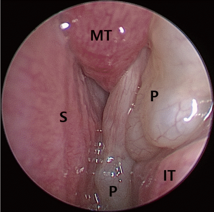
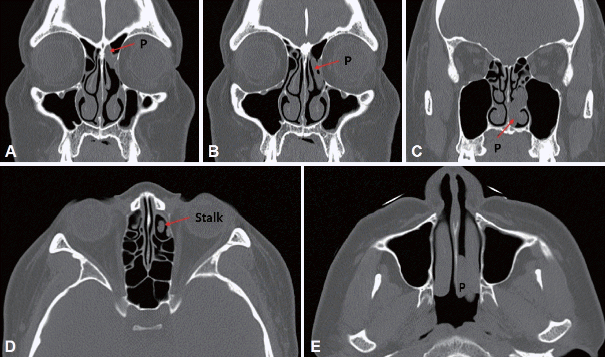
A preoperative endoscopic examination showed a large polyp filling the left nasal cavity, obstructing the middle meatus and common cavity (Fig. 1). Inferior turbinate hypertrophy was observed in the right nasal cavity. In the Korean version of the Sniffin’ Stick test, normosmia was confirmed with a score of 36 points (threshold: 12/16, discrimination: 12/16, and identification: 12/16). A preoperative blood test showed a total immunoglobulin E level of 325.0 IU/mL, a blood eosinophil percentage of 1.3%, and a C-reactive protein level <0.3 mg/dL. Five antigens were confirmed positive in the UniCAP allergy test (Dermatophagoides pteronyssinus class 2, D. farinae class 3, Alternaria tenuis class 2, common ragweed class 3, and mugwort class 2). Contrast-enhanced computed tomography (CT) of the paranasal sinus (PNS) demonstrated a soft tissue opacity in the left anterior ethmoid and an elongated non-enhanced mass extending through the middle meatus to the nasal cavity and choana. There were no specific findings in the left posterior ethmoid, maxillary, frontal, sphenoid, or contralateral PNSs (Fig. 2).
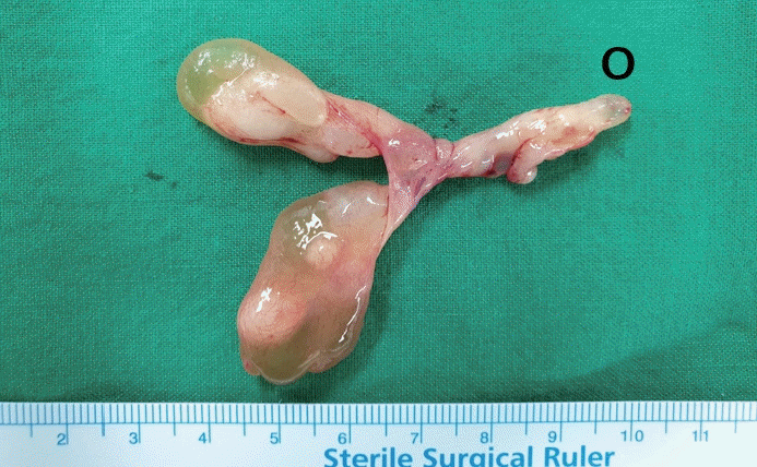
Under general anesthesia, left endoscopic sinus surgery and right inferior turbinoplasty were performed for left chronic paranasal sinusitis and right inferior turbinate hypertrophy, respectively. After resection of the uncinate process and bulla ethmoidalis, polyps were found in the anterior ethmoid mucosa around the opening of the frontal sinus. The polyp originated in the anterior ethmoid mucosa and extended through the middle meatus into the nasal cavity and choana. The mucosa of the left anterior ethmoid sinus remained relatively intact. The polyp was carefully excised along with the mucosal periosteum at the origin. The ethmochoanal polyp was approximately 6.5 cm long and revealed a pedunculated structure. The stalk was about 3.5 cm, and each branch was about 3 cm long (Fig. 3).
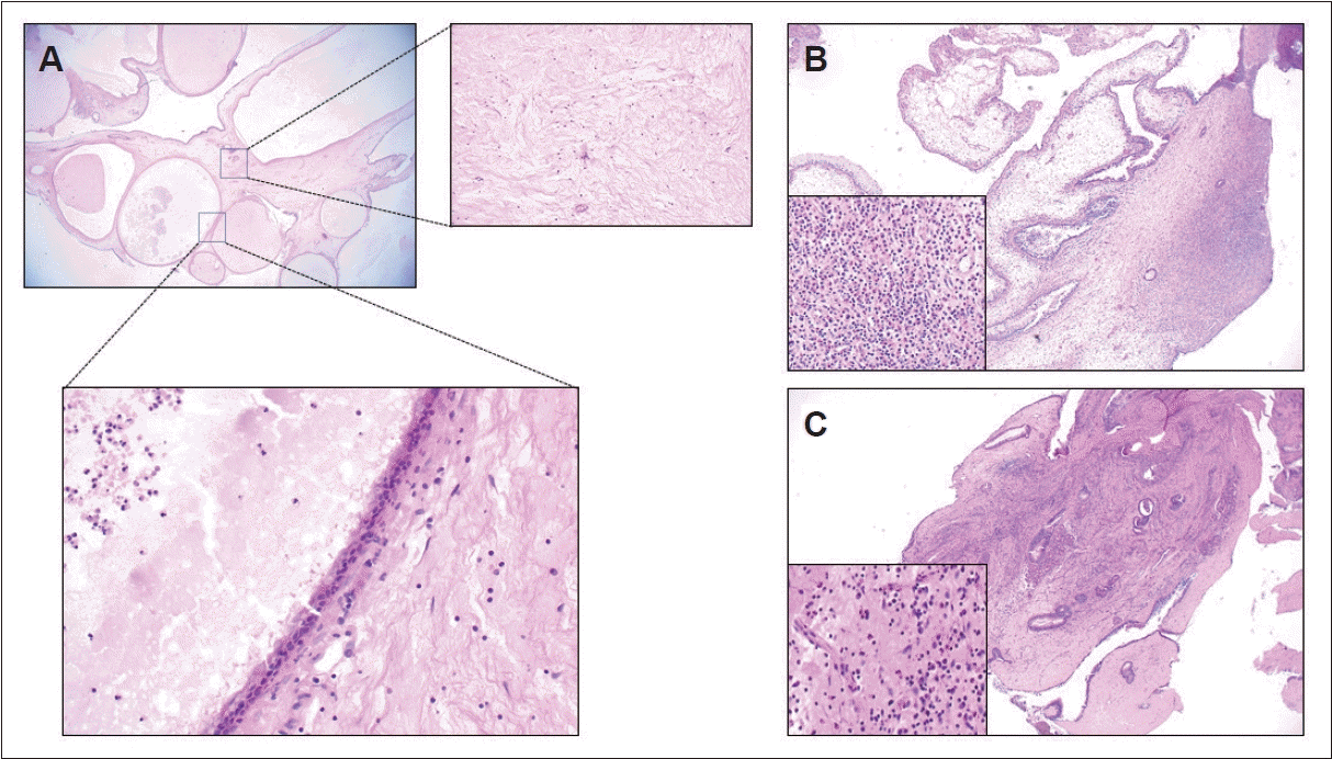
A histopathological examination revealed that the surface of the ethmochoanal polyp was lined by benign respiratory epithelium. The ethmochoanal polyp showed numerous dilated cystic structures lined by columnar epithelium and filled with mucus within the stroma. The stroma of the ethmochoanal polyp showed an edematous or loosely myxoid appearance, with no infiltration of mixed inflammatory cells. Ethmochoanal nasal and anthrochonal polyps revealed prominent inflammatory cells composed of a mixture of lymphocytes, plasma cells, eosinophils, and neutrophils and no dilated cystic structure in the stroma (Fig. 4). After surgery, the patient’s symptoms improved, and an endoscopic examination showed no recurrence at a 3-month follow-up visit.
DISCUSSION
Nasal polyps are most commonly found in the middle meatus and ethmoid sinus. A nasal polyp is a persistent inflammatory lesion that originates from the mucosa of the nasal sinus. According to Stammberger [4], nasal polyps are classified into five types based on their clinical characteristics and histological variations: 1) antrochoanal polyps; 2) large, isolated (choanal) polyps; 3) non-eosinophil dominated polyps with chronic paranasal sinusitis; 4) eosinophil-dominated polyps with chronic paranasal sinusitis; and 5) polyps due to underlying diseases. Nasal polyps are found in approximately 2.5% of Korean adults [12]. In contrast, choanal polyps are uncommon, accounting for about 4%–10% of all nasal polyps in adults and 33% in children [1,6,9]. Various studies have reported that 15%–40% of adolescents and 60%–85% of adults were affected by choanal polyps. The maxillary sinus was the most common site of origin of choanal polyps (55%–91%), followed by the sphenoid sinus (1%–15%) and the ethmoid sinus (1%–5%). Choanal polyps can manifest as solitary lesions in the middle turbinate, nasal septum, and inferior turbinate [1,2,6-9]. Although there have been reports of bilateral cases, these are unusual. Moreover, several studies have reported that ethmochoanal polyps are rare. Kizil et al. [1] found that only 1% of 98 participants had choanal polyps originating from the ethmoid sinus. In a study by Aydin et al. [7] that included 53 patients, only one choanal polyp had an ethmoid origin. Lopatin et al. [2] reported that 5 of 20 patients with choanal polyps had anterior and posterior ethmoid origins. Hong et al. [8] reported three cases of ethmochoanal polyps in South Korea in 2002.
The most typical presenting symptoms, as in this case, are nasal blockage and discharge. In this case, the patient complained of facial pain and had a mild pulling sensation (2 points on the numeric rating scale). After surgery, the patient’s facial pain was relieved. As in this case, facial pain can be a complaint in patients with nasal polyps accompanied by sinusitis with discharge [13]. Therefore, if a patient complains of facial pain, nasal endoscopy is advised to confirm sinus findings such as post-nasal drip and discharge.
In most cases, nasal endoscopy and various radiologic examinations are required to diagnose choanal polyps. Choanal polyps are shown on PNS CT images as soft tissue occupying the middle meatus and posterior choana. Depending on the origin of the polyps, soft tissue density is also seen in the maxillary, ethmoid, and sphenoid sinuses. If a stalk similar to the one in this case exists, the origin of the choanal polyp may be determined by tracing its course. PNS magnetic resonance imaging (MRI) is an additional radiological examination that is used infrequently in our treatment algorithm. Nonetheless, it may help differentiate between choanal polyps and other entities, such as inverted papillomas or malignant neoplasms. On PNS MRI, choanal polyps show radiologic findings including hypointensity on T1-weighted images, hyperintensity on T2-weighted images, and peripheral rim enhancement [14,15]. The differential diagnosis of choanal polyps should include various sinonasal neoplasms such as unilateral nasal polyps, inverted papillomas, squamous cell carcinomas, juvenile angiofibromas, and meningoencephaloceles [1,9,16].
Histopathologically, the surface of polyps is bordered by benign respiratory epithelium and edematous stroma infiltrated with inflammatory cells. Ethmochoanal polyps show numerous dilated cystic structures lined by columnar epithelium and filled with mucus within the stroma. In this study, the ethmochoanal polyp had fewer inflammatory lymphocytes, plasma cells, eosinophils, and neutrophils than nasal polyps. Other studies have found that ethmochoanal polyps contain fewer inflammatory cells, consistent with our findings. However, some studies have reported inconsistent outcomes [2,3,5-7,9,17]. When comparing representative cases of antrochoanal and ethmochoanal polyps in this hospital, no dilated cystic spaces were identified in antrochoanal polyps, unlike ethmochoanal polyps. However, an endoscopic, radiographic, and surgical inspection of the antrochoanal polyps showed evidence of a cystic portion.
Moreover, in a previous study, Berg et al. [18] observed cystic portions in the antrochoanal polyps on a histopathological scale. Therefore, we assume that these pathological differences are probably due to variations in surgical approaches. During the middle meatus antrostomy procedure, leaving all cystic components of choanal polyps that pass through the narrow natural ostium of the maxillary sinus may be challenging. Therefore, an antrochoanal polyp will likely be injured during surgery, and any cystic fluid may be drained. Consequently, the cystic portions of the final sample would be smaller than that of an ethmochoanal polyp.
In the surgical approach to choanal polyps, it is essential to reach the origin site. Antrochoanal polyps frequently pass the natural ostium of the maxillary sinus. When middle meatus antrostomy is performed, the origin site can be located in a relatively spacious maxillary sinus region compared to the ethmoid. However, because of the broad and complicated surgical space, it may be difficult to access the origin site of an ethmochoanal polyp because bony ethmoid structures from the anterior to posterior ethmoid sinus remain as obstacles and possible origin sites. Consequently, during ethmochoanal polyp surgery, a more cautious ethmoidectomy procedure is required.
Endoscopic sinus surgery has recently become the preferred treatment for choanal polyps. After endoscopic excision of the antrochoanal polyp, the recurrence rates range from 3.1% to 26.0% [1,6,7,10]. As in this case, there is a potential of leaving an ethmochoanal polyp if the origin of the polyp is not determined before or during surgery. An adequate surgical plan must be devised to reduce the recurrence rate, considering locations such as the ethmoid and sphenoid sinus, nasal septum, and turbinates, which are uncommon but may occur in patients with a choanal polyp.
 XML Download