

 PDF
PDF Citation
 Print
Print


INTRODUCTION
The sphenoid sinus is a complex structure, in contact with the skull base and close to the optic nerve, internal carotid artery, and cavernous sinus. Therefore, bony defects in this area should be assessed and addressed carefully. Several reports have documented the presence of bony defects on the lateral recess of the sphenoid sinus; these defects are frequently associated with spontaneous cerebrospinal fluid (CSF) leaks. The term “Sternberg’s canal” has been applied to such defects [1]. However, because of their anatomical relationships with the supraorbital fissure and foramen rotundum [2,3], considerable disagreement exists regarding the comparison of such bony defects with Sternberg’s canal, which is a developmental anomaly first observed by Sternberg in 1888. In his research, Sternberg described the course of the canal as running along the medial angle of the superior orbital fissure through the sphenoid body and ending at the vaginal process of the nasopharynx [4]. Therefore, theoretically, Sternberg’s canal is present medial to the superior orbital fissure and maxillary division of the trigeminal nerve (V2) [5]. As bony defects in the lateral recess are located lateral to V2, it would be inappropriate to refer to them as persistent Sternberg’s canals.
Non-iatrogenic, non-traumatic defects located in the lateral wall of the sphenoid sinus have been reported in cases of sphenoid fungal ball (SFB) [6,7]. We recently observed cases of such defects medial to the superior orbital fissure and V2, which we assumed to be instances of persistent Sternberg’s canal. In this study, we attempted to determine the prevalence of such defects according to pathological and associated clinical factors. We also attempted to determine the clinical implications of these defects in inflammatory sphenoid sinus disease.
Go to : 
METHODS
Subjects
We retrospectively reviewed patients aged >18 years who underwent endoscopic sinus surgery or endoscopic endonasal skull base surgery at a tertiary center between January 2014 and January 2019. The patients were categorized into the following three groups: 1) patients with sphenoid SFB, 2) patients with bilateral primary chronic rhinosinusitis (CRS), and 3) normal control patients. The diagnosis of SFB was based on the presence of dense material in the sphenoid sinus and dichotomous branching of fungal hyphae on Gomori methenamine silver (GMS)- and periodic acid Schiff (PAS)- stained samples. Among the SFB cases, those with dichotomous branching of hyphae, invading the mucosal layer on GMS- and PAS-stained samples, were regarded as cases of invasive fungal sinusitis. The diagnosis of primary CRS was based on the European Position Paper on Rhinosinusitis (EPOS) criteria [8]. Patients who underwent septoplasty or endoscopic endonasal skull base surgery for skull base pathologies without sinonasal involvement were regarded as control patients. First, patients’ demographic data, including age and sex, were collected. As SFB was the disease of interest, patients with pathologically proven SFB were selected from the entire study population. It was further determined that 43 of the 1,811 patients included had SFB. Of these 43 patients, four patients in whom the canal could not be visualized because of extensive lateral wall destruction were excluded. Finally, 39 patients were included in the analysis. Previous studies have documented the overall age and sex predilection of patients with fungal balls in Korea [9,10]. Moreover, bony structural changes because of conditions such as osteitis could affect Sternberg’s canal in the paranasal sinus; these changes are also associated with aging in patients with CRS [11]. Thus, we performed age and sex matching for patients with primary CRS and control individuals. For primary CRS, 1:1 matching was possible, but only 2:1 matching could be carried out for normal patients because of the skewed age distribution. The pediatric population was also investigated, as Sternberg’s canal is known to result from incomplete fusion of bony compartments constituting the sphenoid bone during the developmental process. Therefore, patients under the age of 12 years who were recommended to undergo head and neck computed tomography (CT) scans, but without any evidence of sinonasal disease during the study period, were also evaluated. In total, 805 pediatric patients were identified, from whom 39 patients were randomly selected.
Neurological symptoms, if present, and findings of gadolinium-enhanced magnetic resonance imaging (MRI) (performed during the same period as that of the CT scans) were reviewed to determine the clinical implications of these defects.
Patients with CT images with slices of more than 2-mm thickness, those who underwent a previous surgery along the middle cranial base, and those with extensive lateral wall erosion due to the presence of infiltrative lesions were excluded.
This study was approved by the Institutional Review Board of the Seoul National University Bundang Hospital (IRB No. B-2004/607-101). Patient consent has been waived from the IRB. All procedures were performed in accordance with the 1964 Helsinki Declaration and its later amendments or comparable ethical standards.
Radiologic parameters
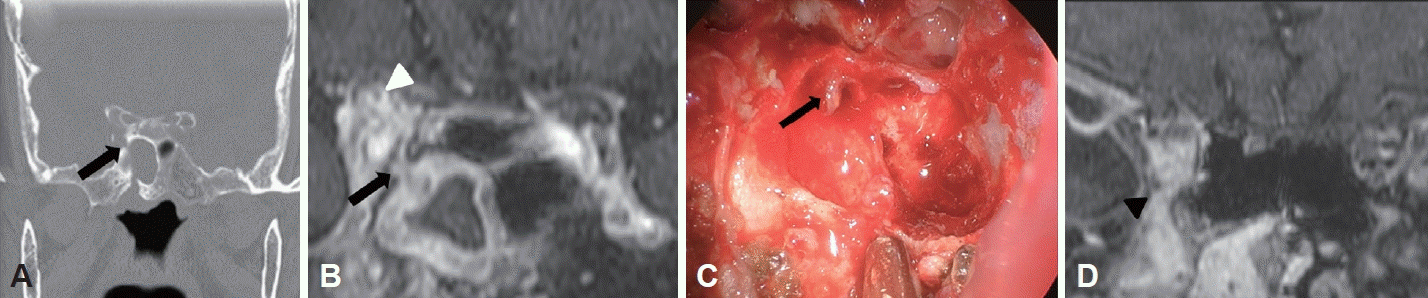
CT images of all patients were reviewed using the INFINITT PACS® (INFINITT Healthcare, Seoul, South Korea). We examined canal defects located medial to the foramen rotundum and superior orbital fissure, just anterior to the opticocarotid recess (Fig. 1), because this was regarded as the location of Sternberg’s canal. The presence of canal wall defects was evaluated with a window level of 0 Hounsfield unit (HU) and width of 2,000 HU.
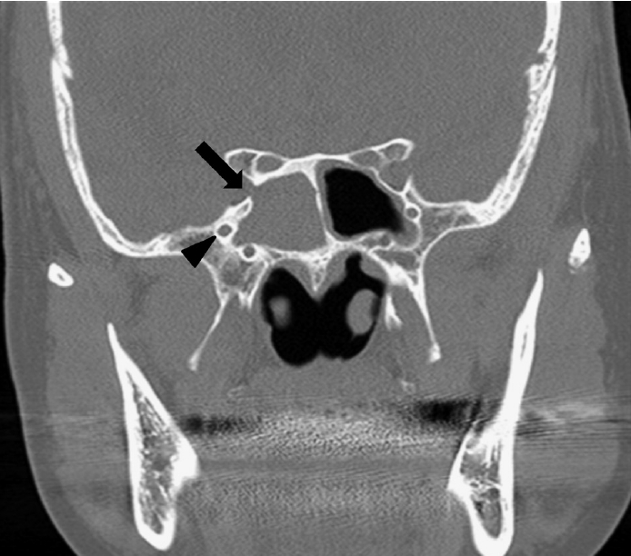
The images were double-checked by inversion of the grayscale display [12], wherein the defect was seen as a radiolucent line between the sphenoid sinus and the inside of the skull base. The presence of defects was ascertained when a definitive line could be visualized in both displays (Fig. 2).
The canal width, Lund–Mackay score [13], osteitis grade, and skull base involvement were recorded. Each patient with a sinonasal pathology in the sphenoid sinus was evaluated. When both sphenoid sinuses were affected, we selected the site with the defect or the site with the larger defect size, as appropriate. In cases with no defect (such as in control patients), the site of evaluation was selected randomly. The presence of osteitis was analyzed as follows: grade 0, no osteitis; grade I, involving less than 50% of the sinus and/or <3 mm of the osteitic wall width; and grade II, involving >50% of the sinus wall and/or 3 mm of the osteitic wall width [14].
Skull involvement was evaluated in the SFB group, as invasive fungal sinusitis is often associated with the presence of a fungal ball [15], suggesting the potential transformation of SFB into an invasive form. Soft tissue enhancement along the skull base on gadolinium-enhanced MRI, along with positive neurologic symptoms, including visual loss, diplopia, and/or severe headache, was regarded as indicating skull base involvement [16-18].
Statistical analysis
Statistical analyses were performed using SPSS 22.0 (IBM Corp., Armonk, NY, USA). Cross-table analysis with the chi-square test or the Fisher’s exact test was used for categorical variables, whereas the Kruskal–Wallis test was used to compare mean values. Logistic regression analysis (enter method) was performed to estimate odds ratios (ORs) and 95% confidence intervals (CIs) for a combination of factors in patients over 18 years of age. The threshold for statistical significance was set at p<0.05. Continuous parametric variables are presented as mean±standard deviation.
Go to :
RESULTS
Clinical characteristics of the patients
In total, 137 patients, including patients with SFB (n=39), age- and sex-matched CRS patients (n=39), controls (n=20), and children under the age of 12 years (n=39), were included in the study. The demographics of the patients are shown in Table 1. The mean age of the adult patients was 68.52±11.42 years, and they showed female predominance (78.6%). The mean age of the pediatric patients was 7.11±3.93 years, and the male-to-female ratio was 27:12.
Prevalence of Steinberg’s canal
The prevalence of Steinberg’s canal was significantly different among the groups. It was highest in patients with SFB (n=22, 56.4%), followed by patients with CRS (n=8, 20.5%), and then control patients (n=2, 10.0%) (Table 2). A significant difference was observed in the prevalence of Steinberg’s canal according to the presence of sphenoid sinus pathology (p<0.001). Children under the age of 12 years had a significantly higher prevalence of Steinberg’s canal (n=18, 46.2%) than control patients over the age of 18 years (p<0.001). In children, 44.4% of Sternberg’s canals appeared to be continuous from the lateral wall to the vaginal process of the sphenoid bone (Fig. 3). The mean size of Steinberg’s canal was 0.96±0.52 mm, and no significant differences were observed between the groups (Table 2).
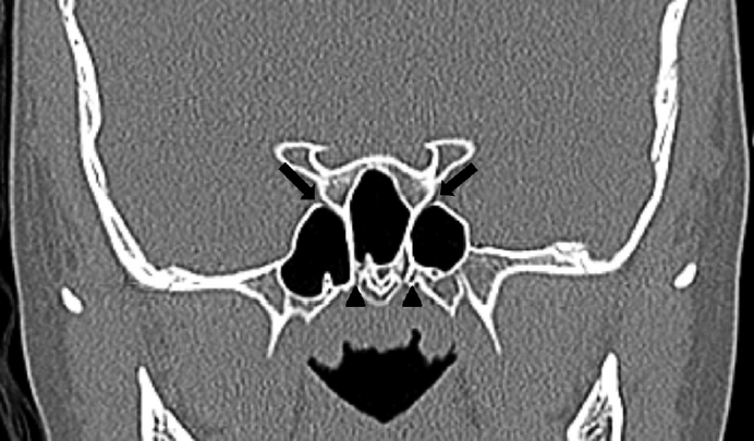 | Fig. 3.A representative CT image of children under the age of 12 years. A CT image of a continuous Sternberg’s canal (arrows) in a 10-year-old boy. Sternberg’s canal appeared to be continuous from the lateral wall to the vaginal process of the sphenoid bone (arrowheads). CT, computed tomography.
|
Table 2.
Radiologic characteristics
![]()
Prevalence of osteitis
The prevalence of osteitis (grade I or grade II) was significantly different among the groups (p<0.001). As expected, osteitis was only observed in patients in the SFB and CRS groups. In the SFB group, 10.3% of the patients had grade I osteitis and 71.8% had grade II osteitis. In the CRS group, 23.1% of the patients had grade I osteitis and 33.3% had grade II osteitis (Table 2).
Factors associated with Sternberg’s canal
Logistic regression analysis revealed that the presence of osteitis, grade I or II, was associated with a higher risk of Sternberg’s canal (grade I: OR=7.757; 95% CI, 1.006–59.787; grade II: OR=6.648; 95% CI, 1.150–38.442). Age, sex, pathology, and the Lund–Mackay scores of the involved side were not associated with the presence of Sternberg’s canal (Table 3).
Table 3.
Logistic regression analysis to identify independent variables associated with canal defects
![]()
Clinical implications: CSF leak and skull base involvement
None of the patients with Sternberg’s canal (n=50) had CSF leaks. Among the 39 patients in the SFB group, brain MRI with gadolinium enhancement was performed in 25 (64.1%). Among these 25 patients, three showed clinical skull base involvement, while also presenting with Sternberg’s canal. However, the presence of Sternberg’s canal was not significantly associated with skull base involvement (p=0.230) (Table 4).
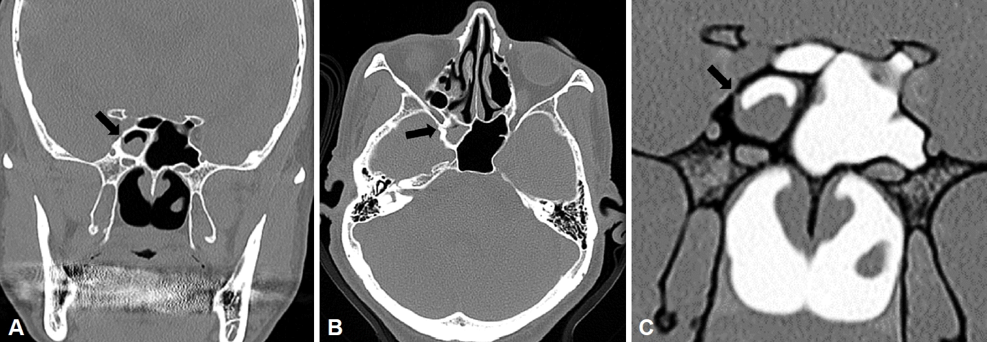
Of the three patients with skull base involvement, two were diagnosed with invasive fungal sinusitis, and the third was diagnosed with Tolosa–Hunt syndrome and SFB. All three patients presented concomitant SFB and soft tissue enhancement along Sternberg’s canal, connecting the sphenoid and cavernous sinus. In the two patients with invasive fungal sinusitis, this canal could have been a route for skull base invasion. However, both patients also showed infiltration of the pterygopalatine fossa extending to the orbital apex, and skull base invasion could have also taken place through this route (Fig. 4).
 | Fig. 4.A representative magnetic resonance image. A: A CT image of Sternberg’s canal (arrow). B: Enhancement along the Sternberg’s canal (arrow) to the orbital apex (arrowhead) on a T1-weighted magnetic resonance image. C: Endoscopic findings after peeling off the mucosa of the lateral wall. Soft tissue along Sternberg’s canal (arrow). D: Enhancement of pterygopalatine fossa (arrowhead). CT, computed tomography.
|
Go to :
DISCUSSION
In our study, patients with SFB had the highest prevalence of Sternberg’s canal, followed by those with CRS, and then control patients without sinonasal inflammatory disease. However, the presence of osteitis was the only radiologic parameter significantly associated with the presence of the canal. The prevalence of Sternberg’s canal was higher in children without any evidence of sinonasal disease than in the control adults.
During the development of the sphenoid sinus, bony fusion between the greater wings and the presphenoid/basisphenoid compartments is completed during childhood. However, some children may present with incomplete fusion manifesting as a lateral craniopharyngeal canal or Sternberg’s canal. Hence, we defined Sternberg’s canal as a bony defect located medial to the foramen rotundum and superior orbital fissure, just anterior to the opticocarotid recess; this definition was followed because the location of the fusion is between the greater wing of the sphenoid and sphenoid body. This location corresponds to that described in the cadaver study by Schick et al. [19], which was reported as Sternberg’s canal.
Bone is a dynamic tissue that responds to a variety of pathologic, mechanical, and physiologic stimuli by altering and repairing its structure through remodeling. Mucosal inflammation in CRS can activate osteoclastic and osteoblastic activities [20]. Therefore, in cases of sphenoidal disease accompanied by osteitis, bone remodeling around the remnants of Sternberg’s canal may result in widening of the canal, rendering it detectable on CT scans. This is thought to explain why Sternberg’s canal was observed in CRS and SFB patients at a significantly higher frequency than in the control group.
In his original report, Sternberg stated that this canal was present in all children aged 3–4 years and in just 4% of adults. In our study, 46.2% of children under the age of 12 years and 10% of control adults had the canal. Differences in the methods of detection of Sternberg’s canal and in age distribution may have resulted in this discrepancy. The mean age of the children included in the study was 7.11 years, which is higher than the age of 3–4 years, when the fusion between the sphenoid bony compartment is reported to begin [21]. This discrepancy could explain the prevalence of Sternberg’s canal observed in the pediatric population in this study. In our study, Steinberg’s canal was considered to be present if the lesion was visible on a high-resolution CT scan. Therefore, some defects may not have been detected in the past, leading to a higher prevalence in adults.
Defects in the lateral recess of the sphenoid sinus are frequently associated with spontaneous CSF leaks and increased intracranial pressure. They are also associated with the presence of arachnoid pits and empty sella, with a predilection for female sex, middle age, and obesity [3]. These lesions could result from altered CSF dynamics in aberrant arachnoid granulations. When CSF pressure increases, CSF drainage from the arachnoid granulation to the dural venous sinus may be impaired, leading to progressive enlargement and scalloping of the underlying bone [22]. These defects are located in the lateral recess of the sphenoid sinus, and the suggested pathophysiology is different from that of Sternberg’s canal. Therefore, Sternberg’s canal might not be an appropriate term for a defect in the lateral recess. In our study, no patients with Sternberg’s canal presented with CSF leaks.
The current study attempted to determine the clinical implications of Sternberg’s canal. Unlike primary CRS, there is a risk of SFB progressing into an invasive form. Therefore, the presence of Sternberg’s canal can be misdiagnosed as an invasive form of SFB, as the canal may mimic a bony destructive lesion, which is seen in cases of invasive fungal sinusitis. Although the presence of Sternberg’s canals was higher in the CRS and SFB groups, the association between skull base involution and Sternberg’s canal presence was not significant in the SFB group. Therefore, the correlation between the presence or absence of canals and skull base involvement is unknown. Regardless of whether Sternberg’s canal is present, patients suspected of having an invasive fungal infection should always receive a comprehensive evaluation, including past medical history, mucosal biopsy, and physical and radiologic evaluations.
All three patients with clinical evidence of skull base involvement presented with Sternberg’s canal. These cases all showed soft tissue enhancement connecting the skull base with the sphenoid sinus. This enhancement is associated with an active granulomatous reaction in invasive fungal sinusitis [23]. Soft tissue enhancement in the skull base along with the mucosa of the sphenoid sinus connected by Sternberg’s canal may therefore imply the spread of the inflammatory reaction against fungal invasion. However, this enhancement may also have resulted from sustained bony irritation due to osteitis, rather than direct fungal invasion [24]. In addition, other routes may be possible, as some patients also presented with infiltration of the pterygopalatine fossa. Therefore, further evaluation is needed to ascertain whether Sternberg’s canal can be a route for skull base invasion.
This study has several limitations. First, there is a possibility of selection bias. In this study, we sought to confirm the pathologic associations of Steinberg’s canal. Hence, patients who underwent surgery were primarily selected. In addition, only age- and sex-matched patients were evaluated. Therefore, our patients may not have represented the entire population, and further studies with larger cohorts are necessary. Second, Sternberg’s canal was defined radiologically; hence, the correspondence between this finding and actual Sternberg’s canal should be further validated through cadaveric studies.
Go to :
 XML Download