Journal List > Brain Tumor Res Treat > v.11(2) > 1516083458

Mackel, Orrego-Gonzalez, and Vega: Awake Craniotomy and Intraoperative Musical Performance for Brain Tumor Surgery: Case Report and Literature Review

Abstract

Music experience and creation is a complex phenomenon that involves multiple brain structures. Music mapping during awake brain surgery, in addition to standard speech and motor mapping, remains a controversial topic. Music function can be impaired selectively, despite overlap with other neural networks commonly tested during direct cortical stimulation. We describe the case of a 34-year-old male patient presenting with a glioma located within eloquent cortex, who is also a professional musician and actor. We performed an awake craniotomy (AC) that mapped the standard motor and speech areas, while the patient played guitar intraoperatively and sang. Outcomes were remarkable with preservation of function and noted improvements in his musical abilities in outpatient follow-up. In addition, we performed a review of the literature in which awake craniotomies were performed for the removal of brain tumors in patients with some background in music (e.g., score reading, humming/singing). To date, only 4 patients have played a musical instrument intraoperatively during an AC for brain tumor resection. Using awake cortical mapping techniques and paradigms for preserving speech function during an intraoperative musical performance with singing is feasible and can yield a great result for patients. The use of standard brain mapping over music processing mapping did not yield a negative outcome. More experience is needed to understand and standardize this procedure as the field of brain mapping continues to grow for tumor resections.

INTRODUCTION

The ability to process, prepare, and produce music in its instrumental and vocal forms is a complex function that demands the coordination of multiple domains from both cerebral hemispheres [1]. As described by Dalla Bella and Berkowska [2]; these neural network entails severe cortical networks including the auditory cortex, motor cortex, and basal ganglia. The output utilizes faculties critical to speech and language, including pitch, prosody, melody, rhythm, and syntax [3]. In addition, functional neuroimaging demonstrates the substantial overlap between music and language [4].
Awake craniotomy (AC) has proven to be very helpful in preserving eloquent speech and motor cortical regions while performing cortical and subcortical dissections. While motor and speech stimulation testing is standard and widely studied in the medical literature, music mapping is still not widely performed and remains a controversial topic, as it is not known if this later approach will add meaningful information beyond standard mapping during surgery. Clinical experience teaches that musical function and language/speech function are domains that, despite the overlap, can also be selectively impaired [56]. There have been instances of amusia without aphasia, as in the case of the resection of right Heschl gyrus gliomas [6]. Conversely, there have been instances of aphasia without amusia, such as a case of a talented French musician that has been reported [5]. Furthermore, refinement in functional neuroimaging demonstrates that even within regions of broad overlap between music and speech/language, distinct activation patterns may still be extrapolated [7].
It may be reasonable to include musical mapping during AC for brain tumor resection in patients who require their musical skills for personal or professional reasons. Recently, Kappen et al. [8] performed a systematic review of patients performing music during AC which showed that mapping music is feasible. In this case report, we explore our experience with AC for brain tumor resection in a patient that played an instrument (guitar) and sang simultaneously during surgery. In these cases, music processing mapping is not fully standardized or understood yet, as we will show from the data within this review. We did not perform music processing mapping and we intend to give our insights about the use of standard mapping in these cases in comparison.

CASE REPORT

A 34-year-old male professional guitarist and singer with a past medical history of depression and 1 year of impaired word-finding and mild cognitive changes presented to the emergency department after a generalized seizure. MRI revealed a non-enhancing mass predominantly located within the left temporal lobe with extension into the insula (Fig. 1). Given the patient’s young age as well as the size, eloquent location and concern for an underlying infiltrative glial tumor, the patient opted for surgical resection via an awake left pterional craniotomy with brain mapping. Before undergoing the procedure, surgical planning included functional magnetic resonance imaging (fMRI) (Fig. 2), using the blood-oxygen-level dependent (BOLD) technique, and diffusion tensor imaging (DTI) (Fig. 3). During fMRI, the patient performed our standard language and tongue motor protocol, demonstrating activation of the left hemisphere and adjacent regions to the tumor in the posterior superior temporal gyrus (pSTG) and inferior motor homunculus in the precentral gyrus.
Resection was planned using the asleep-awake-asleep method with intraoperative speech mapping and electrocorticography. One gram of levetiracetam was administered intraoperatively. Following the craniotomy, the patient was awoken, and the cortical grid was placed to localize the central sulcus. Phase reversal was negative, indicating the motor cortex was posterior to the resection area. With bipolar stimulation, a negative map for speech arrest was obtained and corticectomy was performed. The speech was preserved with continuous testing during the surgery, including singing, as was motor function with guitar playing (Fig. 4, Supplementary Video 1 in the online-only Data Supplement). A near-total resection was obtained and the final pathology was reported as WHO Grade III anaplastic astrocytoma (IDH1 mutant).
Postoperatively, the patient surprisingly displayed neologisms, phonemic and semantic paraphasia. The Western Aphasia Battery showed transcortical sensory aphasia. This aphasia was attributed to postoperative changes and swelling from the resection (Fig. 3). His aphasia began to improve by the second postoperative day and returned to his baseline, and at the 2-month follow-up, he had further improvement from baseline with reported improvement in music and singing performance. He completed 30 sessions of 59.4 Gy of fractionated external beam radiation with concomitant temozolomide. The patient has since returned to professional musical activity and there have been no signs of tumor progression now 18 months postoperation.

DISCUSSION

Awake mapping studies have been built out of a diversity of cortical areas where stimulation specifically impacts musical function while sparing non-music motor/language tasks. This is important for cases where it is important to preserve musicality. Music itself is a complex task that involves several components (e.g., pitch, melody, score reading) located in different regions of the brain [9]. The complexity of music processing and variability among patients underscores the potential benefit of awake music mapping for those patients for whom its preservation is critical.
To date, 20 patients have been reported in the literature of AC for brain tumor resection to map different forms of musical function, not including ours. Furthermore, there is only 1 review of the literature, which encased craniotomies for all causes [8]. In this review, we specifically focus on brain tumor resections to analyze the benefits of this approach in this patient population. In Table 1, we summarize the demographic and clinical characteristics of patients who underwent AC with standard musical tasks for intraoperative assessment. Only 4 patients were assessed intraoperatively while they played a musical instrument (Table 2).
Only 3 patients underwent standardized music testing (15%), while 8 patients were evaluated by a trained third party (40%), and 9 were self-evaluated (45%); 20 cases reported preoperative assessment. Intraoperative testing of musical abilities included singing on 11 patients (52.4%), humming on 3 patients (14.3%), melody recognition tasks on 2 patients (9.5%), score reading on 7 patients (33.33%), and instrument playing on 4 patients (19.04%). Ten studies reported the size of tumor resection. Of these, a gross total resection was achieved in 6 cases (60%), near-total resection in 3 patients (30%), and subtotal resection in only 1 (10%). Outcomes of musical ability postoperatively were reported in 15 cases; 12 of them (80%) had a musical ability that was comparable to preoperative assessment, and in 3 cases (20%), these skills were improved including our case. Only our patient (6.7%) had transcortical sensory aphasia after surgery with complete recovery during follow-up, among all cases with reported outcomes (15 cases). Only 1 patient was reported to have passed away at the end of follow-up [10].
Overall, outcomes following awake mapping are favorable. In all cases, postoperative assessments demonstrated a postoperative interval where the musical function was comparable to the preoperative baseline or superior. We found in our review that music mapping in some of the patients were located in areas close to or within regions that are critical for language and motor tasks. For instance, our review shows that in 4 patients (19%) the pSTG was activated during the musical task; in 6 patients (28.6%) the precentral gyrus was activated, while in 1 patient (4.8%) the supplementary motor area (SMA), and 4 patients (19%) the inferior frontal gyrus (IFG) both were activated [41112131415161718]. pSTG and IFG gyrus are the locations of Wernicke’s and Broca’s area, respectively; while precentral gyrus and SMA are motor areas. These results suggest either a close proximity or overlap of both music with motor and language functions. In 5 patients (23.8%), other areas non-related to motor and speech were related to music production (middle temporal gyrus and postcentral gyrus) [118]. These results point out that music involves a heterogeneous number of brain areas, which can overlap with others related to motor and speech.
In our review, 3 cases (14.3%) including ours, were not specifically mapped for music processing, instead, they focused on the assessment of speech and motor tasks [1315]. Good outcomes in 3 patients could be explained by the overlap mentioned above, hence evaluation of speech could protect the music faculty. An alternative for future cases like these is performing intraoperative functional testing with consistent rotation between language and music assessment. Furthermore, only 4 patients (19%) in the literature have undergone AC while playing an instrument intraoperatively for brain tumor resection including ours (Table 2) [101519]. In addition, we combined singing and guitar strumming, a method of assessment, which has not been done before. Playing an instrument involves multiple skills including coordination, spatial intelligence, musical notation, and rhythm. Hence, we would like to propose performing this type of assessment in future cases when feasible and indicated. With more cases added to the literature, it will be possible to determine if this assessment is the best approach for these types of cases within this patient population.
Despite not undergoing music processing mapping, our case had a postoperative self-assessment with an improvement in musical skills with the guitar and singing. This phenomenon has been attributed in previous reports to the alleviation of brain edema and compression after tumor resection [4]. In addition, neural plasticity and recruitment of adjacent zones play an important role according to image studies [420]. This leads to the question if specific music mapping is necessary for having better outcomes on AC for brain tumor resection. According to the limited experience provided by the literature, we can assume that standard AC for motor and speech functions can be safely done in these cases where it is fundamental to preserve musical abilities. Moreover, in these cases playing an instrument intraoperatively could be enough to preserve the functionality of the patient without requiring complex music mapping. Importantly, we provide evidence that “negative mapping” may be sufficient for music preservation when operating in cerebrum dense with musical function as an excellent functional outcome can be obtained without positively stimulating eloquent musical sites. This strategy of negative mapping for music aligns with the current evolution in language mapping that de-emphasizes positive site control and instead focuses on identifying negative areas that allow a more precisely tailored surgery [21]. These approaches should be addressed when there are more cases in the literature, enough to provide statistical evidence about what is the best procedure in these cases. Furthermore, the role of preoperative imaging (fMRI and DTI) in these cases needs to be elucidated. Similar to language testing, it remains an outstanding question whether high-quality noninvasive functional imaging could obviate the need for intraoperative testing.
Another caveat of AC for brain tumor resection in musicians that need to be addressed is the lack of a standardized assessment of musical capabilities, unlike speech. Only 3 cases in our review were evaluated with a musical scale the Montreal Battery of Evaluation of Amusia (MBEA), whereas the majority were subjectively assessed by themselves or a third party [22]. Objective assessments eliminate the risk of bias and could help reduce morbidity after AC, as suggested by Papatzales et al. [22], during AC for language mapping. A potential limitation in our case was our reliance on the patient’s subjective assessment of their musical function pre- and postoperatively. On the one hand, we may have missed subtle benefit or injury from surgery if measured by objective markers; on the other hand, if the goal of surgery is preserving a function subjectively important to the patient, it is not clear that objective musical function test are needed pre- and post-surgery so long as the patient retains the ability to appraise their own function and is pleased with the outcome. We recommend that the degree of pre- and postoperative testing to assess impact on musical function should be decided on a case-by-case basis but advocate consideration should be given intraoperatively as to the role of a trained musicologist intraoperatively similar to trained administrators of speech testing using a standardized test such as the MBEA.
In this study, we present a case of awake cortical mapping in which we performed standard mapping for brain tumor resection in a professional musician. Our approach did not provide worse outcomes than musical mapping and helped to preserve music function in these patients with outstanding results. The literature review showed that there are few cases of intraoperatively instrument playing during AC for brain tumor resection, to date. In addition, other aspects of the technique need to be addressed. More experience is needed to understand and standardize this procedure. More cases should be added to the literature to carry on prospective studies with higher evidence in the future. This will allow the neurosurgeons to better understand if music mapping is necessary and provides better outcomes for patients in these selected cases.

Notes

Ethics Statement: This report was conducted according to the guidelines of the Declaration of Helsinki for biomedical research, and was deemed exempt from patient consent by the Institutional Review Board (2021P000600). Patient consent was obtained for pictures and operative videos utilized in this manuscript.

Author Contributions:

  • Conceptualization: Rafael A. Vega.

  • Investigation: all authors.

  • Project administration: Rafael A. Vega.

  • Resources: Eduardo E. Orrego-Gonzalez.

  • Supervision: Rafael A. Vega.

  • Validation: Rafael A. Vega.

  • Writing—original draft: Charles E. Mackel, Eduardo E. Orrego-Gonzalez.

  • Writing—review & editing: all authors.

Conflicts of Interest: The authors have no potential conflicts of interest to disclose.

Funding Statement: None

Availability of Data and Material

The datasets generated or analyzed during the study are available from the corresponding author on reasonable request.

References

1. Saito Y, Ishii K, Yagi K, Tatsumi IF, Mizusawa H. Cerebral networks for spontaneous and synchronized singing and speaking. Neuroreport. 2006; 17:1893–1897. PMID: 17179865.
2. Dalla Bella S, Berkowska M. Singing and its neuronal substrates: evidence from the general population. Contemp Music Rev. 2009; 28:279–291.
3. Zarate JM. The neural control of singing. Front Hum Neurosci. 2013; 7:237. PMID: 23761746.
4. Zhang J, Lu JF, Wu JS, Yao CJ, Zhuang DX, Qiu TM, et al. A unique case of Chinese language and music dissociation with tumor located in Broca’s area: multimodal mapping for tumor resection and functional preservation. Clin Neurol Neurosurg. 2013; 115:2230–2233. PMID: 23910999.
5. Fisher CA, Larner AJ. Jean Langlais (1907-91): an historical case of a blind organist with stroke-induced aphasia and Braille alexia but without amusia. J Med Biogr. 2008; 16:232–234. PMID: 18952998.
6. Russell SM, Golfinos JG. Amusia following resection of a Heschl gyrus glioma. Case report. J Neurosurg. 2003; 98:1109–1112. PMID: 12744373.
7. Rogalsky C, Rong F, Saberi K, Hickok G. Functional anatomy of language and music perception: temporal and structural factors investigated using functional magnetic resonance imaging. J Neurosci. 2011; 31:3843–3852. PMID: 21389239.
8. Kappen PR, Beshay T, Vincent AJPE, Satoer D, Dirven CMF, Jeekel J, et al. The feasibility and added value of mapping music during awake craniotomy: a systematic review. Eur J Neurosci. 2022; 55:388–404. PMID: 34894015.
9. Scerrati A, Labanti S, Lofrese G, Mongardi L, Cavallo MA, Ricciardi L, et al. Artists playing music while undergoing brain surgery: a look into the scientific evidence and the social media perspective. Clin Neurol Neurosurg. 2020; 196:105911. PMID: 32505870.
10. Scerrati A, Mongardi L, Cavallo MA, Labanti S, Simioni V, Ricciardi L, et al. Awake surgery for skills preservation during a sensory area tumor resection in a clarinet player. Acta Neurol Belg. 2021; 121:1235–1239. PMID: 32372400.
11. Bass DI, Shurtleff H, Warner M, Knott D, Poliakov A, Friedman S, et al. Awake mapping of the auditory cortex during tumor resection in an aspiring musical performer: a case report. Pediatr Neurosurg. 2020; 55:351–358. PMID: 33260181.
12. Garcea FE, Chernoff BL, Diamond B, Lewis W, Sims MH, Tomlinson SB, et al. Direct electrical stimulation in the human brain disrupts melody processing. Curr Biol. 2017; 27:2684–2691.e7. PMID: 28844645.
13. Herbet G, Lafargue G, Almairac F, Moritz-Gasser S, Bonnetblanc F, Duffau H. Disrupting the right pars opercularis with electrical stimulation frees the song: case report. J Neurosurg. 2015; 123:1401–1404. PMID: 26140496.
14. Nathoo N, Nair D, Phillips M, Vogelbaum MA. Mapping prosody: correlation of functional magnetic resonance imaging with intraoperative electrocorticography recordings in a patient with a right-sided temporooccipital glioma. Case illustration. J Neurosurg. 2005; 103:930. PMID: 16305000.
15. Piai V, Vos SH, Idelberger R, Gans P, Doorduin J, Ter Laan M. Awake surgery for a violin player: monitoring motor and music performance, a case report. Arch Clin Neuropsychol. 2019; 34:132–137. PMID: 29490011.
16. Riva M, Casarotti A, Comi A, Pessina F, Bello L. Brain and music: an intraoperative stimulation mapping study of a professional opera singer. World Neurosurg. 2016; 93:486.e13–486.e18.
17. Roux FE, Borsa S, Demonet JF. “The mute who can sing”: a cortical stimulation study on singing. J Neurosurg. 2009; 110:282–288. PMID: 18821833.
18. Roux FE, Lubrano V, Lotterie JA, Giussani C, Pierroux C, Demonet JF. When “abegg” is read and (“A, B, E, G, G”) is not: a cortical stimulation study of musical score reading. J Neurosurg. 2007; 106:1017–1027. PMID: 17564174.
19. Leonard MK, Desai M, Hungate D, Cai R, Singhal NS, Knowlton RC, et al. Direct cortical stimulation of inferior frontal cortex disrupts both speech and music production in highly trained musicians. Cogn Neuropsychol. 2019; 36:158–166. PMID: 29786470.
20. Cargnelutti E, Ius T, Skrap M, Tomasino B. What do we know about pre- and postoperative plasticity in patients with glioma? A review of neuroimaging and intraoperative mapping studies. Neuroimage Clin. 2020; 28:102435. PMID: 32980599.
21. Sanai N, Berger MS. Operative techniques for gliomas and the value of extent of resection. Neurotherapeutics. 2009; 6:478–486. PMID: 19560738.
22. Papatzalas C, Fountas K, Kapsalaki E, Papathanasiou I. The use of standardized intraoperative language tests in awake craniotomies: a scoping review. Neuropsychol Rev. 2022; 32:20–50. PMID: 33786797.

Supplementary Materials

The online-only Data Supplement is available with this article at https://doi.org/10.14791/btrt.2023.0002.

Video 1

Awake craniotomy with musical performance for brain tumor surgery. This narrated operative video demonstrates the technical steps and nuances of our approach in a case involving a lesion in the left temporal lobe.
btrt-11-145-s001.mov
Fig. 1

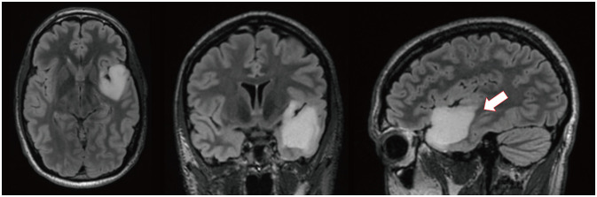
Representative T2/fluid-attenuated inversion recovery MRI on admission (axial, coronal, and sagittal, from left to right, respectively). These images show a hyperintense non-enhancing mass in the left temporal lobe, measuring 4.1×3.6×3.4 cm with extension to posterior superior temporal gyrus (arrow).

btrt-11-145-g001
Fig. 2

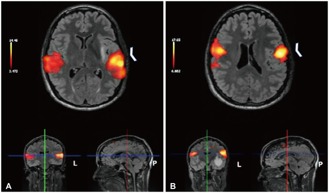
Functional magnetic resonance imaging (fMRI). A: Axial fMRI shows activation of language areas during word-generation/sentence completion tasks in bilateral cerebral hemispheres in temporal lobes (left greater than right; arrowhead). B: Axial fMRI shows blood-oxygen-level dependent activation can be seen superior to the mass during the movement of the tongue in the precentral gyrus bilaterally (arrowhead).

btrt-11-145-g002
Fig. 3

Diffusion tensor imaging demonstrates tracts from the arcuate fasciculus and inferior longitudinal fasciculus partially displaced from the mass effect of the tumor. This also demonstrates how close the fibers are associated with the lesion, which would benefit from an awake craniotomy.

btrt-11-145-g003
Fig. 4

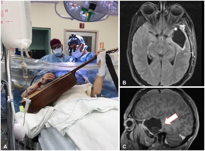
Resection of brain tumor with speech mapping. A: Intraoperative image of the patient playing his guitar during the awake craniotomy. B and C: Postoperative axial (B) and sagittal (C) fluid-attenuated inversion recovery MRI showing resection cavity with a small residual portion of blood products and granulation tissue at the base (arrow).

btrt-11-145-g004
Table 1

Awake craniotomy for music mapping in brain tumor resection: demographic, clinical, and operative characteristics for cases with standard music tasks
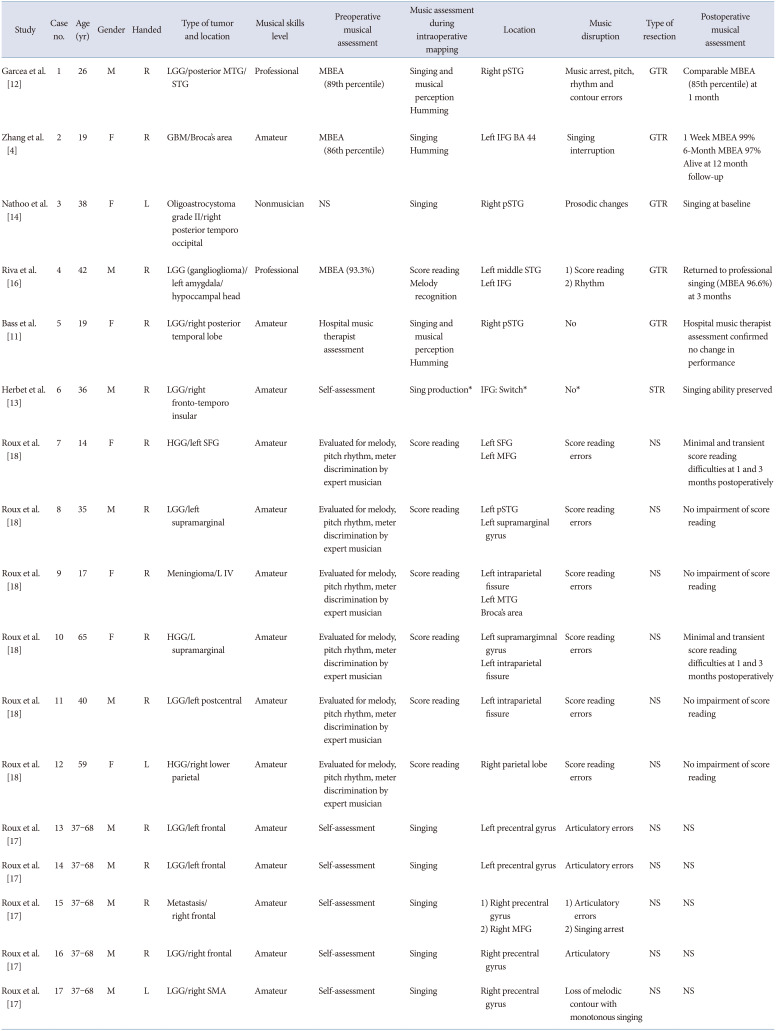
btrt-11-145-i001
Study Case no. Age (yr) Gender Handed Type of tumor and location Musical skills level Preoperative musical assessment Music assessment during intraoperative mapping Location Music disruption Type of resection Postoperative musical assessment
Garcea et al. [12] 1 26 M R LGG/posterior MTG/STG Professional MBEA (89th percentile) Singing and musical perception
Humming
Right pSTG Music arrest, pitch, rhythm and contour errors GTR Comparable MBEA (85th percentile) at 1 month
Zhang et al. [4] 2 19 F R GBM/Broca’s area Amateur MBEA (86th percentile) Singing
Humming
Left IFG BA 44 Singing interruption GTR 1 Week MBEA 99%
6-Month MBEA 97%
Alive at 12 month follow-up
Nathoo et al. [14] 3 38 F L Oligoastrocystoma grade II/right posterior temporo occipital Nonmusician NS Singing Right pSTG Prosodic changes GTR Singing at baseline
Riva et al. [16] 4 42 M R LGG (ganglioglioma)/left amygdala/hypoccampal head Professional MBEA (93.3%) Score reading
Melody recognition
Left middle STG
Left IFG
1) Score reading
2) Rhythm
GTR Returned to professional singing (MBEA 96.6%) at 3 months
Bass et al. [11] 5 19 F R LGG/right posterior temporal lobe Amateur Hospital music therapist assessment Singing and musical perception
Humming
Right pSTG No GTR Hospital music therapist assessment confirmed no change in performance
Herbet et al. [13] 6 36 M R LGG/right fronto-temporo insular Amateur Self-assessment Sing production* IFG: Switch* No* STR Singing ability preserved
Roux et al. [18] 7 14 F R HGG/left SFG Amateur Evaluated for melody, pitch rhythm, meter discrimination by expert musician Score reading Left SFG
Left MFG
Score reading errors NS Minimal and transient score reading difficulties at 1 and 3 months postoperatively
Roux et al. [18] 8 35 M R LGG/left supramarginal Amateur Evaluated for melody, pitch rhythm, meter discrimination by expert musician Score reading Left pSTG
Left supramarginal
Score reading errors NS No impairment of score reading
Roux et al. [18] 9 17 F R Meningioma/L IV Amateur Evaluated for melody, pitch rhythm, meter discrimination by expert musician Score reading Left intraparietal fissure
Left MTG
Broca’s area
Score reading errors NS No impairment of score reading
Roux et al. [18] 10 65 F R HGG/L supramarginal Amateur Evaluated for melody, pitch rhythm, meter discrimination by expert musician Score reading Left supramargimnal gyrus
Left intraparietal fissure
Score reading errors NS Minimal and transient score reading difficulties at 1 and 3 months postoperatively
Roux et al. [18] 11 40 M R LGG/left postcentral Amateur Evaluated for melody, pitch rhythm, meter discrimination by expert musician Score reading Left intraparietal fissure Score reading errors NS No impairment of score reading
Roux et al. [18] 12 59 F L HGG/right lower parietal Amateur Evaluated for melody, pitch rhythm, meter discrimination by expert musician Score reading Right parietal lobe Score reading errors NS No impairment of score reading
Roux et al. [17] 13 37–68 M R LGG/left frontal Amateur Self-assessment Singing Left precentral gyrus Articulatory errors NS NS
Roux et al. [17] 14 37–68 M R LGG/left frontal Amateur Self-assessment Singing Left precentral gyrus Articulatory errors NS NS
Roux et al. [17] 15 37–68 M R Metastasis/right frontal Amateur Self-assessment Singing 1) Right precentral gyrus
2) Singing arrest
1) Articulatory errors
2) Right MFG
NS NS
Roux et al. [17] 16 37–68 M R LGG/right frontal Amateur Self-assessment Singing Right precentral gyrus Articulatory NS NS
Roux et al. [17] 17 37–68 M L LGG/right SMA Amateur Self-assessment Singing Right precentral gyrus Loss of melodic contour with monotonous singing NS NS

*Switch from normal speech to singing. LGG, low grade glioma; MTG, middle temporal gyrus; STG, superior temporal gyrus; MBEA, Montreal Battery of Evaluation of Amusia; pSTG, posterior superior temporal gyrus; GTR, gross total resection; GBM, glioblastoma multiforme; IFG; inferior frontal gyrus; BA, Brodmann area; NS, non-specified; HGG, high grade glioma; SFG, superior frontal gyrus; MFG, middle frontal gyrus; SMA, supplementary motor area; STR, subtotal resection; NTR, near total resection

Table 2

Cases of awake craniotomy and intraoperative instrument playing: demographic, clinical, and operative characteristics
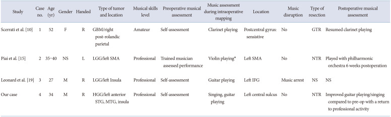
btrt-11-145-i002
Study Case no. Age (yr) Gender Handed Type of tumor and location Musical skills level Preoperative musical assessment Music assessment during intraoperative mapping Location Music disruption Type of resection Postoperative musical assessment
Scerrati et al. [10] 1 52 F R GBM/right post-rolandic parietal Amateur Self-assessment Clarinet playing Postcentral gyrus: sensistive No GTR Resumed clarinet playing
Piai et al. [15] 2 35–40 NS L LGG/left SMA Professional Trained musician assessed performance Violin playing* Left SMA No NTR Played with philharmonic orchestra 6 weeks postoperation
Leonard et al. [19] 3 27 M R LGG/left Insula Professional Self-assessment Guitar playing Left IFG Music arrest NS NS
Our case 4 34 M R HGG/left anterior STG, MTG, insula Professional Self-assessment Singing, guitar playing Left central sulcus No NTR Improved guitar playing/singing compared to pre-op with a return to professional activity

*Seizure arrested intraoperative assessment. GBM, glioblastoma multiforme; GTR, gross total resection; LGG, low grade glioma; SMA, supplementary motor area; NTR, near total resection; IFG, inferior frontal gyrus; NS, non-specified; HGG, high grade glioma; STG, superior temporal gyrus; MTG, middle temporal gyrus

TOOLS
Similar articles