

 PDF
PDF Citation
 Print
Print


INTRODUCTION
Immunoglobulin G4-related disease (IgG4-RD) is a systemic immune-mediated fibroinflammatory disease. The disease can be characterized by the elevated level of IgG4 in the serum. On the other hand, a high IgG4 level is not diagnostic because it can be associated with many other conditions, such as allergic, infectious, and malignant diseases.1 Furthermore, it can present differently in many organs: the pancreas, bile ducts, gallbladder, thyroid, lungs, pericardium, salivary glands, kidneys, mesentery, periorbital tissues, prostate, skin, and retroperitoneum.1,2 Nevertheless, the small bowel involvement of IgG4-RD is rare. This paper presents a case of IgG4-RD involving the small bowel, particularly the distal ileum.
CASE REPORT
An 81-year-old female was admitted to the authors’ hospital complaining of intermittent abdominal pain and dyspepsia accompanied by hematochezia for one month. She did not have any systemic diseases except hypertension for 25 years. She denied any history of using alcohol and tobacco, or other recreational substances.
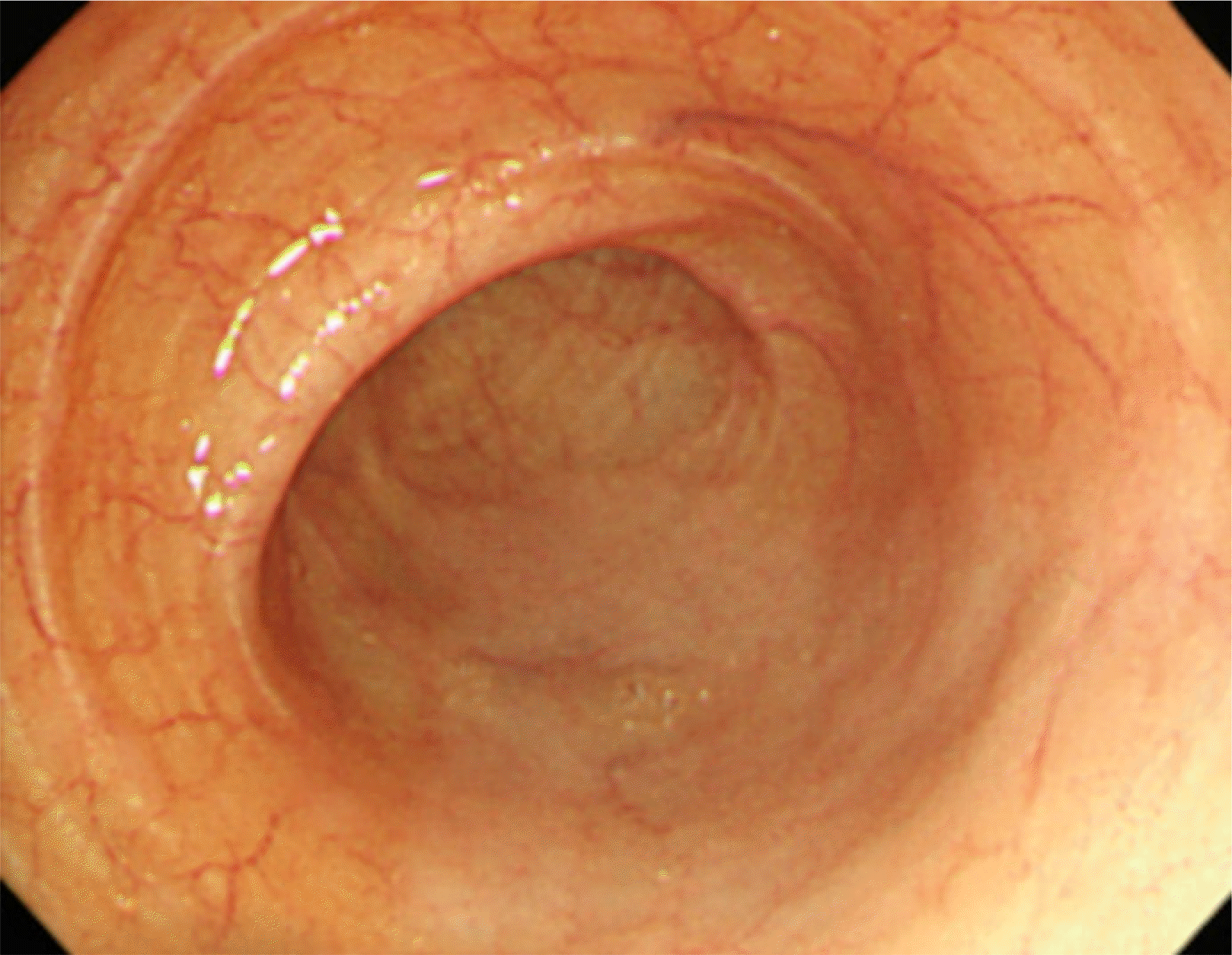
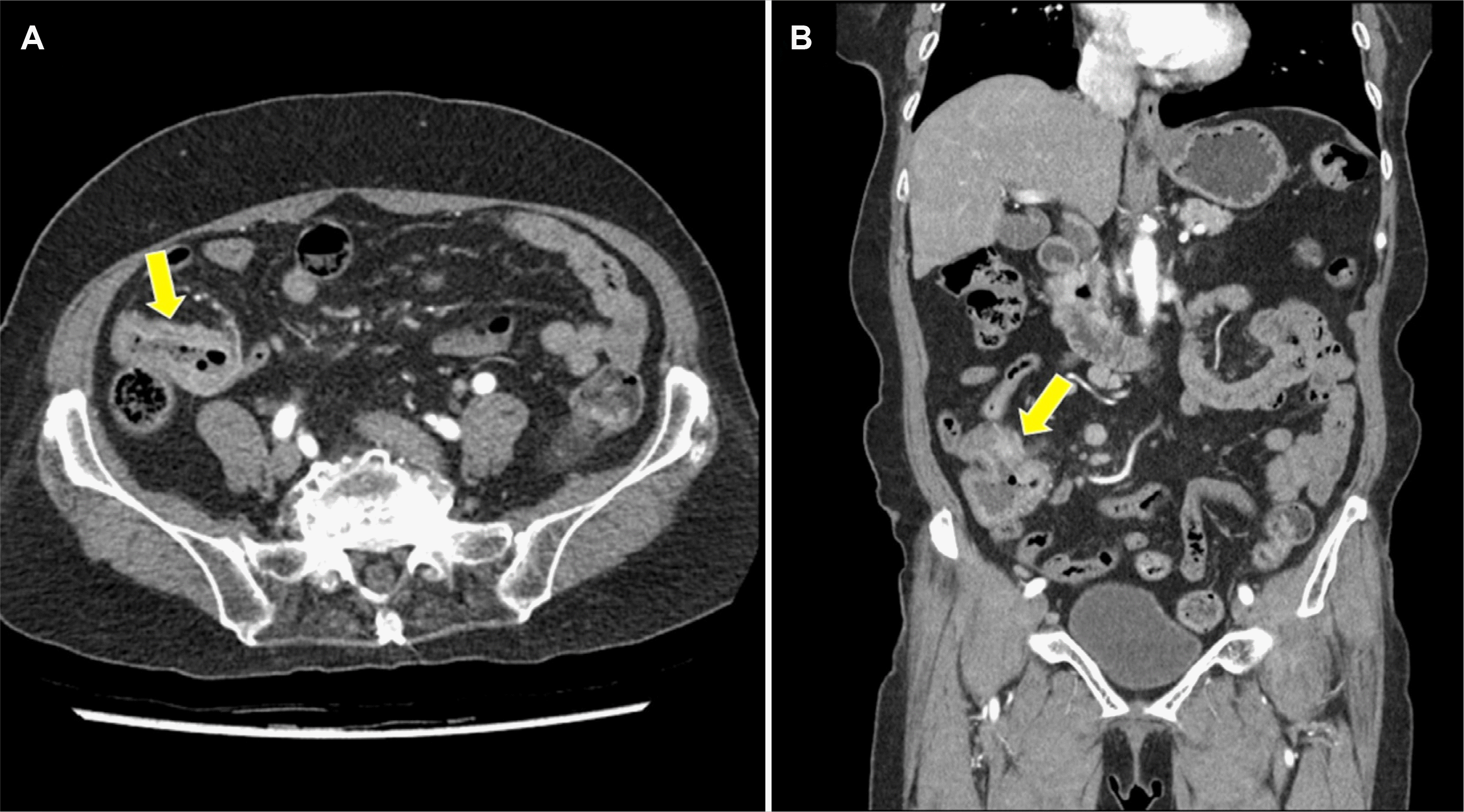
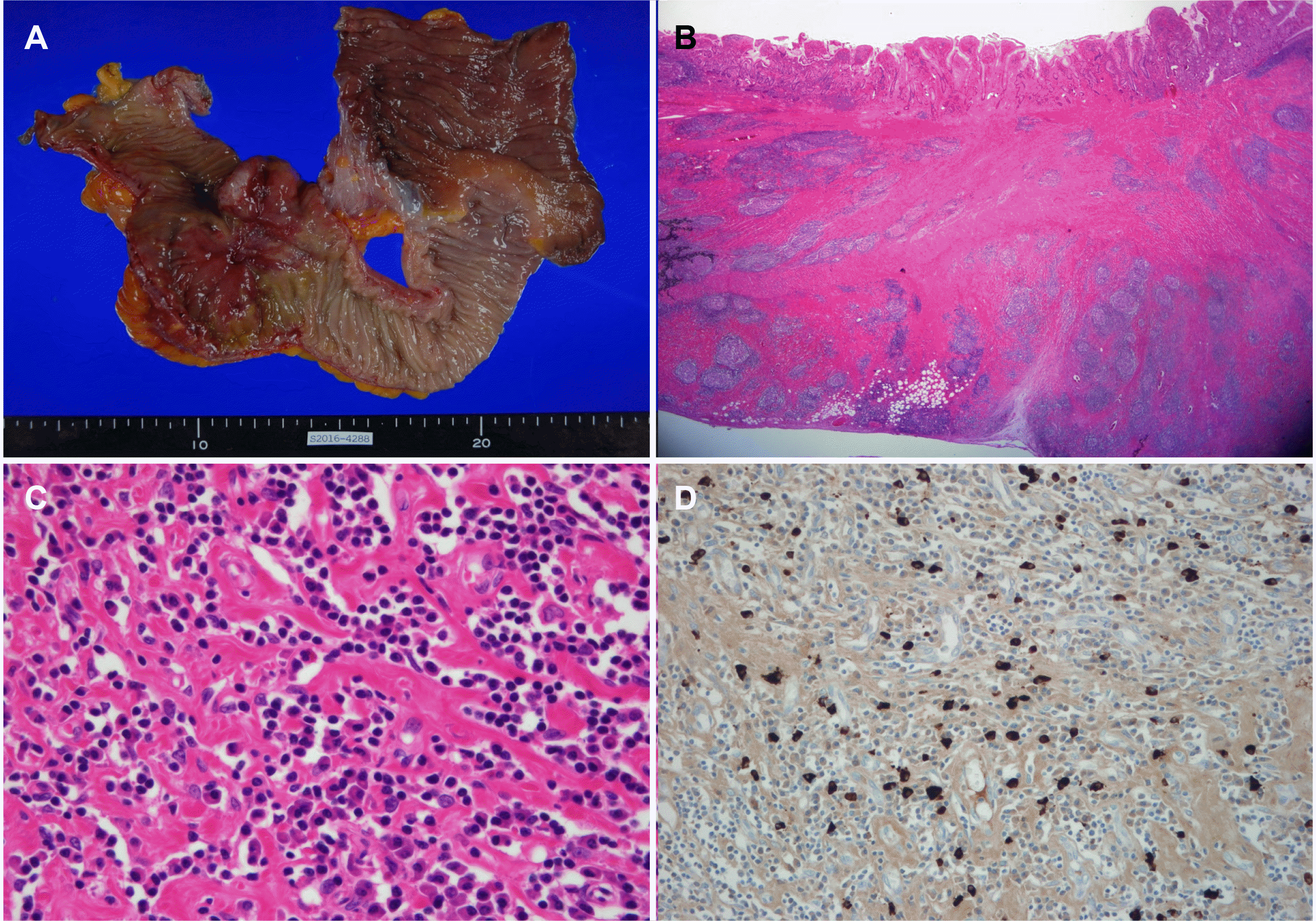
Upon admission, the blood pressure, pulse rate, and body temperature were 125/74 mmHg, 64/min, and 36.5℃, respectively. The laboratory data were as follows: white blood cell count 4,730/mm3, hemoglobin 9.8 g/dL, hematocrit 30.7%, platelet count 351,000/mm3, aspartate aminotransferase 17 IU/L, alanine aminotransferase 15 IU/L, alkaline phosphatase 84 IU/L, lactate dehydrogenase 203 U/L, amylase 38 U/L, C-reactive protein 0.3 mg/dL, Carcinoembryonic antigen 1.4 ng/mL, CA19-9 7.4 U/mL, IgG1 533.2 mg/dL, IgG2 166.1 mg/dL, IgG3 30.5 mg/dL, and IgG4 18.9 mg/dL. A colonoscopy did not show any abnormal findings in the terminal ileum (Fig. 1). Abdominal computed tomography revealed segmental aneurysmal dilatation and irregular wall thickening in the distal ileum (Fig. 2). Based on these clinical and radiology findings, malignant disease, such as small bowel lymphoma was suspected as a presumptive diagnosis. The patient underwent an exploratory laparoscopy. An approximately 4 cm mass encircling the distal ileum was observed in the surgical field. Therefore, an extensive resection and lymph node dissection were performed under the suspicion of a malignancy. The patient underwent a right hemicolectomy, which involved removing the cecum and ascending colon (9 cm in length), with a distal ileal resection (28 cm in length) and lymph node dissection. The surgical gross specimen showed a 4×4 cm ulceroinfiltrative lesion of the distal ileum (Fig. 3A). The ulceroinfiltrative lesion was located 7 cm and 11 cm apart from the ileal resection margin and ileocecal valve, respectively. The lymph nodes, the remaining ileal mucosa, and the colonic mucosa were intact. A histological examination revealed chronic ileitis with ulceration and transmural lymphoid aggregates (Fig. 3B), dense lymphoplasmacytic infiltration, and storiform fibrosis (Fig. 3C). Immunohistochemistry revealed many IgG4-positive plasma cells (>50 per high power field [HPF]) in the distal ileum, indicative of IgG4-RD (Fig. 3D). Therefore, the patient was finally diagnosed with IgG4-RD involving the distal ileum. The patient was discharged on postoperative day 10 without postoperative complications and was followed up via the outpatient clinic for five years without the recurrence of IgG4-RD.
DISCUSSION
Since IgG4-RD was recognized as a disease entity 20 years ago, it is now accepted that IgG4-RD could be manifested as many mimics of inflammatory or malignant diseases. This pleomorphic feature of IgG4-RD makes a differential diagnosis difficult, particularly when presenting as a mass-like lesion. Moreover, the diagnostic difficulty of IgG4-RD leads to the development of several guidelines and diagnostic criteria from several study groups.3-6 As IgG4-RD is a systemic autoimmune-related disease, the treatment strategies and disease prognosis differ according to the organs involved. Therefore, a new guideline focusing on the involvement of the digestive tract was published in 2020 by the European Society of Gastroenterology & Surgery groups.7 The European guideline highlights the need for a comprehensive work-up. The HISORt criteria is used as diagnostic criteria, named by the initials of the following words: “h”istopathological and “i”maging features, “s”erum IgG4 elevation, other “o”rgan involvement, and “r”esponse to steroid “t”herapy. Furthermore, a definite diagnosis of IgG4-RD is made when fulfilling the organ-specific diagnostic criteria given the established organ-specific diagnostic criteria.4,6
In laboratory tests, the serum IgG and IgG4 elevation, autoantibodies including antinuclear antibody, and rheumatoid factor can help diagnose IgG4-RD, even though such laboratory findings are not specific for the diagnosis. The histological examination revealed abundant IgG4+ plasma cells within the affected tissues as distinct features: proposed diagnostic criteria of >50 IgG4 + plasma cells per HPF and an IgG4 +/IgG + ratio > 40%.1,4,7 Other important pathological features include storiform fibrosis, lymphoplasmacytic infiltration, and phlebitis. In the present case, although the serum IgG4 was within the normal range, storiform fibrosis, dense lymphoplasmacytic infiltration, and IgG4+ plasma cells >50/HPF were demonstrated in the resected tissue, making a definite diagnosis of IgG4-RD involving the distal ileum of the small bowel. In addition, this case showed that long-term remission without relapse had been maintained after a surgical resection for approximately five years. Watanabe et al. reported that a surgical resection without steroid maintenance therapy could achieve the long-term remission of IgG4-RD.8 IgG4-RD has a high relapse rate of approximately 50% after the discontinuation of the steroid, even though steroid is a very effective agent for the treatment of IgG4-RD,9 and it can develop into malignant disease, particularly for IgG4-related pancreatitis (also known as autoimmune pancreatitis type 1).10-12 Therefore, a surgical resection for IgG4-RD can be a treatment option, especially when the IgG4-RD manifests as a localized lesion of the gastrointestinal tract with concern for possible malignancy as a differential diagnosis.
IgG4-RD can involve various organs, such as the pancreas, bile ducts, gallbladder, thyroid, liver, kidneys, and lungs. Although IgG4-RD can manifest only in a single organ, as in the present case, the involvement of multiple organs simultaneously is a distinct feature of IgG4-RD. Therefore, when IgG4-RD is suspected, it is important to determine if the other organs are involved.
The pancreas and bile ducts are the most frequently affected organs in IgG4-related pancreatitis and IgG4-related cholangitis. On the other hand, the other gastrointestinal tracts are rarely involved, even though a few reports of IgG4-RD involving the esophagus or stomach have been published. Furthermore, small bowel involvement has been scarcely reported.8,13-18
The symptoms of IgG4-RD can differ according to the involved organs. In particular, in hepatobiliary involvement (IgG4-related cholangitis or IgG4-related pancreatitis), IgG4-RD usually presents with obstructive jaundice, weight loss, and abdominal discomfort. On the other hand, the symptoms related to IgG4-RD involving the small bowel have not been well reported. Therefore, considering the previous reports of IgG4-RD of the small bowel, the symptoms may include recurrent abdominal pain with/without vomiting or bloating and obscure bleeding without definite lesions on routine examinations.
In elderly patients presenting with abdominal pain, dyspepsia, and hematochezia, it is crucial to differentiate these symptoms from infectious enterocolitis, ischemic colitis, and gastrointestinal tract malignancy. In the present case, there were no signs of infection, such as fever, and the symptoms were not acute, which helped to exclude infectious enterocolitis and ischemic colitis.
Regarding the radiology findings of IgG4-RD involving the small bowel, Ko et al.14 described the following radiology findings of a computed tomography scan: losses of mural stratification, irregularly thickened wall, and segmental aneurysmal dilatation, which are similar findings of our case.
In summary, IgG4-RD can mimic lymphoproliferative disorders, even in the distal ileum of the small bowel. Furthermore, to the best of our knowledge, this is the first case report demonstrating the long-term clinical outcomes after radical surgery for IgG4-RD solely involving the distal ileum of the small bowel. This case suggests that a radical resection without maintenance therapy can be a treatment option, particularly when the IgG4-RD manifests as a localized gastrointestinal tract lesion.
 XML Download