Abstract
Background/Aims
The Reflux Symptom Index (RSI) is a questionnaire that evaluates the severity of extra-esophageal symptoms and is one of the most widely used measures to evaluate LPR. This study assessed the validity and reliability of the RSI questionnaire in Bahasa Indonesia and investigated the association between each extra-esophageal symptom reported in the questionnaire and the severity of erosive esophagitis as determined by endoscopic findings.
Methods
85 adult patients with GERD symptoms had an upper endoscopy examination and were asked to complete the translated RSI. The validity and reliability of the questionnaire were assessed.
Results
The construct validity of the RSI translated into Bahasa Indonesia was verified with the r value of each question being higher than the crucial table value (r>0.213, p<0.05). Our questionnaire had a Cronbach alpha value of 0.81, which indicates an acceptable level of internal consistency. At least one extra-esophageal symptom was seen in 91.7% of patients with Los Angeles (LA) grade B or higher-grade esophagitis. In addition, the presence of extra-esophageal symptoms was associated with significant mucosal erosion (p=0.20). The symptoms of cough after eating or lying down and chronic cough were associated with the severity of esophageal mucosal erosion (p<0.05).
Conclusions
The version of RSI translated into Bahasa Indonesia is a valid and reliable tool for assessing extra-esophageal GERD symptoms. The occurrence of extra-esophageal symptoms in patients with typical GERD symptoms is associated with endoscopic findings of LA grade B or erosive esophagitis of higher severity.
Gastroesophageal reflux disease (GERD) is characterized by the reflux of the stomach contents into the esophagus, resulting in symptoms and complications.1 The reported prevalence of GERD in the adult population worldwide was 13.9%.2 There are substantial differences in the prevalence of GERD between regions and countries, with Indonesia reporting a prevalence rate of 57.6%.3 In addition to classic symptoms such as heartburn and regurgitation, GERD may also manifest extra-esophageal symptoms, including chronic cough, throat clearing, hoarseness, asthma, and laryngitis. It is estimated that almost a third of GERD patients have extra-esophageal symptoms, with atypical chest pain being most frequently reported, followed by dysphagia and hoarseness.4,5
Extra-esophageal GERD symptoms are believed to be induced by the microaspiration of gastric contents resulting in a direct injury to the larynx, which is referred to as laryngoesophageal reflux (LPR). Stimulation of the esophageal-tracheobronchial reflex by the vagus nerve is proposed as another underlying mechanism.4 LPR and other extraesophageal manifestations of GERD are evaluated via laryngoscopy. LPR is characterized by posterior commissure hypertrophy, laryngeal and arytenoid inflammation, vocal cord edema, and endolaryngeal mucus.6
The most common objective test for evaluating GERD symptoms is upper endoscopy. According to a recent consensus, a diagnosis of GERD is made when a patient presents with heartburn and regurgitation in addition to Los Angeles (LA) grade B erosive esophagitis on endoscopy. In addition, an endoscopic finding of LA grade C or D erosive esophagitis is conclusive evidence of GERD.4 The presence and severity of erosive esophagitis determine the treatment strategy.
On the other hand, the diagnosis of extra-esophageal GERD is hard to establish since the symptoms are elusive and overlap with those of other diseases.7 Therefore, in patients with symptoms of extra-esophageal GERD, endoscopic results of LA grade B or higher are considered clinically significant erosive esophagitis. Usually, 24-hour reflux monitoring is required to identify the causality of GERD with its extra-esophageal manifestations.7 However, such monitoring facilities are not readily available at many health centers in Indonesia. Furthermore, Indonesia has limited endoscopy centers despite being the fourth most populous country in the world. Moreover, the prevalence of extra-esophageal symptoms in GERD has not been studied in Indonesia.
The Reflux Symptom Index (RSI) is a questionnaire that focuses on extra-esophageal symptoms and is one of the most widely used measures to evaluate LPR. The RSI questionnaire comprises nine subdomains, with scores ranging from 0 to 45. Patients with scores ≥13 are considered positive for LPR.8 Translations of the RSI questionnaire in several languages, including Spanish, Brazilian-Portuguese, Turkish, and Polish have been published.9-12 Indonesia is a country with diverse ethnicity and cultural backgrounds. Hence, it is essential to consider cross-cultural aspects when translating the RSI questionnaire to Bahasa Indonesia. Valid and reliable measures to evaluate extra-esophageal symptoms are still required since most of these symptoms are not specific. The primary objective of our study was to evaluate the validity and reliability of the RSI questionnaire translated to Bahasa Indonesia. Our secondary objective was to determine the prevalence of extra-esophageal symptoms of GERD and the factors associated with them.
The process of translating the RSI questionnaire to Bahasa Indonesia was done using the method suggested by Acquadro et al.13 Two professional translators (N.B. and H.P.) worked independently to generate the first two versions of the forward translation (v1 and v2). Differences were discussed with two gastroenterology experts (M.M. and T.S.) and one senior otolaryngologist (R.F.P.) until consensus was achieved (v3). This version (v3) was handed to another licensed translator with a medical background (I.K.) for backward translation. At this step, no modifications were made since the back-translation was relatively similar to the original. Ten patients of different ethnic backgrounds (4 Javanese, 3 Madurese, 1 Sundanese, and 1 Mongoloid) were then subjected to a pre-test cognitive interview to confirm that the translated questionnaire was understandable. Consensus was reached on the final version of the questionnaire (v4) after considering participant feedback. The main structure of the original questionnaire was retained during the translation process.
This was a cross-sectional study conducted in the gastro-enterology clinics at Dr. Soetomo General Hospital, Surabaya, and Siti Khadijah Sepanjang Hospital Sidoarjo. Eighty-five adults (aged ≥18 years old) who were evaluated via endoscopy for GERD were enrolled in our study. We included patients with symptoms of heartburn and/or regurgitation at least twice a week over the previous 3 months. The exclusion criteria included patients with a history of autoimmune disease, cirrhosis, chronic rhinosinusitis, malignancy, asthma, cardiovascular disease, and pregnant women. Patients with normal esophageal mucosal findings on esophagogastroduodenoscopy (EGD) but demonstrating other gastroduodenal pathologies were also excluded. All study participants were recruited consecutively by two gastroenterology experts and were not allowed to take acid-suppressant medication four weeks prior to endoscopy. Each participant provided informed consent following ethical clearance by the Medical Research Ethics Committee of the Dr. Soetomo General Hospital.
The data collected included sex, age, marital status, ethnicity, BMI, and changes in the esophageal mucosa on EGD. The LA classification system was used to describe the extent of visible mucosal breaks as follows: LA Grade A, one (or more) mucosal break(s) ≤5 mm not extending between the tops of two mucosal folds; LA grade B, one (or more) mucosal break(s) >5 mm not extending between the tops of two mucosal folds; LA grade C, one (or more) mucosal break(s), continuous between the tops of two or more mucosal folds but involving <75% of the circumference; LA grade D, One (or more) mucosal break(s) involving ≥75% of the circumference.14
Descriptive statistics were analyzed using the mean (±standard deviation), number (n), and percentage (%). We used the Pearson product-moment correlation test to evaluate the construct validity of the RSI by comparing the r value to the critical table value for each item in the overall survey (r>0.213, p<0.05).15 Reliability was measured using Cronbach’s alpha internal consistency measurement, with alpha values classified as follows: excellent (>0.9), good (>0.8), acceptable (>0.7), questionable (>0.6), poor (>0.5), unacceptable (<0.5).16 The ability of the translated version of the RSI to discriminate LA grade B or higher esophagitis was evaluated using receiver operating characteristic (ROC) analysis. The association between each extra-esophageal symptom presented in the RSI and the esophagitis grade was assessed using the Spearman ρ coefficient. All data were analyzed using IBM SPSS Statistics version 25.0 (IBM Co., Armonk, NY, USA).
Table 1 presents the clinical characteristics of the 85 participants in this study. The study population had a mean age of 41 years. Among the subjects, 69.4% were female and 68.2% were married. In terms of ethnicity, 72.9% of the study population was Javanese, 17.6% Madurese, 5.9% Chinese, and 3.6% Sundanese. The average BMI was 23.8 kg/m2. EGD investigation revealed that 35.3% of the subjects had no mucosal break, 38.8% had LA grade A esophagitis, 23.5% had LA grade B esophagitis, and 2% had LA grade C esophagitis.
The construct validity of the version of RSI questionnaire translated to Bahasa Indonesia was verified with the r value of each question being higher than the crucial table value (r>0.213, p<0.05) (Table 2). The questionnaire had a Cronbach alpha value of 0.81, which indicates an acceptable level of internal consistency.
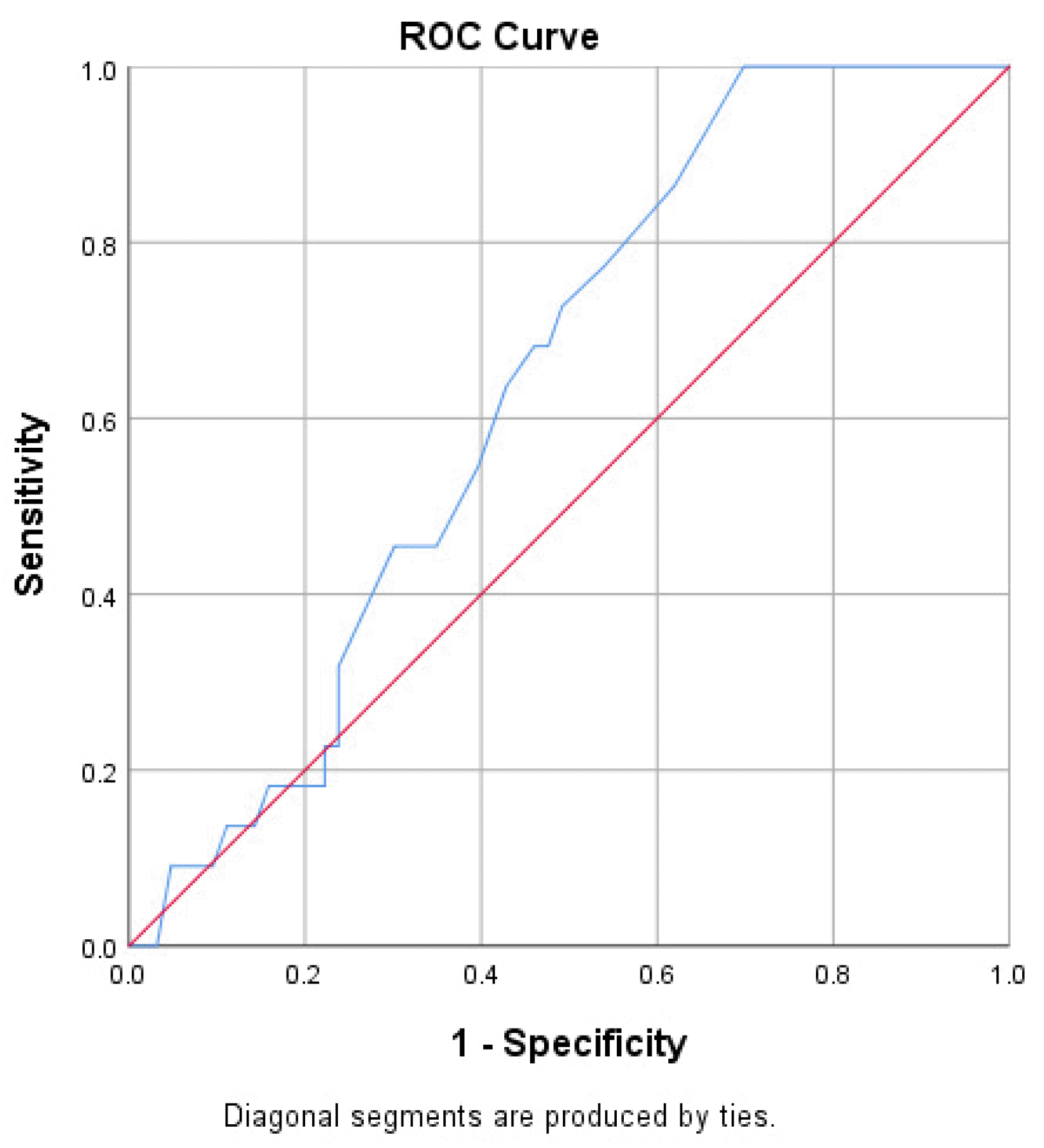
The ROC curve analysis of the translated version of the RSI questionnaire revealed an area under the curve (AUC) of 0.647 (0.518-0.755) (Fig. 1). However, a diagnostic cut-off point could not be determined since the AUC was <0.7.
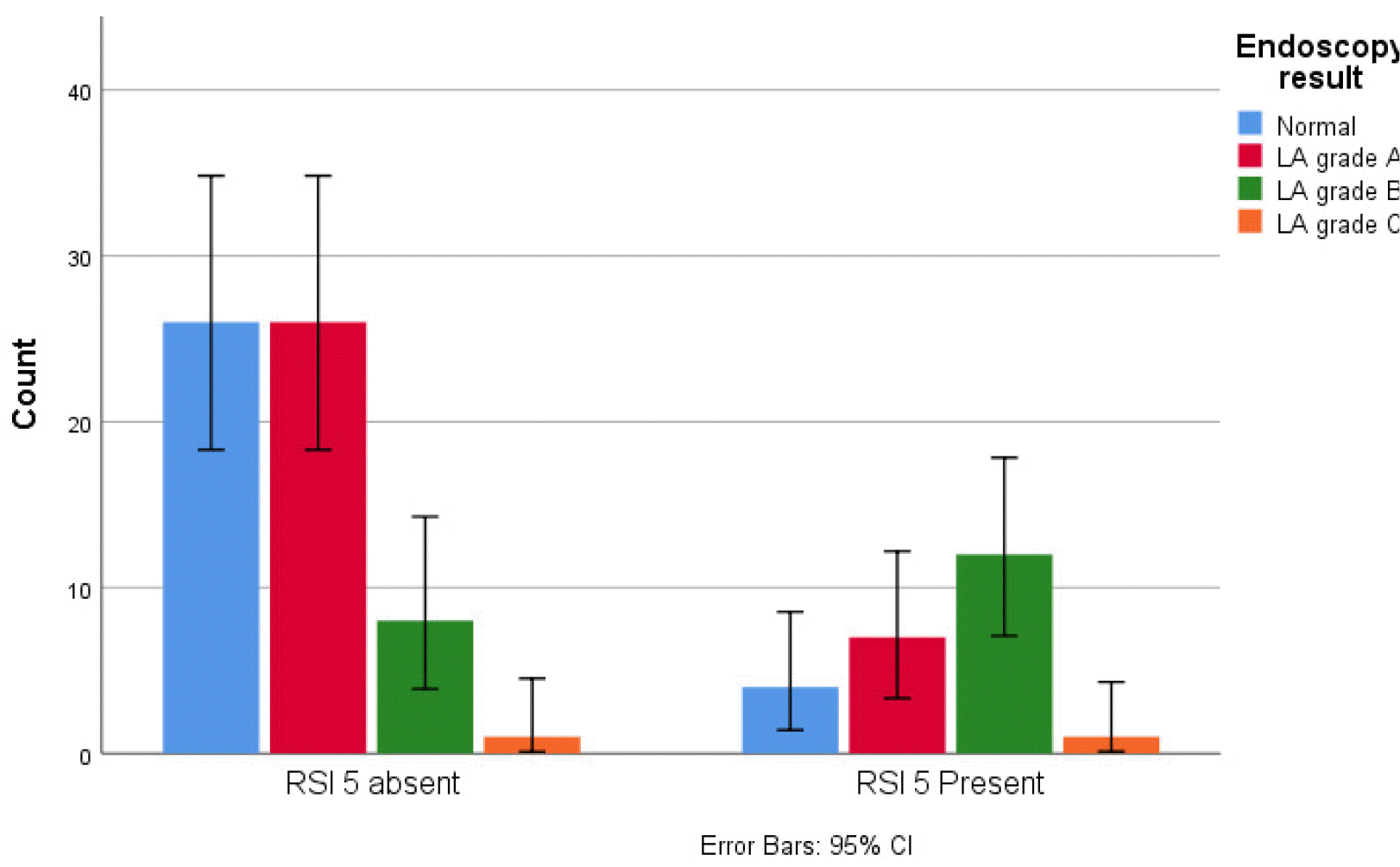
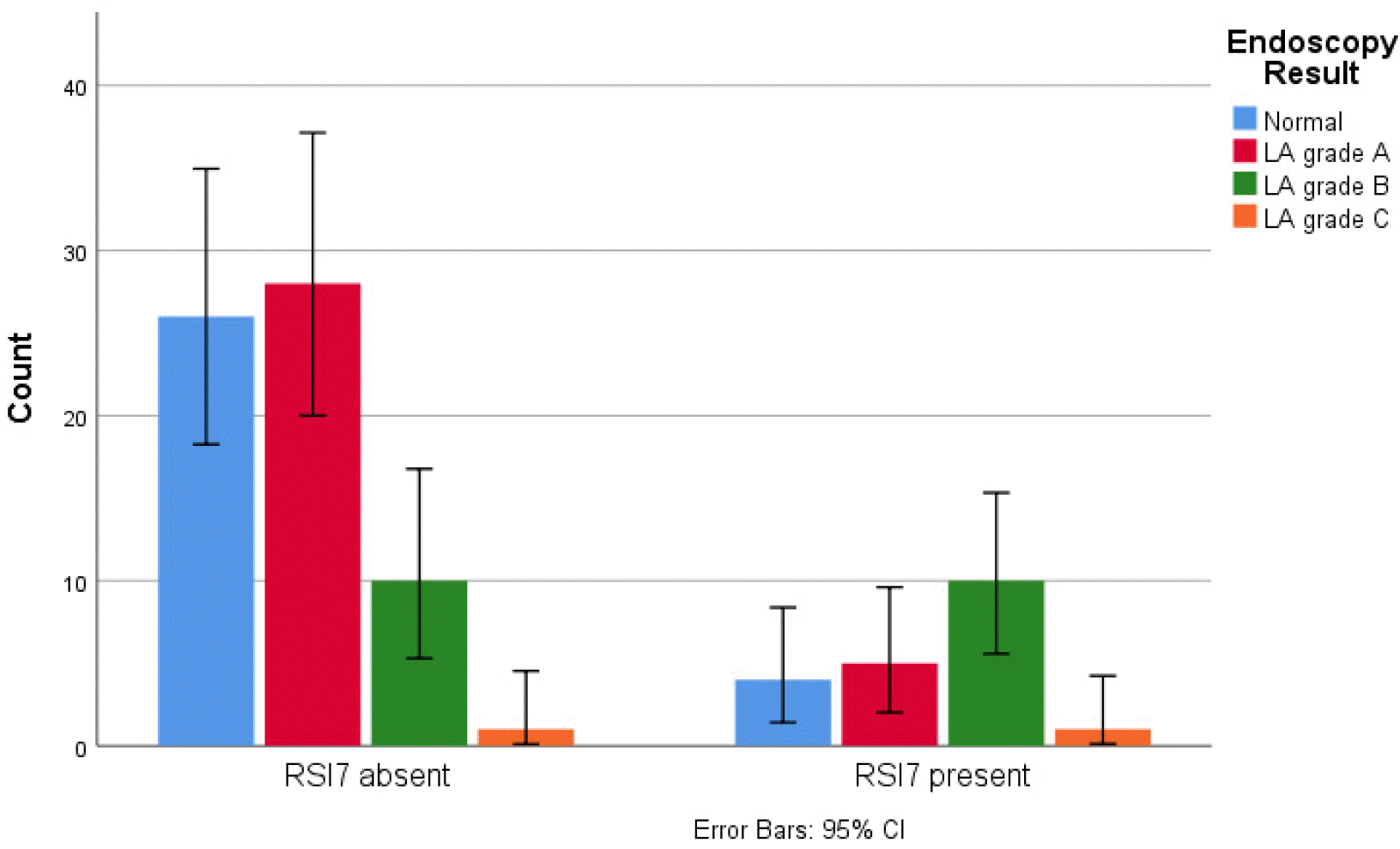
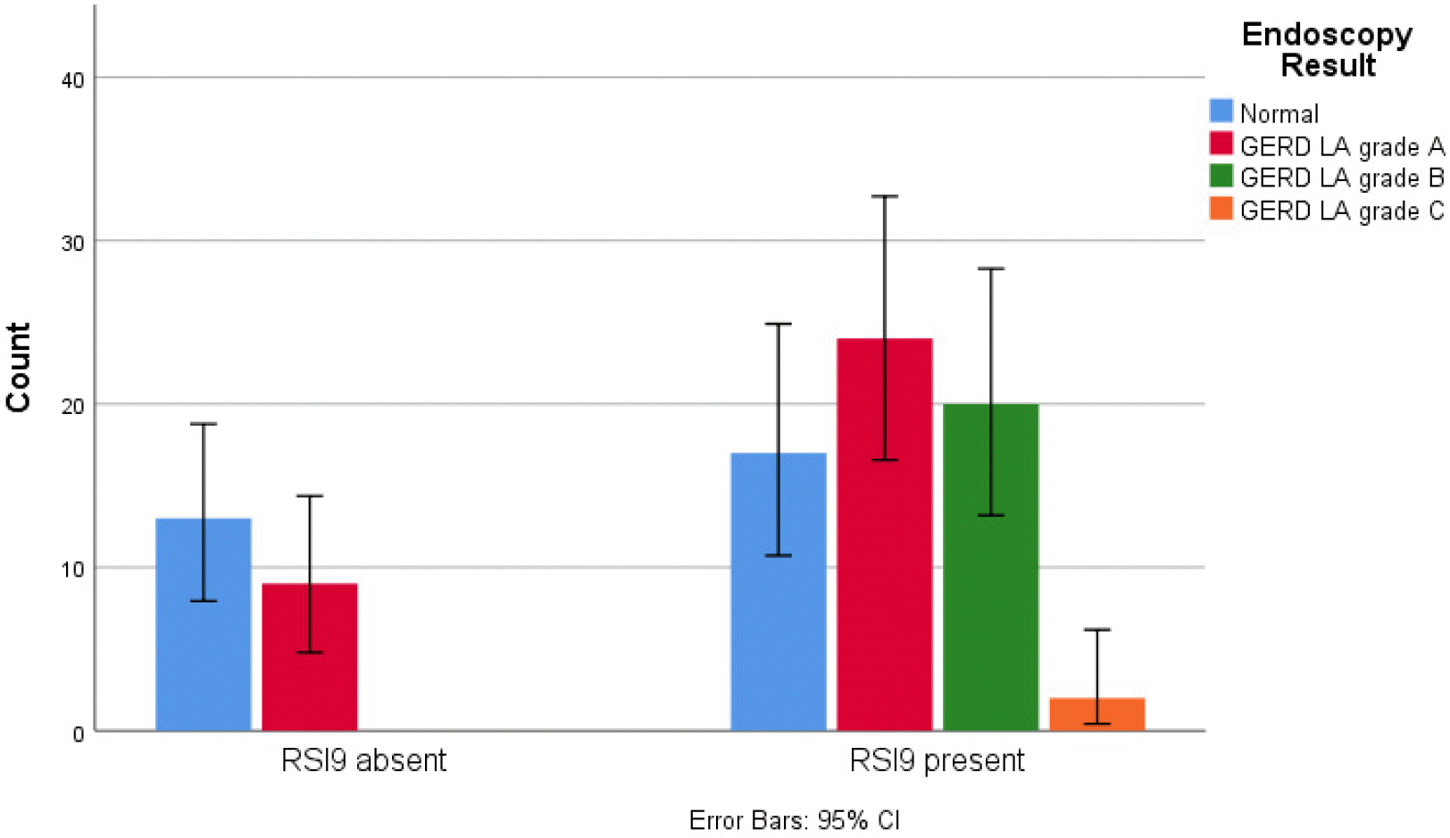
The prevalence of extra-esophageal symptoms in the study was 83.53%. Among patients with LA grade B or higher esophagitis, 91.7% had at least one extra-esophageal symptom. The presence of extra-esophageal symptoms was found to be associated with significant mucosal erosion (p=0.20) (Table 3). A further breakdown of the extra-esophageal symptoms showed that the prevalence of chronic cough in subjects with LA grade B or higher esophagitis was 50%. In contrast, the prevalence was only 14.3% in those not suffering from LA grade B or higher esophagitis. The prevalence of cough after eating or lying down was 59.1% in subjects with significant erosive esophagitis, while it was only present in 17.5% of those without significant erosive esophagitis. The correlation analysis demonstrated that questions 5 (cough after eating or lying down), 7 (chronic cough), and 9 (heartburn, chest pain, indigestion, or reflux symptoms) in the RSI questionnaire were associated with severity of esophageal mucosal erosion (p<0.05) (Figs. 2-4). In addition, Table 4 presents the correlation between each extra-esophageal symptom and the LA grade of erosive esophagitis.
In this study, the validity and reliability of the RSI questionnaire translated into Bahasa Indonesia for the assessment of the extra-esophageal symptoms of GERD were demonstrated. The reliability analysis showed that the translated questionnaire had an alpha value of 0.81, which was classified as good reliability.17 The results are in line with previous studies that evaluated the translation of the RSI questionnaire into multiple languages.9-12 The pre-final version of the translated RSI questionnaire was tested first on ten patients to ensure that it was comprehensible to individuals from various cultural backgrounds. Since Javanese, Sundanese, and Madurese are three of the five most common ethnic groups in Indonesia, the participants of the study were an accurate representation of the national population.18 The AUC of the translated questionnaire for predicting significant erosive esophagitis was 0.647 (range 0.518-0.755). However, the low value of the AUC was expected since the questionnaire was initially developed to evaluate extra-esophageal symptoms and predict LPR.
In the study, 83.53% of the subjects showed extra-esophageal symptoms in GERD. This number is higher than those of previous studies and may be attributed to the small sample size and the recruitment method in our study.5,19 This study revealed that extra-esophageal symptoms were more prevalent in patients with LA grade B or higher esophagitis compared to those without mucosal erosion or with LA grade A esophagitis. A significant association between the presence of extra-esophageal symptoms and higher grade esophageal erosion was also demonstrated (Table 3). Several earlier studies have also reported similar findings.5,20,21 The ProGERD study showed that extra-esophageal symptoms are significantly more prevalent in patients with erosive reflux disease (ERD) compared with patients with non-erosive reflux disease (NERD).5 The study demonstrated LA grade C/D esophagitis to be one of the risk factors for extra-esophageal symptoms. A deeper analysis showed that the extra-esophageal symptoms associated with erosive esophagitis were chronic cough and cough after lying down. No significant correlation was found for the other extra-esophageal symptoms. However, one study done in 2008 reported the absence of a significant relationship between extra-esophageal symptoms and the grade of esophagitis.19 The discrepancies between this and the current study may be explained by the differences in recruitment criteria and the methods used for the assessment of extra-esophageal GERD.
The latest guideline of the American College of Gastroenterology (ACG) on GERD states that the role of endoscopy in the diagnosis of extra-esophageal GERD is not yet clear.7 While the presence of significant esophageal erosion during endoscopy indicates GERD, the causality of the reflux and esophageal symptoms is still hard to establish. Endoscopic findings are also not specific and may be found even in asymptomatic individuals.22 It should be noted, however, that the study documenting this finding included the presence of LA grade A esophagitis as a definite diagnosis of GERD. According to the current paradigm LA grade A esophagitis is not a reliable finding and should not be used to for the diagnosis of GERD.7 Similarly, laryngoscopy findings have been shown to have a low correlation with extra-esophageal symptoms and pH-impedance monitoring.6,7 Currently, 24-hour-pH monitoring or impedance monitoring is more useful to diagnose extra-esophageal GERD, as these tests can record the temporal association between reflux and the symptoms and establish causality.7 Our findings may imply that RSI accompanied by significant esophageal erosion via endoscopic findings may indicate extra-esophageal GERD more definitively. However, a confirmed diagnosis still requires reflux monitoring.
We recognize that the association between extra-esophageal symptoms and ERD in our study might represent coexisting pathology and may not be caused by GERD alone. In fact, this is one of the challenges faced in the diagnosis of extra-esophageal GERD as there is no gold standard to establish the causality of the symptoms to the reflux. The study has a cross-sectional design that also implies that no causal relationship may be inferred from this association. Unfortunately, we had only two participants who had LA grade C esophagitis and no participant with grade D esophagitis. The absence of sufficient patients in each grade of severity of esophagitis was a drawback as symptoms and esophagitis severity could not be correlated in their entirety. We did not perform a laryngoscopy to identify the laryngeal damage due to LPR. The recruitment method of our study may have introduced bias since the subjects were from a gastroenterology clinic in the referral hospitals. Finally, we also recognize the relatively small sample size as another weakness in our study.
The version of the RSI questionnaire translated to Bahasa Indonesia was found to be valid and reliable for assessing extra-esophageal GERD symptoms. The occurrence of extra-esophageal symptoms in patients with typical GERD symptoms is associated with endoscopic findings of LA grade B or higher erosive esophagitis.
REFERENCES
1. Vakil N, van Zanten SV, Kahrilas P, Dent J, Jones R. Global Consensus Group. 2006; The Montreal definition and classification of gastroesophageal reflux disease: a global evidence-based consensus. Am J Gastroenterol. 101:1900–1920. quiz 1943DOI: 10.1111/j.1572-0241.2006.00630.x. PMID: 16928254.
2. Nirwan JS, Hasan SS, Babar ZU, Conway BR, Ghori MU. 2020; Global prevalence and risk factors of gastro-oesophageal reflux disease (GORD): Systematic review with meta-analysis. Sci Rep. 10:5814. DOI: 10.1038/s41598-020-62795-1. PMID: 32242117. PMCID: PMC7118109.
3. Syam AF, Sobur CS, Hapsari FCP, Abdullah M, Makmun D. 2017; Prevalence and risk factors of GERD in indonesian population- An internet-based study. Advanced Science Letters. 23:6734–6738. DOI: 10.1166/asl.2017.9384.
4. Durazzo M, Lupi G, Cicerchia F, et al. 2020; Extra-esophageal presentation of gastroesophageal reflux disease: 2020 update. J Clin Med. 9:2559. DOI: 10.3390/jcm9082559. PMID: 32784573. PMCID: PMC7465150.
5. Jaspersen D, Kulig M, Labenz J, et al. 2003; Prevalence of extra-oesophageal manifestations in gastro-oesophageal reflux disease: an analysis based on the ProGERD Study. Aliment Pharmacol Ther. 17:1515–1520. DOI: 10.1046/j.1365-2036.2003.01606.x. PMID: 12823154.
6. Belafsky PC, Postma GN, Koufman JA. 2001; The validity and reliability of the reflux finding score (RFS). Laryngoscope. 111:1313–1317. DOI: 10.1097/00005537-200108000-00001.
7. Katz PO, Dunbar KB, Schnoll-Sussman FH, Greer KB, Yadlapati R, Spechler SJ. 2022; ACG clinical guideline for the diagnosis and management of gastroesophageal reflux disease. Am J Gastroenterol. 117:27–56. DOI: 10.14309/ajg.0000000000001538. PMID: 34807007. PMCID: PMC8754510.
8. Belafsky PC, Postma GN, Koufman JA. 2002; Validity and reliability of the reflux symptom index (RSI). J Voice. 16:274–277. DOI: 10.1016/S0892-1997(02)00097-8. PMID: 12150380.
9. Eckley CA, Tangerina R. 2021; Sensitivity, Specificity, and reproducibility of the Brazilian Portuguese version of the reflux symptom index. J Voice. 35:161.e15–161.e19. DOI: 10.1016/j.jvoice.2019.08.012. PMID: 31586513.
10. Calvo-Henríquez C, Ruano-Ravina A, Vaamonde P, et al. 2019; Translation and validation of the reflux symptom index to Spanish. J Voice. 33:807.e1–807.e5. DOI: 10.1016/j.jvoice.2018.04.019. PMID: 30076096.
11. Akbulut S, Aydinli FE, Kuşçu O, et al. 2020; Reliability and validity of the Turkish reflux symptom index. J Voice. 34:965.e23–965.e28. DOI: 10.1016/j.jvoice.2019.05.015. PMID: 31248727.
12. Włodarczyk E, Jetka T, Miaśkiewicz B, Skarzynski PH, Skarzynski H. 2022; Validation and reliability of Polish version of the reflux symptoms index and reflux finding score. Healthcare (Basel). 10:1411. DOI: 10.3390/healthcare10081411. PMID: 36011068. PMCID: PMC9408310.
13. Acquadro C, Conway K, Christelle G, Mear I. 2012. Linguistic validation manual for health outcome assessments. Mapi Institute;Downey, CA:
14. Lundell LR, Dent J, Bennett JR, et al. 1999; Endoscopic assessment of oesophagitis: clinical and functional correlates and further validation of the Los Angeles classification. Gut. 45:172–180. DOI: 10.1136/gut.45.2.172. PMID: 10403727. PMCID: PMC1727604.
15. Sigle D. 2022. Critical values of the Pearson product-moment correlation coefficient. Neag Center for Gifted Education and Talent Development University of Connecticut;Available from: https://researchbasics.education.uconn.edu/r_critical_value_table/. cited 2023 Jan 24.
16. George D, Mallery P. 2003. SPSS for windows step by step: A simple guide and reference. 11.0 update. 4th ed. Allyn & Bacon;Boston:
17. Cho E, Kim S. 2015; Cronbach's coefficient alpha: Well known but poorly understood. Organizational Research Methods. 18:207–230. DOI: 10.1177/1094428114555994.
18. Na'im A SH. 2010. The 2010 Census of the Indonesian Population: Citizenship, Ethnicity, Religion, and Daily Language. Badan Pusat Statistik;Jakarta:
19. Lai YC, Wang PC, Lin JC. 2008; Laryngopharyngeal reflux in patients with reflux esophagitis. World J Gastroenterol. 14:4523–4528. DOI: 10.3748/wjg.14.4523. PMID: 18680233. PMCID: PMC2731280.
20. Smit CF, van Leeuwen JA, Mathus-Vliegen LM, et al. 2000; Gastropharyngeal and gastroesophageal reflux in globus and hoarseness. Arch Otolaryngol Head Neck Surg. 126:827–830. DOI: 10.1001/archotol.126.7.827. PMID: 10888993.
21. Sadiku E, Hasani E, Këlliçi I, et al. 2021; Extra-esophageal symptoms in individuals with and without erosive esophagitis: a case-control study in Albania. BMC Gastroenterol. 21:76. DOI: 10.1186/s12876-021-01658-z. PMID: 33593300. PMCID: PMC7885502.
22. Lei WY, Yu HC, Wen SH, et al. 2015; Predictive factors of silent reflux in subjects with erosive esophagitis. Dig Liver Dis. 47:24–29. DOI: 10.1016/j.dld.2014.09.017. PMID: 25308612.