INTRODUCTION
Orthodontic treatment aims not only to merely relocate the maxillomandibular dentition but also improve facial esthetics. Therefore, orthodontists should be able to predict the pattern of changes in the relationship between teeth and facial soft tissues when establishing a diagnosis and treatment plan.
1
Numerous studies, which were conducted on the changes in the facial soft tissues attributed to orthodontic treatment, can help clinicians predict treatment outcomes. However, the association between hard and soft tissues remains controversial. Caplan and Shivapuja
2 reported that when the anterior teeth of adult patients with bimaxillary protrusion were retracted, the ratio of anterior teeth retraction to lip retraction were 1.75:1 and 1.2:1 in maxilla and mandibular, respectively. Sohn and Park
3 reported that the ratios of anterior teeth retraction to the lip retraction were 2.84:1 and 1.45:1 in maxilla and mandibular, respectively. However, some studies have found no significant association between the changes in dentition and soft tissue profile changes.
4,5 A systematic review implied that soft tissue changes were negligible, whereas individual variations in the response were large.
6
Burstone
7 stated that one of the major problems in the creation of orthodontic treatment plans is determining the anterior-posterior positioning of the anterior teeth. It is also important to consider the shape of the surrounding soft tissues and position of the lips. Even young edentulous patients who lacked tooth support experienced a small amount of posterior lip compression. This is because even without the support of teeth, a protruding look remains in cases of lip are fullness. Therefore, when establishing the location of the anterior teeth and formulating a treatment plan, the presence or absence of lip incompetence should be considered.
Lip incompetence has various causes, including imbalance of the maxillofacial structure, lip strain, short upper lip length, and increased anterior facial height.
8 Nevertheless, large retraction of the anterior teeth does not necessarily result in large posterior movement of the lips. Several studies have examined the changes in soft tissues following the retraction of the anterior teeth during orthodontic treatment in extraction cases. However, few studies have assessed the relationship between lip incompetence and patterns of lip retraction.
The aim of the present study was to 1) compare changes in hard tissue and soft tissue after the four first premolars were extracted with anterior teeth retraction according to the presence or absence of lip incompetence and 2) examine the correlation between them. We hypothesized that there would be no difference between the presence and absence of lip incompetence with respect to the soft tissue changes.
Go to :

RESULTS
Table 1 showed the demographic characteristics of the sample population. No significant differences in the age or treatment duration were observed between the competent and incompetent groups. The means and standard deviations for skeletal, dental, and soft tissue measurements in both groups at T1, T2, and T2–T1 are shown in
Table 2. There was no significant difference in skeletal measurements and dental measurements between the two groups at T1, excluding the angle between the Frankfort horizontal and mandibular planes at T2. No statistically significant differences were found between the two groups in the skeletal changes after treatment (
Table 2).
Table 2
Comparison of the skeletal changes between the competent and incompetent groups
|
Variable |
T1 |
|
T2 |
|
T2–T1 |
|
Competent group |
Incompetent group |
p-value |
Competent group |
Incompetent group |
p-value |
Competent group |
p-value |
Incompetent group |
p-value |
Intergroup comparison, p-value |
|
Skeletal |
|
|
|
|
|
|
|
|
|
|
|
|
|
|
SNA (°) |
82.11 ± 3.55 |
82.48 ± 3.27 |
0.732 |
|
80.74 ± 3.79 |
81.98 ± 3.34 |
0.280 |
|
−1.37 ± 1.70 |
0.002**,c
|
−0.50 ± 1.01 |
0.039*,c
|
0.059 |
|
SNB (°) |
76.34 ± 3.19 |
76.41 ± 2.98 |
0.945 |
|
76.11 ± 3.45 |
76.34 ± 3.11 |
0.823 |
|
−0.24 ± 1.08 |
0.339 |
−0.07 ± 1.05 |
0.764 |
0.904 |
|
ANB (°) |
5.76 ± 1.63 |
6.07 ± 1.53 |
0.787 |
|
4.53 ± 1.76 |
5.60 ± 2.15 |
0.094 |
|
−1.23 ± 1.69 |
0.004**,c
|
−0.47 ± 1.21 |
0.108 |
0.110 |
|
Wits appraisal (mm) |
1.56 ± 1.86 |
1.21 ± 3.47 |
0.693 |
|
−0.63 ± 2.28 |
−0.12 ± 3.27 |
0.576 |
|
−2.18 ± 2.56 |
0.001**,c
|
−1.33 ± 2.61 |
0.034*,c
|
0.303 |
|
Occlusal plane to GoMe (°) |
17.89 ± 3.59 |
19.81 ± 2.93 |
0.072 |
|
15.14 ± 4.30 |
17.59 ± 3.08 |
0.063 |
|
−2.75 ± 3.35 |
0.002**,c
|
−2.21 ± 3.04 |
0.004**,c
|
0.659 |
|
SN-GoMe (°) |
37.91 ± 4.65 |
40.16 ± 2.87 |
0.073 |
|
37.23 ± 5.10 |
39.78 ± 2.91 |
0.060 |
|
−0.68 ± 1.46 |
0.053 |
−0.38 ± 1.41 |
0.242 |
0.518 |
|
Occlusal plane to SN (°) |
20.01 ± 4.74 |
20.35 ± 3.32 |
0.796 |
|
22.09 ± 4.07 |
21.77 ± 4.49 |
0.816 |
|
2.07 ± 3.14 |
0.008**,c
|
1.42 ± 3.38 |
0.076 |
0.738 |
|
FMA (°) |
29.11 ± 5.79 |
32.23 ± 4.10 |
0.057 |
|
28.27 ± 5.73 |
32.29 ± 4.13 |
0.015*,a
|
|
−0.85 ± 2.88 |
0.204 |
0.06 ± 2.02 |
0.892 |
0.255 |
|
Dental |
|
|
|
|
|
|
|
|
|
|
|
|
|
|
IMPA (°) |
101.45 ± 5.92 |
101.05 ± 5.15 |
0.725 |
|
94.92 ± 4.89 |
90.02 ± 6.66 |
0.012*,a
|
|
−6.53 ± 5.59 |
< 0.001***,d
|
−11.03 ± 6.82 |
< 0.001***,c
|
0.104 |
|
U1 to SN (°) |
106.48 ± 7.98 |
109.42 ± 5.34 |
0.200 |
|
98.76 ± 6.89 |
97.41 ± 4.68 |
0.474 |
|
−7.72 ± 5.41 |
< 0.001***,c
|
−12.00 ± 5.65 |
< 0.001***,c
|
0.126 |
|
Interincisal angle (°) |
110.08 ± 20.67 |
109.39 ± 6.13 |
0.247 |
|
129.12 ± 7.52 |
132.64 ± 7.97 |
0.159 |
|
19.04 ± 20.75 |
< 0.001***,d
|
23.25 ± 7.89 |
< 0.001***,c
|
0.402 |
|
U1 to HRP (°) |
53.50 ± 4.99 |
50.23 ± 3.83 |
0.877 |
|
60.80 ± 5.12 |
61.90 ± 5.04 |
0.296 |
|
7.30 ± 5.34 |
< 0.001***,c
|
11.67 ± 5.76 |
< 0.001***,c
|
0.910 |
|
L1 to HRP (°) |
60.69 ± 5.33 |
59.16 ± 5.22 |
0.430 |
|
68.32 ± 5.77 |
70.74 ± 7.96 |
0.993 |
|
7.63 ± 6.81 |
< 0.001***,c
|
11.58 ± 7.35 |
< 0.001***,c
|
0.993 |
|
U1 to HRP (mm) |
1.14 ± 0.69 |
0.87 ± 0.68 |
0.433 |
|
1.63 ± 1.06 |
2.15 ± 1.50 |
0.209 |
|
0.49 ± 1.00 |
0.043*,c
|
1.28 ± 1.59 |
0.002**,d
|
0.065 |
|
L1 to HRP (mm) |
1.08 ± 0.69 |
0.83 ± 0.69 |
0.455 |
|
1.24 ± 0.99 |
1.75 ± 1.50 |
0.209 |
|
0.31 ± 1.47 |
0.896 |
1.12 ± 1.91 |
0.029*,d
|
0.142 |
|
U1 to VRP (mm) |
25.28 ± 5.95 |
28.04 ± 4.78 |
0.120 |
|
17.49 ± 6.04 |
19.33 ± 4.93 |
0.298 |
|
−7.79 ± 1.48 |
< 0.001***,c
|
−8.71 ± 1.67 |
< 0.001***,d
|
0.072 |
|
L1 to VRP (mm) |
19.16 ± 6.52 |
22.89 ± 5.52 |
0.058 |
|
13.12 ± 5.80 |
15.23 ± 4.53 |
0.207 |
|
−6.04 ± 1.68 |
< 0.001***,c
|
−7.65 ± 2.47 |
< 0.001***,d
|
0.074 |
|
Soft tissue |
|
|
|
|
|
|
|
|
|
|
|
|
|
|
Upper lip thickness (mm) |
12.76 ± 2.07 |
12.30 ± 2.07 |
0.491 |
|
15.16 ± 2.42 |
14.17 ± 2.35 |
0.060 |
|
2.40 ± 2.20 |
< 0.001***,c
|
1.87 ± 1.20 |
< 0.001***,d
|
0.350 |
|
Lower lip thickness (mm) |
15.60 ± 2.32 |
15.64 ± 1.72 |
0.946 |
|
15.84 ± 2.20 |
15.54 ± 1.65 |
0.583 |
|
0.25 ± 2.04 |
0.595 |
−0.10 ± 1.52 |
0.737 |
0.565 |
|
Nasolabial angle (°) |
102.27 ± 9.91 |
93.22 ± 12.03 |
0.008**,b
|
|
113.42 ± 11.35 |
106.14 ± 11.95 |
0.028*,a
|
|
11.15 ± 8.99 |
< 0.001***,c
|
12.91 ± 6.45 |
< 0.001***,d
|
0.480 |
|
Ls to HRP (mm) |
10.16 ± 2.71 |
12.65 ± 2.96 |
0.007**,a
|
|
8.62 ± 2.55 |
10.67 ± 2.80 |
0.020*,a
|
|
−1.54 ± 1.47 |
< 0.001***,c
|
−1.98 ± 2.34 |
0.001**,a
|
0.482 |
|
Li to HRP (mm) |
5.12 ± 2.13 |
6.97 ± 2.80 |
0.024*,a
|
|
5.49 ± 2.12 |
5.79 ± 2.37 |
0.676 |
|
0.37 ± 1.61 |
0.315 |
−1.17 ± 3.02 |
0.098 |
0.053 |
|
Ls to VRP (mm) |
33.97 ± 6.22 |
37.31 ± 4.90 |
0.174 |
|
32.01 ± 6.20 |
33.18 ± 5.06 |
0.517 |
|
−2.88 ± 1.46 |
< 0.001***,c
|
−4.13 ± 1.76 |
< 0.001***,a
|
0.019*,a
|
|
Li to VRP (mm) |
34.28 ± 6.51 |
37.56 ± 6.02 |
0.038*,b
|
|
30.00 ± 6.49 |
31.98 ± 5.44 |
0.321 |
|
−4.28 ± 1.60 |
< 0.001***,c
|
−5.57 ± 2.23 |
< 0.001***,d
|
0.030*,a
|
|
Stms to HRP (mm) |
2.19 ± 1.56 |
4.24 ± 2.28 |
0.002**,a
|
|
1.94 ± 1.79 |
2.60 ± 1.96 |
0.314 |
|
−0.24 ± 1.16 |
0.364 |
−1.64 ± 2.36 |
0.011*,d
|
0.052 |
|
Stmi to HRP (mm) |
1.95 ± 1.57 |
2.07 ± 1.25 |
0.583 |
|
1.78 ± 1.34 |
2.11 ± 1.79 |
0.883 |
|
−0.17 ± 1.15 |
0.525 |
0.04 ± 1.40 |
0.888 |
0.445 |
|
Stms to VRP (mm) |
28.23 ± 8.25 |
32.23 ± 5.02 |
0.049 |
|
26.64 ± 6.33 |
27.49 ± 4.87 |
0.639 |
|
−1.59 ± 5.25 |
0.005**,d
|
−4.75 ± 1.57 |
< 0.001***,a
|
0.002**,d
|
|
Stmi to VRP (mm) |
27.48 ± 8.31 |
30.81 ± 5.70 |
0.152 |
|
24.90 ± 6.23 |
25.95 ± 5.12 |
0.561 |
|
−2.58 ± 5.08 |
0.002**,d
|
−4.85 ± 2.18 |
< 0.001***,d
|
0.063 |
|
Stms to Stmi (mm) |
0.33 ± 0.11 |
4.12 ± 1.57 |
< 0.001***,b
|
|
0.28 ± 0.18 |
0.34 ± 0.25 |
0.416 |
|
−0.04 ± 0.19 |
0.953 |
−3.78 ± 1.54 |
< 0.001***,d
|
< 0.001***,d
|

No significant intergroup difference in the extent of retraction and vertical movement of the upper and lower incisior was observed. The extent of retraction of the upper and lower incisors were –7.79 ± 1.48 mm and –6.04 ± 1.68 mm, respectively in the competent group (p < 0.001), and –8.71 ± 1.67 mm and –7.65 ± 2.47 mm, respectively in the incompetent group (p < 0.001).
The labrale superioris (Ls) and labrale inferioris (Li) moved more posteriorly and horizontally in the incompetent group compared to in the competent group (
p < 0.05). In addition, the vertical movements of the stomion superius (Stms) and stomion inferius (Stmi) demonstrated no significant differences. However, a greater decrease in the Stms horizontal movement was observed following treatment in the incompetent group compared to in the competent group (
p = 0.002) (
Table 2). The mean ratio of both, in the competent group, when the anterior teeth were retracted, the ratios of anterior teeth retraction to lip retraction were 2.70:1 and 1.42:1 in the maxilla and mandible, respectively in the competent group, and 2.11:1 and 1.37:1 in the maxilla and mandible, respectively in the incompetent group (
Table 3).
Table 3
Ratio of the amount of incisor retraction to lip retraction
|
Variable |
Competent group |
Incompetent group |
|
U1:Ls |
2.70:1 |
2.11:1 |
|
L1:Li |
1.42:1 |
1.37:1 |

The relationship between incisor and lip retraction is shown in
Table 4. A strong positive correlation was observed between the posterior movement of the Ls (Ls to VRP) and upper central incisor retraction (U1 to VRP) (
p < 0.01) in both groups, especially the incompetent group (r = 0.767). In contrast, a positive correlation between the posterior movement of the Li (Li to VRP) and the lower central incisor (L1 to VRP) was only observed in the incompetent group (r = 0.837;
p < 0.01). Moreover, in the incompetent group the positive correlation was observed between the posterior movement of Ls with the change in the thickness of the upper lip.
Table 4
Pearson’s correlation coefficients between the soft tissue and dental changes
|
Variable |
Competent group |
Incompetent group |
|
Ls to VRP |
U1 to VRP
0.579**
|
U1 to VRP
0.767**
|
|
Upper lip thickness
0.010 |
Upper lip thickness
0.691*
|
|
Li to VRP |
L1 to VRP
0.223 |
L1 to VRP
0.837**
|
|
Lower lip thickness
0.332 |
Lower lip thickness
0.273 |

Multiple regression analysis was performed to identify the variables that could significantly predict changes in the soft tissues of the lips. Retraction of the upper lip was mostly influenced by the retraction of the upper incisors In the competent group (competent group: β = 0.660;
p < 0.001/incompetent group: β = 0.477;
p < 0.05). However, the posterior movement of the Li was most influenced by the lower central incisor, which was only observed in the incompetent group (
Table 5).
Table 5
Multiple regression models for the lips and anterior teeth of the competent and incompetent group
|
Variable |
|
Competent group |
|
Incompetent group |
|
Dependent |
Independent |
B |
SE |
β |
t
|
p-value |
B |
SE |
β |
t
|
p-value |
|
Ls to VRP |
U1 to VRP |
|
0.654 |
0.137 |
0.660 |
4.782 |
< 0.001***
|
|
0.503 |
0.223 |
0.477 |
2.258 |
0.042*
|
|
L1 to VRP |
|
0.089 |
0.157 |
0.102 |
0.563 |
0.583 |
|
0.046 |
0.137 |
0.065 |
0.337 |
0.742 |
|
U1 to SN |
|
−0.037 |
0.051 |
−0.136 |
−0.715 |
0.487 |
|
−0.018 |
0.062 |
−0.057 |
−0.287 |
0.779 |
|
Upper lip thickness (initial) |
|
0.165 |
0.283 |
0.235 |
0.585 |
0.566 |
|
0.242 |
0.231 |
0.285 |
1.046 |
0.310 |
|
Lower lip thickness (initial) |
|
−0.317 |
0.253 |
−0.503 |
−1.254 |
0.227 |
|
−0.152 |
0.278 |
−0.149 |
−0.548 |
0.591 |
|
Stms to Stmi |
|
0.203 |
1.108 |
0.031 |
0.183 |
0.858 |
|
0.136 |
0.176 |
0.120 |
0.777 |
0.451 |
|
Li to VRP |
U1 to VRP |
|
0.244 |
0.248 |
0.257 |
0.981 |
0.344 |
|
0.043 |
0.315 |
0.032 |
0.135 |
0.895 |
|
L1 to VRP |
|
0.301 |
0.232 |
0.363 |
1.294 |
0.218 |
|
0.555 |
0.194 |
0.617 |
2.859 |
0.013*
|
|
U1 to SN |
|
−0.083 |
0.076 |
−0.321 |
−1.090 |
0.296 |
|
−0.041 |
0.088 |
−0.105 |
−0.471 |
0.646 |
|
Upper lip thickness (initial) |
|
−0.032 |
0.281 |
−0.048 |
−0.114 |
0.910 |
|
−0.120 |
0.299 |
−0.111 |
−0.400 |
0.694 |
|
Lower lip thickness (initial) |
|
−0.096 |
0.251 |
−0.160 |
−0.382 |
0.707 |
|
−0.052 |
0.359 |
−0.040 |
−0.145 |
0.887 |
|
Stms to Stmi |
|
1.445 |
1.638 |
0.231 |
0.882 |
0.394 |
|
0.235 |
0.248 |
0.163 |
0.947 |
0.361 |

Go to :

DISCUSSION
This study evaluated the change of lip-facial profile following the retraction of anterior teeth in skeletal Class II malocclusion, with the hypothesis that there was no difference in soft tissue changes between the presence or absence of lip incompetence. To avoid potential confounding effects influencing the changes in the lip facial profile, many factors were considered, such as dentofacial morphology, age, sex, and soft tissue thickness (Aniruddh et al.
12). Additionally, lip growth reportedly continues until around 17 years of age
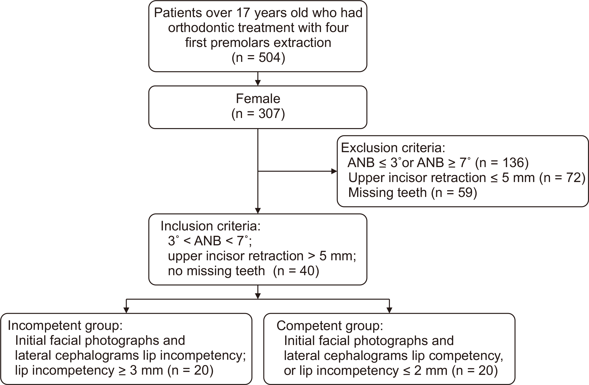
13; thus, many individuals with lip incompetence at 13 years may develop spontaneous lips-together posture at rest by 17 years of age. Therefore, this study included adult female patients aged > 17 years with no significant between-group differences in the dentofacial morphology or soft tissue thickness (
Tables 1 and
2).
Various superimposition methods have been developed using different reference planes.
14,15 Among them, Bjork’s method is regarded as highly reproducible. However, Bjork’s method is incompatible with computer-based cephalometrics. The SN superimposition method has been widely used as a computer-compatible superimposition method. Little or no difference in the accuracy and reproducibility was observed in subsequent studies on the differences between the Bjork’s and SN superimposition methods.
14,16
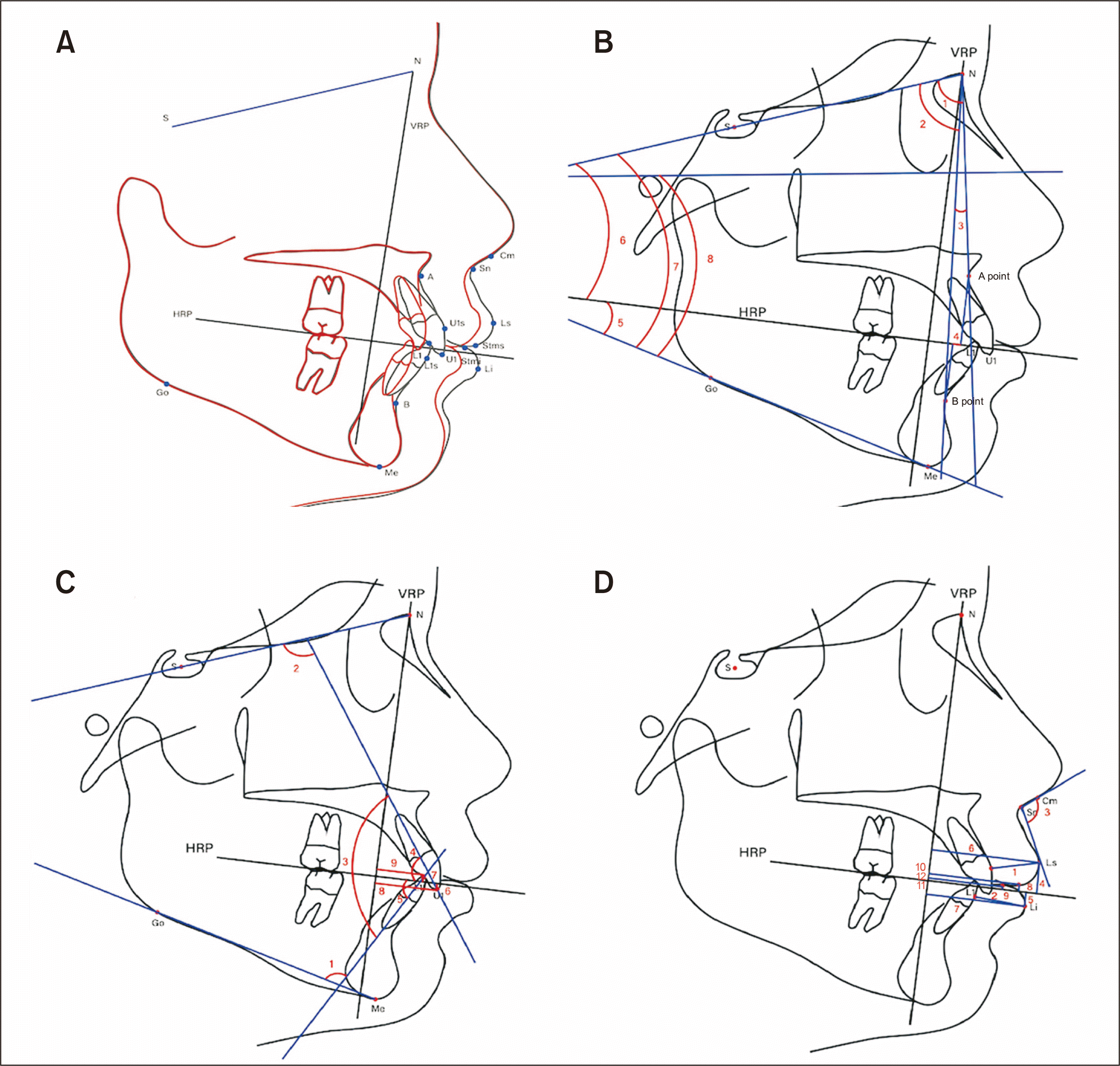
An occlusal plane with good reproducibility was set as the horizontal reference line, whereas the VRP was set as the plane passing through the nasion and perpendicular to the HRP. In clinical practice, a relaxed lip position is less reliable in cephalogram testing, unless electromyography is used. Nevertheless, the use of such positions should not be avoided if clinically helpful information can be obtained.
7
Many studies have reported the ratio of lip retraction to the corresponding retraction of the anterior teeth, and most have reported lower lip retraction to be more sensitive than incisor retraction.
2,17,18 Considering the reason for the poor response of the posterior movement of the upper lip to incisor retraction, Burstone
7 mentioned that in evaluating the soft tissue profile, individual variations in the thickness and length should be considered. Hershey
4 reported that the original force per unit area exerted by the lips, variations in the soft tissue, and changes in the intercanine width, which may alter the tension of the buccinator mechanism, should be considered. In the present study, both groups revealed that the movement of the lower lip was more sensitive than that of the upper lip (
Table 2). In both groups, the upper and lower lips were retracted along with tooth movement, but the movement was significantly greater in the incompetent group than in the competent group. There was a significant difference in the horizontal movement of the soft tissue point Stms (
p < 0.01); however, no significant difference was found in the soft tissue point Stmi between groups (
Tables 2 and
3).
In our study, the upper lip thickness significantly increased after treatment in both groups (
Table 2). There was no significant correlation between the extent of movement of the upper and lower anterior teeth (
Table 3). The amount of change in the thickness of the upper lip was similar to that reported in previous studies.
19,20 Increased lip thickness may have been caused by lip eversion.
In this study, a strong and significant positive correlation was observed between the upper lip movement and that movement of the upper anterior teeth, in which Pearson’s correlation coefficient was 0.579 in the competent group and 0.767 in the incompetent group. Whereas, the positive correlation between the movement of the lower lip and that of the lower incisor movement was only observed significantly in the incompetent group with r = 0.837 (
Table 4).
Multiple regression analysis was conducted to determine the predictability of lip changes after retraction of the anterior teeth accompanied by premolar extraction.
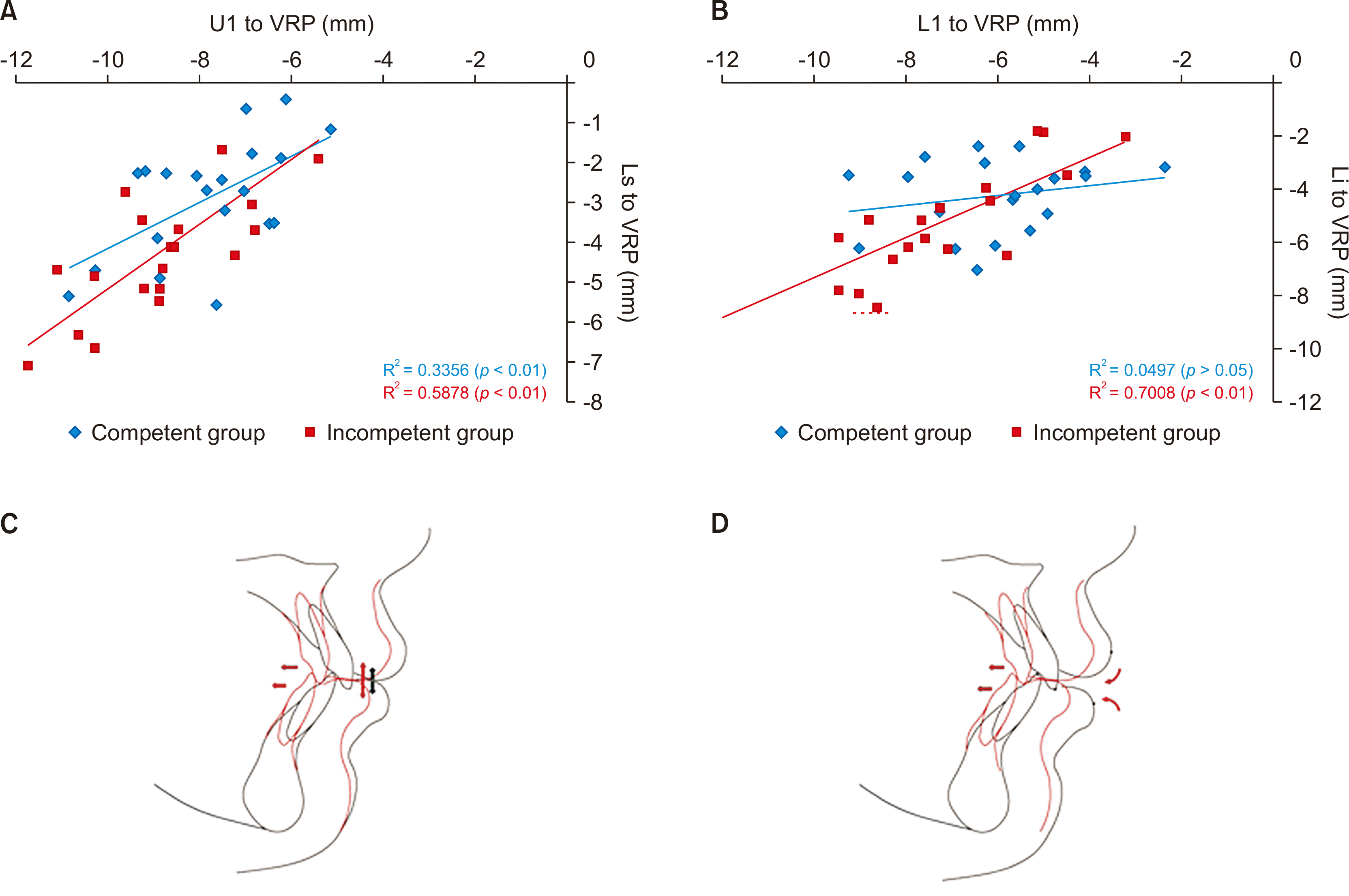
18,19 A positive correlation between the Li posterior movement and the lower incisor was observed only in the incompetent group. Multiple regression analysis demonstrated that predicting the soft tissue changes was challenging even in the competent group. These results indicated that the soft tissue response to an increase in the amount of lower incisor retraction in the competent group was not proportional to the amount of lower incisor retraction. The coefficient of determination for predicting the upper and lower lip based on the retraction of upper and lower incisors respectively were 0.59 and 0.70 in the incompetent group, showing a moderate to high predictability of the lip-profile change, which was higher than that in the competent group (
Figure 3A and B). Therefore, the retraction pattern of the lip-facial profile following the retraction of the anterior teeth seems to be more predictable in the incompetent group than in the competent group.
 | Figure 3
Scatter plot of the hard tissue versus soft tissue changes in both group and comparison of treatment changes between the two groups. A, U1 to VRP versus Ls to VRP in both groups. B, L1 to VRP versus Li to VRP in both groups. C, Lip retraction pattern in the competent group (vertical arrows indicate mutual lip pressure that may resist posterior displacement of the lips according to incisor retraction, indicated by horizontal arrows). D, Lip retraction pattern in the incompetent group (curved arrows represent free lip retraction without resistance).
U1, upper central incisor edge; VRP, vertical reference plane; Ls, labrale superioris; L1, lower central incisor edge; Li, labrale inferioris.

|
The difference between the two groups may not be attributed to the effect of the initial lip thickness but to interference between the upper and lower lips in the competent group.
7 In the competent group, inherent contact between the upper and lower lips may resist lingual displacement of the lips due to retraction of the incisors. In contrast, subsequent retraction may have occurred without resistance in the incompetent group (
Figure 3C and D).
Bloom20 indicated that it is possible to use methods such as regression analysis or scatter plots, because a high correlation exists between the amount of change in the hard and soft tissues. Therefore, simple regression analysis was used to determine the measure of hard tissue changes that had the most influence on the soft tissue changes. The intergroup difference in the pattern of lip retraction following to the retraction of anterior teeth and the difference in the regression analysis indicated that it is essential to evaluate the initial presence of lip incompetence. Changes in the lip facial profile caused by hard-tissue reconstruction were limited and less predictable in the competent group than in the incompetent group. Thus, sufficient explanation should be provided to patients during consultation.
This study has the following implications for developing treatment plans for patients who are dissatisfied with soft tissue esthetics: 1) Since the factors associated with good esthetic outcomes vary among individuals, this study revealed that little or no posterior lip movement is achieved after posterior retraction of the anterior teeth, after the extraction of the premolars in patients with lip competence, through accurate goal setting and reference to previous studies. Therefore, our findings would assist in determining whether extraction is necessary for orthodontic treatment in relieving lip protrusion. 2) In patients with lip incompetence, extraction treatment seems to be beneficial in terms of lip-facial profile improvement, in which a significant posterior movement of the lips follows the retraction of anterior teeth, enabling the prediction of whether esthetic improvement of facial appearance would be significant. To our knowledge, this is the first clinical study to highlight the importance of evaluating lip incompetence.
However, this study has a few limitations that should be considered when applying the results. This study analyzed changes in the teeth and lips in two dimensions, using lateral cephalograms, but more progressive studies recommend the use of three-dimensional analysis tools.
Go to :


 PDF
PDF Citation
 Print
Print


 XML Download