

 PDF
PDF Citation
 Print
Print


INTRODUCTION
Maxillary protraction is an orthopedic treatment for growing patients with maxillary deficiency.1,2 During maxillary protraction, direct and continuous orthopedic forces are imposed on the dentition or maxilla and transmitted to the circummaxillary sutures, resulting in the separation of circummaxillary sutures, increased anabolic rates of sutural cells, and stimulated activities of transcriptional factors inducing osteogenesis.3-6
Forward movement of the maxillary complex depends on the reconstruction of many circummaxillary sutures, including the zygomaticomaxillary suture (ZMS), zygomaticotemporal suture (ZTS), frontomaxillary, and pterygopalatine sutures. As patients age, the circummaxillary sutures gradually start to close, and more interdigitation can obstruct the reconstruction of sutures.7-10 This growth effect may hamper the treatment efficacy, increase undesirable side effects, and even cause failure during maxillary protraction. Therefore, the suture maturation conditions can be used to predict individual responses to maxillary protraction. To better describe the maturation of circummaxillary sutures, Angelieri et al.11 and Angelieri et al.12 proposed a new method to define the maturation stages of the ZMS and midpalatal sutures (MPS) based on cone-beam computed tomography (CBCT) analysis. This method has been increasingly accepted and widely used to explore the relationship between the two sutures and other developmental indexes.13-16
Anatomically, the ZTS, connecting the zygomatic and temporal bones, is long and slender. Since the direction of the protraction force is nearly perpendicular to the axis of the ZTS, the force can be almost fully transmitted to the ZTS. Therefore, the force is directly transmitted to the ZTS without any force dispersion in other directions. Several studies using finite element analysis pointed out the great stress distribution on the ZTS in different methods of maxillary protraction.17-19 Owing to the anatomic characteristics and great stress distribution of the ZTS, significant ZTS separation and reconstruction occur.17-21 A characteristic remodeling pattern of zygomatic and temporal bones was observed with sliding movement, with newly formed cellular bone deposition at the tip of bony processes in the direction of the sliding, resorption in some spots on the side of bony processes, and rest lines along the sutural edges.20,21 The fiber bundles were stretched between the depositional bony processes nearly parallel to the zygomatic arch.21 According to these findings, it is suggested that the ZTS plays an important role in maxillary protraction and that the maturation of the ZTS may directly reflect the reconstruction in response to maxillary protraction. In addition, in patients with wider maxillary sinuses, ZMS maturation stages are difficult to assess, and ZTS could be an alternative to observe.12 As such, ZTS maturation stages are not only reliable indicators for predicting the treatment effect of maxillary protraction but are also able to avoid the anatomical impact of the maxillary sinuses. Therefore, ZTS was selected as the subject of this study. As the development and morphology of ZTS are similar to those of ZMS,7,12,21 we referred to the previous method of ZMS to evaluate the ZTS maturation stages in this study.
This study aimed to define the maturation stages of the ZTS and investigate the correlation between ZTS maturation stages and growth indicators widely used in clinics, including chronological age and cervical vertebral maturation (CVM) stages, to provide evidence for clinical decisions regarding maxillary protraction.
MATERIALS AND METHODS
This study was approved by the independent Ethics Committee of the Shanghai Ninth People’s Hospital affiliated with Shanghai Jiao Tong University School of Medicine (Registration number: SH9H-2022-T346-1).
Cone-beam computed tomography resources
All CBCT images used in this study were obtained from the Oral Radiology Department of Shanghai Ninth People’s Hospital for orthodontic diagnosis and treatment. These CBCT images were obtained using iCAT FLX (KaVo Dental, Biberach, Germany), with a 16 × 13 cm field of view, 20–30 seconds scanning time, and 0.25 mm spatial resolution. The sample selection criteria were as follows: age ranging from 6 to 20 years; Chinese people; no history of previous orthodontic or orthognathic treatment; no history of maxillofacial trauma, tumors, or diseases affecting maxillofacial bone development; and clear CBCT image data.
Cone-beam computed tomography evaluation
All the participants were evaluated using Mimics Research (version 21.0; Materialise NV, Leuven, Belgium) by one evaluator in a dark room. All participants were blinded to the evaluator’s information. Both the left and right ZTS maturation stages were evaluated, and the more mature stage was adopted in this study. After the ZTS maturation stages were assessed, the same participants were re-examined to evaluate the CVM stages.
The procedures to visualize the ZTS image are as follows:
Head position correction: Rotate the vertical, horizontal, and anteroposterior cursors to eliminate the influence of head position deviation during scanning and orient the images to the natural head position.
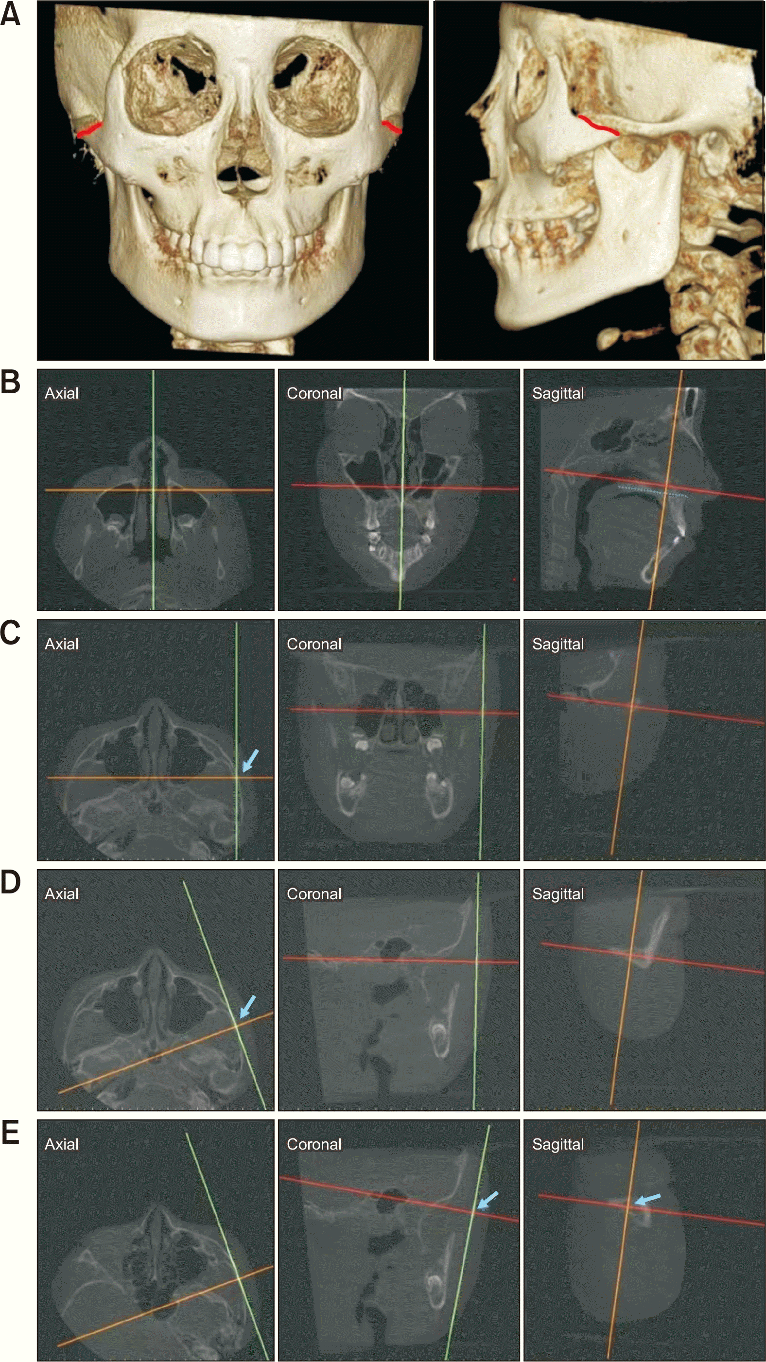
A cross-sectional slice of the ZTS was visualized in this study. To visualize the ZTS (left side as an example) in the sagittal view, several adjustments were made (Figure 1), in which the horizontal cursor (red line) was placed parallel to the palatal plane (Figure 1B). In the axial view, when the ZTS is seen, the vertical cursor (green line) is rotated to traverse through the ZTS (Figure 1C and 1D). In the coronal view, the vertical cursor (green line) is rotated to traverse through the ZTS (Figure 1E), which makes it possible to observe the morphology of the ZTS in the sagittal view.
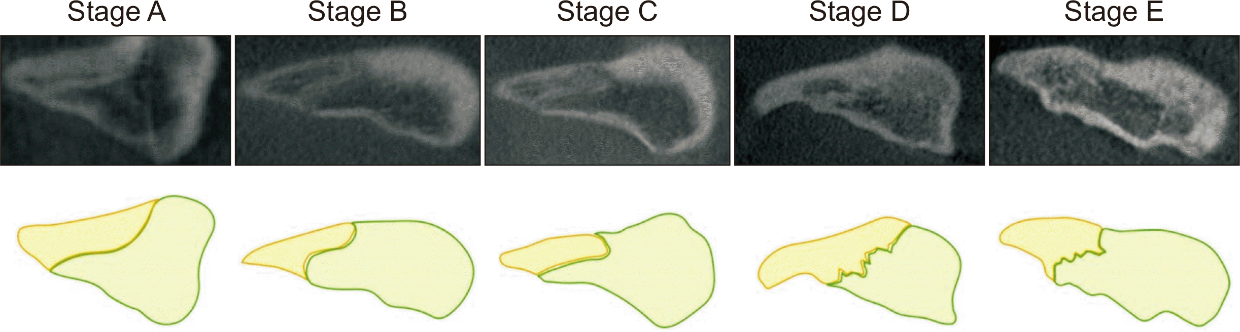
Based on the method followed in the previous studies on ZMS stage evaluation,12 the ZTS stages are defined as follows (Figure 2):
Stage A: The ZTS is visualized as a uniform high-density line with no significant interdigitation.
Stage B: The ZTS presents a thicker, scalloped, high-density line with some interdigitation. In some portions, two thin parallel high-density lines are close to each other with narrow low-density spaces between them.
Stage C: The ZTS can be observed as two parallel high-density lines without intersections. The two lines are close to each other and separated by narrow, low-density spaces.
Stage D: Fusion occurred; however, the two lines are still observed in some portions.
Stage E: The ZTS has completely fused, and lines cannot be observed in any portion of the ZTS.
Lateral cephalograms were obtained from CBCT images to evaluate the CVM stages. The CVM method proposed by Baccetti et al.22 was adopted in this study.
Statistical analysis
A weighted kappa coefficient for intra- and inter-evaluator agreements was calculated. Spearman’s correlation analysis and intraclass correlation coefficient (ICC) were used to evaluate the correlations between the indicators. One-way analysis of variance was used to analyze the relationship between ZTS maturation stages and chronological age. The rank-sum test was used to analyze the distinction between the ZTS maturation stages of male and female populations in the same age group. Likelihood ratios (LRs) were calculated to identify the diagnostic value of chronological age and CVM stage for the ZTS stage. Analyses were performed using Statistical Package for the Social Sciences software version 14.0 (SPSS Inc., Chicago, IL, USA). Statistical significance was set at p < 0.05.
RESULTS
A total of 261 patients were recruited in this study, including 112 males (mean age, 13.1 ± 3.3 years; age range 6.8–20.7 years) and 149 females (mean age, 13.7 ± 3.1 years; age range 7.4–20.6 years). The reproducibility of the ZTS staging method was satisfying (weighted kappa value = 0.887, p < 0.05 for evaluator 1 vs. evaluator 2; weighted kappa value = 0.843, p < 0.05 for evaluator 1 vs. evaluator 3; weighted kappa value = 0.800, p < 0.05 for evaluator 2 vs. evaluator 3). The intra-evaluator reliability for the ZTS maturation and CVM methods was also good (weighted kappa value = 0.892, p < 0.05, ZTS maturation method; weighted kappa value = 0.960, p < 0.05, CVM method).
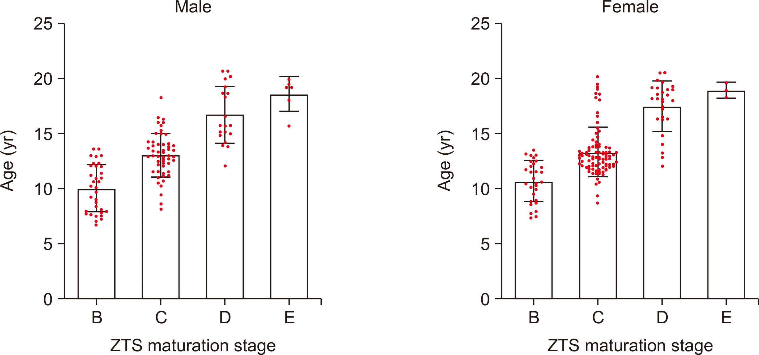
Stage A was not observed in any of the participants in this study. The distribution of participants presenting with various ZTS maturation stages in different age groups is shown in Table 1. Descriptive statistics for the age distribution patterns of the ZTS maturation stages classified by sex are shown in Table 2 and Figure 3. A statistically positive correlation was found between the chronological age and ZTS maturation stage (r = 0.727, ICC = 0.330, p < 0.01), whereas sex had no significant influence on the ZTS maturation stage.
The CVM stages were also evaluated. The distribution of participants presenting with various ZTS maturation stages in the different CVM stage groups is shown in Table 3. A statistically positive correlation was found between the CVM and ZTS maturation stages (r = 0.747, ICC = 0.621, p < 0.01).
The positive LRs of the CVM stages for the identification of the ZTS maturation stages are shown in Table 4 to evaluate the diagnostic value. The LR value of cervical stage 1 (CS1) for the diagnosis of ZTS stage B was > 10, indicating that CS1 has a high diagnostic value for stage B. The LR value of CS2 for the identification of ZTS stage B was > 9, demonstrating a significant increase in the likelihood (40–45%) of ZTS stage B. CS6 showed high positive LRs between 5 and 10, suggesting an increase in the likelihood (30–45%) of ZTS stages D and E.23 The presence of CS3 and CS5 also produced a small increase in the likelihood (15–25%) of stages C and D, respectively.23
To provide more evidence for the diagnostic value of the CVM stages, data from various CVM and ZTS stages were combined. CS1 and CS 2 were both matched with ZTS stage B, CS1–CS3 were matched with ZTS stage B and C, and CS6 was matched with ZTS stage D and E. Four parameters were calculated: sensitivity, specificity, positive predictive value, and LR. The results are summarized in Table 5. In all cases, the LRs were > 10, indicating a significant probability of the respective ZTS stages; therefore, the three parameters (CS1 and CS2, CS1 to CS3, and CS6) could be considered reliable indicators for the identification of the ZTS maturation stages.23
DISCUSSION
Maxillary protraction is a widely used method for the early treatment of maxillary deficiency. Studies reported that maxillary advancement of 2.19–2.90 mm could be achieved by tooth-borne maxillary protraction, whereas 3.78–4.01 mm could be achieved by skeletal anchorage maxillary protraction.24-26 However, the timing of maxillary protraction displays an important influence on the outcome. With aging, sutures become more zigzagged in shape and more complex in structure, with more interdigitation between bones.7,9 Therefore, the reconstruction of sutures can be hampered, which may impair the treatment effects of maxillary protraction during the patient’s growth. To date, studies on the CBCT maturation stages of circummaxillary sutures are limited to the ZMS; however, it is sometimes difficult to assess the maturation stages of the ZMS due to the influence of the maxillary sinuses.12-15 As the ZTS also plays an important role in maxillary protraction, it was selected for evaluation in this study to provide recommendations for clinics, whereas the ZMS maturation stages are not available.
Cone-beam computed tomography images were obtained for analysis in this study and are widely used in orthodontic practice. Compared with conventional computed tomography, CBCT provides a significantly lower radiation dose but still enables an accurate depiction of the craniofacial bone and soft tissues.27,28 On CBCT images, ZTSs were clearly visible, and the aging changes reported in previous studies were observed in this study, including increased complexity, interdigitation, and tortuosity,7 all of which were observed in this study, except for stage A. As the image of ZTS stage A shown in Figure 2 was observed in the CBCT image of a 4-year-old child, a wide distribution of ZTS stage A may be observed in children under 6 years of age. However, owing to poor cooperation and the low necessity of undergoing CBCT, children at such young ages are not required to undergo CBCT, which results in the absence of samples from the younger age group and ZTS stage A.29
As the interdigitation and fusion of circummaxillary sutures have significant negative impacts on the maxillary protraction, studies were designed to reveal the relevance between suture morphology and reconstruction.13,30,31 Angelieri et al.13 demonstrated that patients at stages A and B of ZMS had a more satisfying outcome after maxillary protraction than those at stage C. In contrast, almost no sagittal or vertical effects were observed in patients at stages D and E.13 Similar findings were found in a study of the relationship between rapid maxillary expansion outcomes and MPS maturation stages.30,31 These findings enhance our comprehension of the clinical significance of suture stages, defining stages A, B, and C as appropriate indicators for suture remodeling and stages D and E as contraindications.
Previous studies have demonstrated a large variability in chronological ages for maturation stages of circummaxillary sutures, especially at stage C.32-34 In this study, similar distribution was found; stage C was observed in all age groups, ranging from 8.2 to 20.2 years of age. Stages B, C, and D have been observed across a wide age range. The investigation by Angelieri et al.12 reported that all maturation stages of ZMS were observed in the group aged 10–15 years, while according to this study, stages B to E of ZTS were observed in patients aged 14–16 years, in agreement with the study of ZMS by Ok et al.14 Such findings indicate that age alone may not be reliable for the prediction of maxillary protraction outcomes.
The age groups in this study were similar to those in previous studies; therefore, it is feasible to compare the ZTS stage distribution in similar age groups using the ZMS. In detail, Stage B was the highest in the < 11-year group, Stage C was the highest in the 11–14 and 14–18-year groups, and Stage D was the highest in the > 18-year group. However, previous studies on ZMS reported that it was stages A, B, D, and E.14,31 ZTS may develop faster below 11 years of age, whereas ZMS may develop faster above 14 years of age.
Cervical vertebral maturation is a reliable biological indicator for the assessment of skeletal maturation, which can be obtained using a conventional lateral cephalogram.22 Because of its advantages, including the lower need for cooperation, lower cost, and lower radiation dose, it is widely applied to assess children’s growth stage and their potential for development to predict their response to orthopedic force applications and decide the treatment planning of early skeletal malocclusion. In this study, the correlation between CVM and maturation stages of ZTS was demonstrated to be high, which is in agreement with previous studies on MPS.16,35,36 When examining each CVM stage, the highest ratio was stage D at CS6, stage C at CS3, CS4, and CS5, stage B at CS2 and CS1, respectively. However, according to the MPS data, it was stage A or B at CS1, stage B or C at CS2, stage C at CS3, stage C at CS4, stage D or E at CS5, and stage E at CS6.16,35,36 The fusion of ZTS may occur later than that of MPS. The previous investigation showed that fusion occurred earlier at ZMS than that at MPS; thus, it is suggested that ZTS may close later than ZMS.14
Likelyhood ratio is a parameter for the diagnostic value of a new indicator.23 An LR > 10 can be interpreted as a conclusive increase (over 45%) in the probability of disease and argues for a reliable indicator for the identification of the disease.23 Therefore, LRs were calculated to assess the diagnostic value of CVM stages for ZTS maturation stages. According to the positive LRs of CVM stages for the diagnosis of ZTS stages, CS1 and CS2 indicated stage B, CS1 to CS3 indicated stages B and C, and CS6 indicated stages D and E, similar to previous findings in MPS.35,36 Thus, CS1, CS2, and CS3 are considered reliable indicators for the proper time of maxillary protraction. Cervical stage 6, which suggests that fusion has already occurred partially or completely, can be considered a surgical indication. These CVM stages may provide valuable recommendations for clinics without CBCT scanning or patients who cannot cooperate with the examination. However, at CS4 and CS5, further inspection using CBCT was necessary to confirm ZTS maturation.
Few studies have recommended that the proper age for orthopedic force appliances differs by sex, considering the earlier maturation of females than males.16,31,37 However, in this study, sex displayed no significant influence on ZTS maturation stages, in agreement with the findings of Li et al.15
The present study, along with similar studies focusing on the maturity of circummaxillary sutures, provides a basis for clinical decisions regarding maxillary protraction. However, several questions remain unanswered. First, Liu et al.38 found that the forward movement of the ZTS, but not the ZMS, was associated with ZMS maturation stages by comparing CBCT images before and after maxillary protraction treatment in the clinic. The ZMSs of all the patients ranged from stage B to C, indicating that no ossification occurred in the ZMS.38 However, the forward movement of ZTS is related to the ZMS maturation stage, demonstrating that fusion may have already occurred in ZTS.38 Therefore, it is assumed that ZTS may close earlier than ZMS, contrary to the analysis in the preceding paragraph. It is suggested that there may be differences between populations; therefore, studies comparing the maturation stages of different sutures in the same group of patients are necessary to confirm the development sequence of circummaxillary sutures. Second, the important role of ZTS in maxillary protraction was suggested by the stress distribution and active reaction of ZTS during treatment; therefore, more clinical trials exploring the correlation between the response of maxillary protraction and ZTS maturation stages are necessary in the future. Third, Isfeld et al.39 reported that the maturation stage had no significant influence on the treatment results of the rapid maxillary expansion. Therefore, to verify the reliability of the suture maturation stage method, further investigations comparing the orthopedic treatment effects in patients at various suture maturation stages are necessary.
CONCLUSIONS
The evaluation of the ZTS maturation stages using CBCT images is feasible and should be a reliable supplement to the previous ZMS method. Both chronological age and CVM stages were found to be related to ZTS maturation stages, whereas sex had no significant influence on ZTS maturation stages. The correlation between ZTS maturation and CVM stages may be more significant than that with chronological age. Cervical stage 1, CS2, CS3, and CS6 can be considered indicators for clinical decisions regarding maxillary protraction.
 XML Download