

 PDF
PDF Citation
 Print
Print


INTRODUCTION
Dentoalveolar expansion can correct mild maxillary transverse deficiencies.1-3 However, if the patient's biological limitations are not considered during therapy, the alveolar bone plates may get compromised and result in alveolar defects, such as dehiscences and fenestrations.1,4-7
Cone-beam computed tomography (CBCT) has shown dehiscences and significant buccal alveolar bone thickness reductions in patients with orthodontic arch expansion.5,8-11 Before treatment, a periodontal evaluation should be conducted because many patients have alveolar defects.12,13 Cone-beam computed tomography is the preferred diagnostic imaging method for periodontal bone tissue evaluation, especially in adult patients requiring teeth movement towards the buccal alveolar bone.10,11,14
In recent years, patients concerned with esthetics and comfort have opted for clear aligners, which have been helpful in their improving self-esteem and well-being.15 Contrary to fixed appliances, orthodontic clear aligners have not been extensively studied.16-20 Thus, this study examined the effects of maxillary orthodontic expansion on alveolar bone tissue in adult aligner patients using CBCT images. We also examined whether the amount of arch expansion was safe to prevent bone loss and whether a particular group of teeth was more periodontally affected.
MATERIALS AND METHODS
The present study was approved by the research ethics committee of the School of Dentistry of Ribeirão Preto (FORP), University of São Paulo, according to protocol number CAAE 10113319 1 0000 5419.
Of the total 144 orthodontic records studied, including CBCT images acquired with a voxel size of 0.25 mm, 30 records were included in this retrospective study based on the following inclusion criteria: adult patients older than 21 years treated with clear aligners for maxillary dentoalveolar expansion; good facial esthetics; good oral and general health; no use of drugs affecting bone metabolism in the past year; presence of complete permanent dentition, except third molars; maxillary dental crowding < 3 mm; absence of anterior and posterior crossbites; and absence of supernumerary teeth or dental agenesis. The exclusion criteria were as follows: patients with maxillary dental crowding > 3 mm and/or poor oral health and hygiene.
The obtained CBCT records were of 22 females and 8 males, all Brazilian whites with a mean age of 36 years and 3 months old (ranging from 22 to 55 years), and their malocclusions were classified as follows: 22 records had Class II (A point-nasion-B point [ANB] angle > 4°) and eight records had Class I (ANB angle between 0° and 4°). Considering the facial pattern, 15 records were dolichofacial, 10 mesofacial and 5 brachyfacial.
All the patients underwent orthodontic treatment in a private dental clinic (Clínica de Ortodontia Figueiredo) in the city of Sorocaba (SP), Brazil, between 2012 and 2019. Because this is a retrospective study, the CBCT images were part of the patient’s dental records for diagnosis and treatment planning. All the cases were treated with clear aligners of the Invisalign® system (Align Technology, San José, CA, USA) by the same orthodontist, with an average treatment time of 19 months between the pre- and post-expansion phases. Attachments were used in all the cases and placed depending on the needs established in the virtual plan (ClinCheck®; Align Technology, San Jose, CA, USA). The patients replaced their aligners within 2 weeks and oral hygiene was monitored during the treatment period. The interval between the periodontal appointments was 8 weeks. Interproximal enamel reduction (IPR) in the upper arch was performed in 40% of the sample to manage dental crowding and minimize the black triangle between the incisors when required. The average IPR was 0.83 mm.
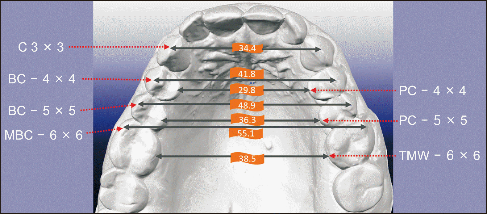
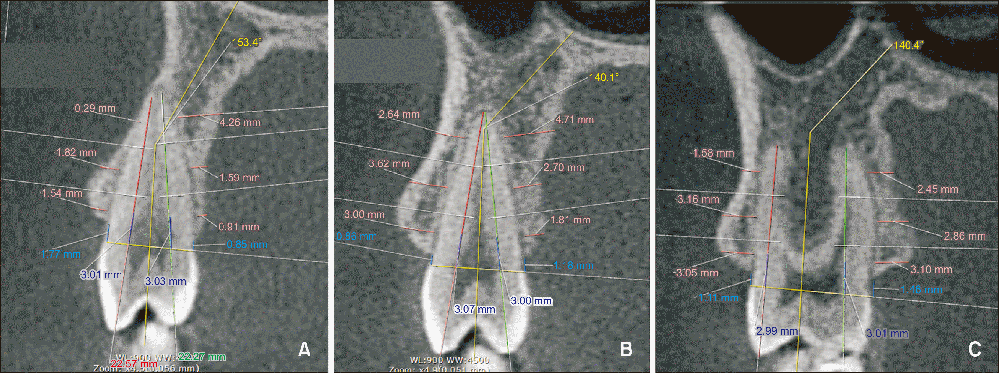
A total of 210 transverse measurements were performed in digital models to quantify the maxillary expansion and correlate the effect of clear aligners on the maxillary alveolar bone by means of CBCT. The iTero Element scanner® (Align Technology) was used to obtain all the models and the OrthoCAD iCast Orthodontic 3D Digital Modeling Study (Align Technology) to perform the measurements. The following landmarks were used: the most buccal point of the cusp of the canines (C3), the most buccal (buccal cusp of first premolar, BC 4; buccal cusp of second premolar, BC 5) and most palatal points (palatal cusp of first premolar, PC 4, palatal cusp of second premolar, PC 5) of the cusps of the premolars, the most buccal point of the mesiobuccal cusp of the first molars (MBC 6), and palatal sulcus adjacent to the gingiva (TMW 6) (Figure 1). The amount of the increase in transverse expansion was evaluated in the digital models.21-23
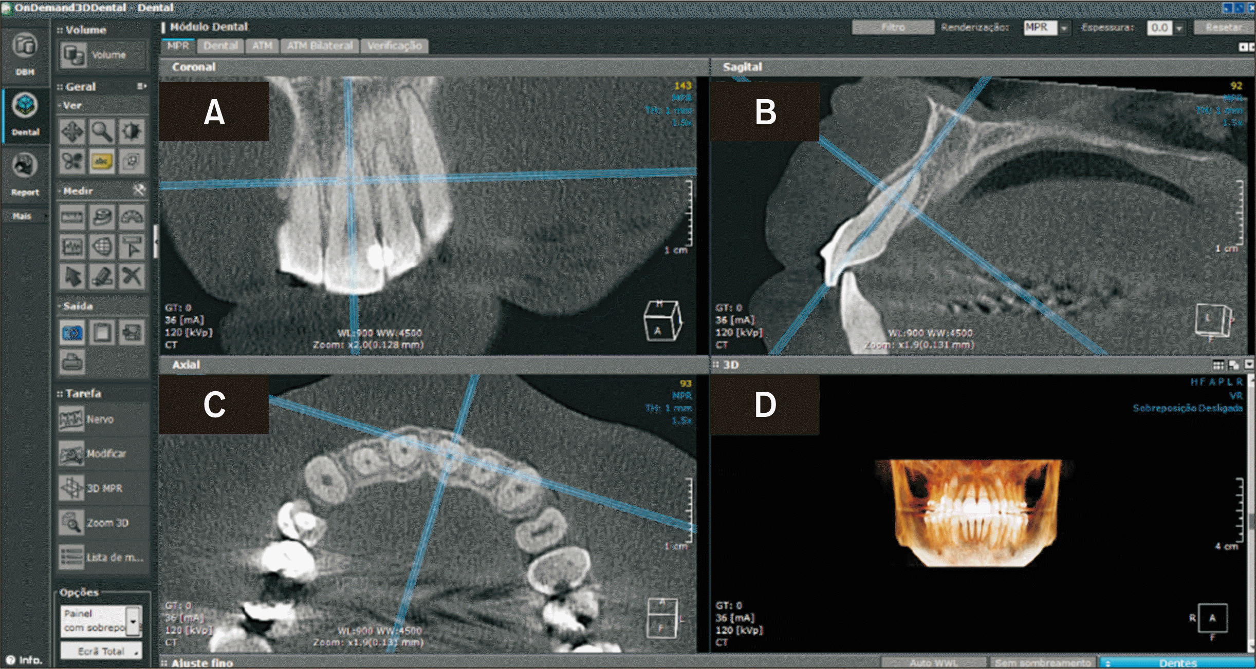
Cone-beam computed tomography was used to quantify the alveolar bone on the buccal and palatal surfaces of 12 maxillary teeth (from the first molar to the first molar) and measure the angle formed by the long axis of the teeth in relation to the palatal plane, thus totaling 2,880 linear measurements and 360 angular measurements, which were compared between T0 (before maxillary expansion) and T1 (after maxillary expansion). The CBCT images were acquired with an i-CAT scanner (Classic 3-D Dental Imaging System; Imaging Sciences International, Hatfield, PA, USA) operating at 120 Kvp, 8 mA, exposure time of 40 seconds, and voxel size of 0.25 mm. The patient was seated with the midsagittal plane and Frankfurt horizontal plane parallel to the floor, in maximal intercuspation position. The resulting data on each patient were recorded in the Digital Imaging and Communications in Medicine (DICOM) format. OnDemand 3D software (Cybermed Inc., Daejeon, Korea) (Figure 2) was used on a notebook (DELL G7, 17”, Intel® CoreTM i7-8750H; Dell Technologies Inc., Round Rock, TX, USA), in which the ruler and angle tools were applied to obtain linear and angular measurements, respectively. After positioning the image, the coronal and sagittal views were used to measure the posterior and anterior teeth, respectively with the vertical and horizontal lines positioned through the long axis of the teeth (Figure 3).
The linear and angular tomographic measurements were made based on the protocol set by Nahás–Scocate and Scocate.10,24
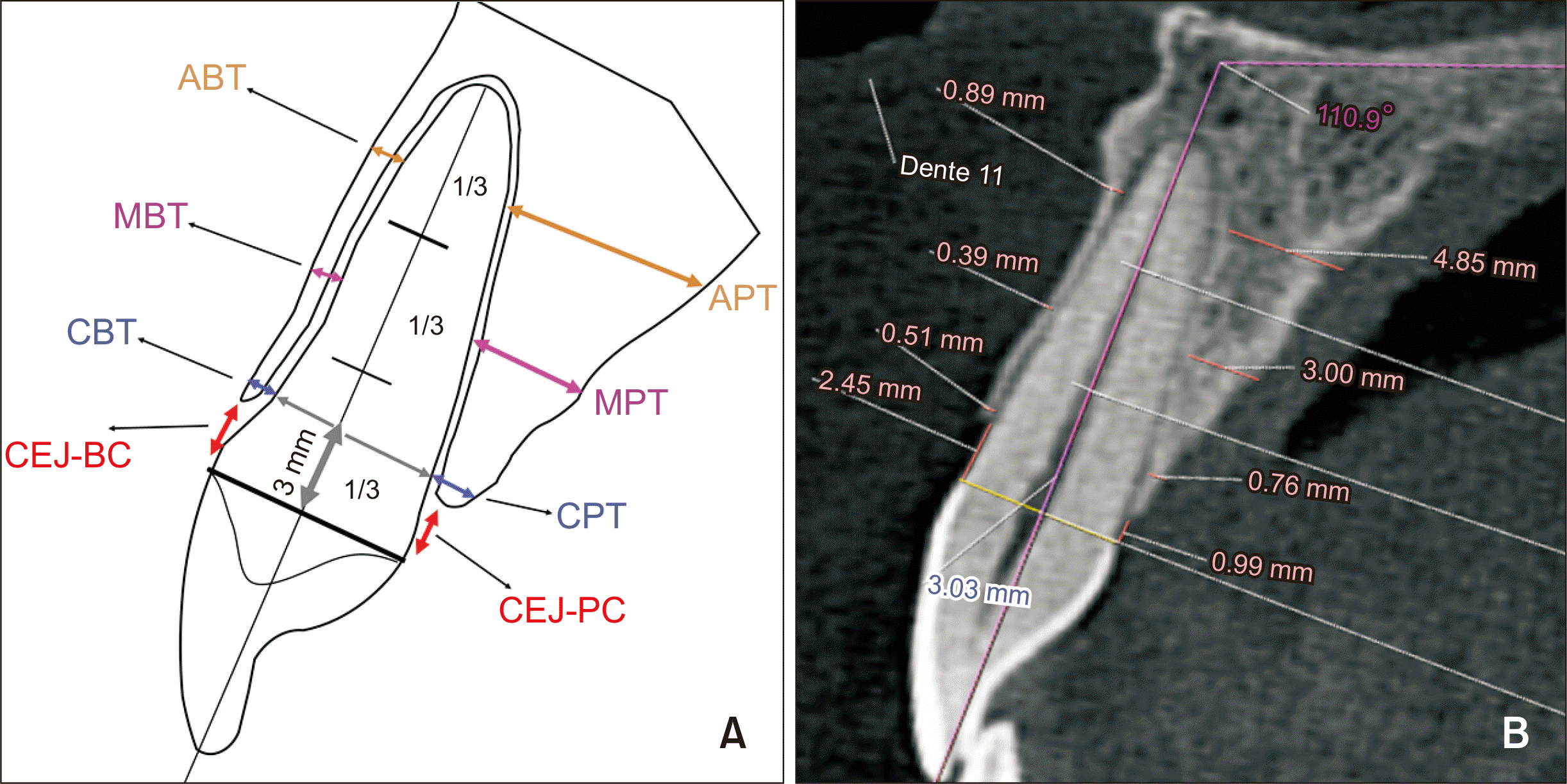
For measuring the alveolar bone of the incisors and canines, a straight line was traced passing through the cementoenamel junction (CEJ) (buccal and palatal) and the root length (from the CEJ to the apex) was measured and divided into three-thirds. The measurement of the first root third (cervical) was based on two aspects: a) height from the CEJ to the alveolar bone crest (CEJ-buccal crest/CEJ-palatal crest) and b) thickness of the alveolar bone crest at a 3-mm distance from the CEJ towards the apex (cervical buccal thickness [CBT]/cervical palatal thickness [CPT]). The middle and apical root thirds were further divided by half to determine the landmarks of the middle (middle buccal thickness [MBT]/middle palatal thickness [MPT]) and apical (apical buccal thickness [ABT]/apical palatal thickness [APT]) thirds of the roots where the bone thickness was measured. All the measurements were made in the buccal and palatal faces by positioning the ruler tool perpendicular to the long axis of the root (i.e., from the external limit of the cortical bone to the root surface) and according to the buccal (CBT, MBT, and ABT) and palatal (CPT, MPT, and APT) bone thicknesses in the cervical, middle, and apical thirds of each tooth (Figure 4).
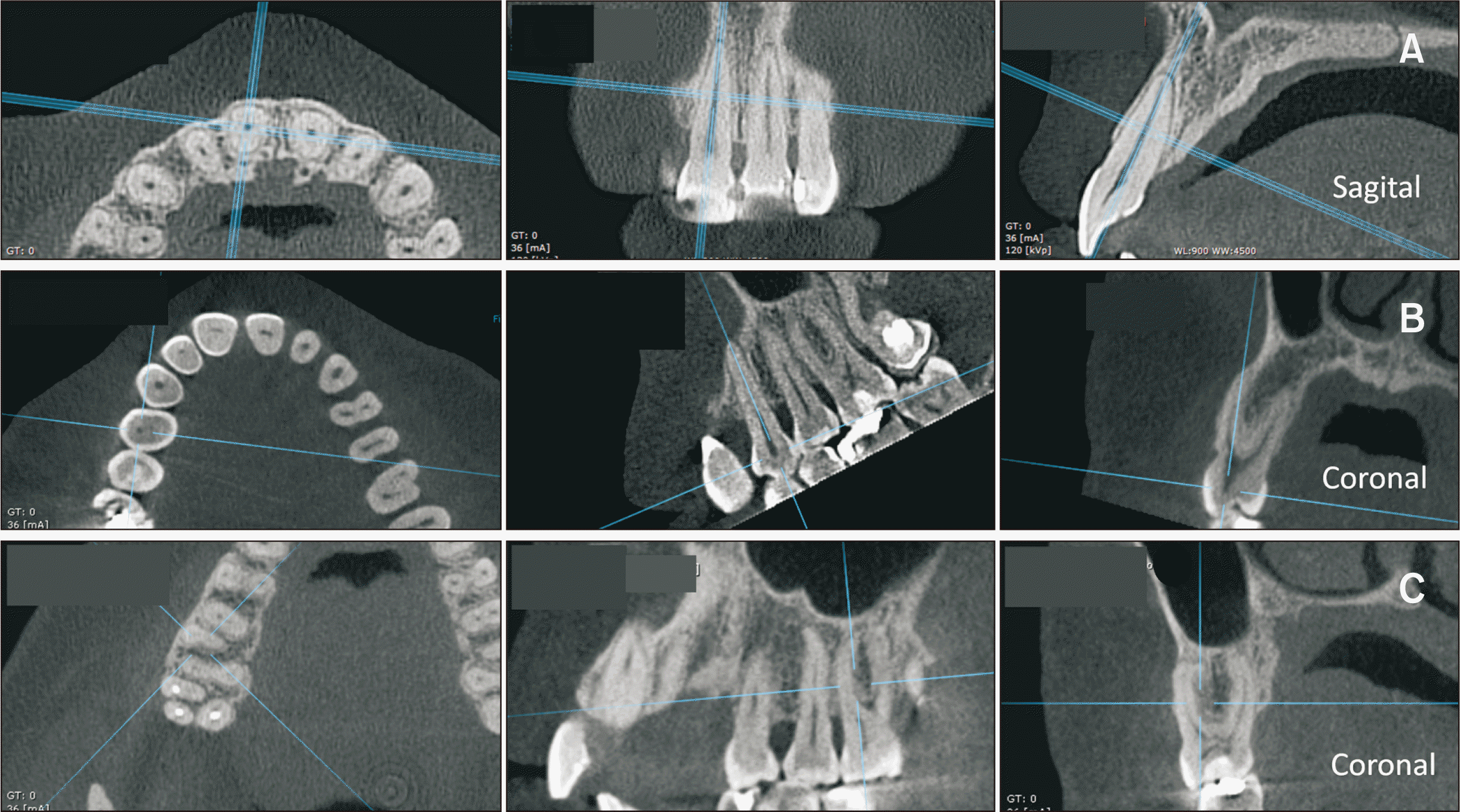
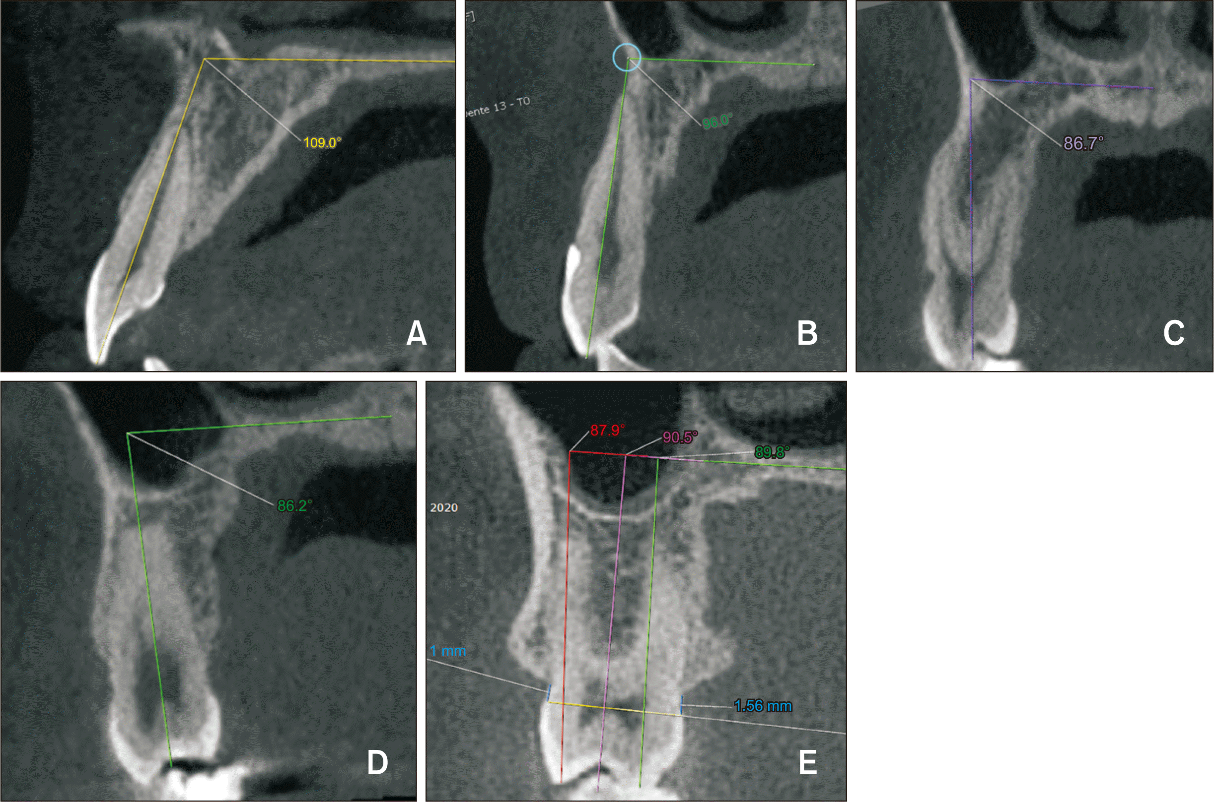
To measure the alveolar bone tissue of the first and second premolars in the coronal view, the same process described above was performed, except for the inclusion of two reference lines traced from the tips of the buccal and palatal cusps along the long axis of the respective roots. The mesiobuccal and palatal roots were used as references in the region of the first molars (Figure 5). After defining the image in the axial view, the coronal reference line automatically formed by the software was rotated with the cursor toward the palatal root for the measurement of the tooth in the coronal view (Figure 3C). The thickness of the buccal and palatal bone plates was measured in accordance with the tooth’s long axis in the sagittal and coronal planes. The thickness of the alveolar bone tissue changes upon measuring the severely rotated tooth. Rotated teeth occupy less space before treatment; thus, a decrease in bone thickness can be expected after treatment, which could result in underestimating the amount of bone tissue before treatment if we used the shape of the arch as a reference in an axial view.
Inclination of the anterior and posterior teeth was considered by means of the angle formed by the palatal plane and long axis of the teeth (tooth.PP) (Figure 6).
Statistical analysis
The intraclass correlation coefficient was used to obtain the measurement error based on nine patients, who were randomly chosen.
Descriptive data were obtained from all the measurements. The comparison between the two times, T0 and T1, was carried out by using Student’s t-test in the model variables and analysis of variance (ANOVA) for repeated measurements in the variables of the tooth.PP. Generalized estimating equation models were used to assess the correlation of the parameters with the tooth.PP measurement. The level of significance adopted was 5%. Correlations were made using the software R version 3.6.0 and Pearson’s correlation coefficient. The sample power for repeated measurements was above 80% for the majority of the variables studied and less than 80% for the distance between cementoenamel junction-buccal bone crest (CEJ-BC) for the anterior teeth and CBT for the molars. Measurement error was performed by using the intraclass correlation coefficient, and the result was close to 1 (lowest value = 0.892), showing excellent reproducibility and reliability of measurements. The interval between the measurements was a minimum of 3 weeks. Student’s t-test was applied to analyze sexual dimorphism, which showed no significant difference between the sexes for all the measurements.
RESULTS
The transverse measurements in the digital models demonstrated a significant increase after the maxillary expansion (Table 1). We may also consider distal rotation to affect arch expansion in the molar region.
In the buccocervical region, the CEJ-BC measurement showed a variation between the teeth before treatment with aligners, in which the central incisors had the smallest measurement, followed by second premolars, lateral incisors, first molars, first premolars, and lastly by canines. The CBT measurement showed a greater amount of bone tissue in the second premolars, followed by the first molars, central incisors, lateral incisors, canines, and first premolars before treatment. After maxillary expansion, no significant decrease was observed in the thickness of the alveolar bone in any of the teeth evaluated on both sides. However, in the cervical region, the CEJ-PC measurement also showed variation between teeth, with the central incisors having the smallest measurement, followed by lateral incisors, second premolars, canines, first premolars, and lastly, first molars before treatment. After arch expansion, no posterior tooth revealed a significant decrease in the alveolar bone thickness as the CEJ-PC measurement decreased for the premolars (both, p = 0.019) and first molars (p = 0.001), whereas the CPT measurement increased significantly in both molars (p = 0.001) as the bone height and thickness improved in the cervical region. Alveolar bone thickness (i.e., CPT) was maintained for the canines and lateral incisors; however, it significantly decreased for the central incisors (p < 0.005), which increased the CEJ-PC measurement (p = 0.001) (Table 2).
In the middle region, buccally the alveolar bone tissue before treatment was greater in the second premolars, followed by the central incisors, lateral incisors, first molars, first premolars, and canines. Importantly, there was an increase in bone thickness in the central incisors (p = 0.013), lateral incisors (p = 0.013), and canines (p = 0.013), and a decrease in the posterior teeth following maxillary expansion with aligners (p = 0.032 for premolars and p = 0.007 for molars). In the middle region, palatally, the amount of alveolar bone tissue before treatment was greater for the canines, followed by the second premolar, central incisors, lateral incisors, first premolars, and first molars. Considering the T0 and T1 periods, a significant difference was observed in the increase of bone tissue for the first molars (p = 0.007) and a decrease for the central incisors (p = 0.032) (Table 3).
In the apical buccal region, the bone plate was thicker in the second premolars, followed by the first molars, central incisors, lateral incisors, canines, and lastly first premolars before treatment. After treatment, there was a significant increase in the alveolar bone thickness for all teeth (p = 0.001 in anterior teeth and p = 0.025 in premolars), except the first molars (as their thickness was kept). In the apical palatal region, the canines had a greater amount of bone tissue before treatment, followed by the second premolars, central incisors, first premolars, lateral incisors, and molars. After arch expansion, no significant changes were observed upon comparing the treatment periods (Table 4).
The tooth.PP measurement increased significantly between the time periods for all the evaluated teeth, except the first molars (as no significant inclination was observed) (Table 5).
DISCUSSION
Scientific studies investigating the effects of maxillary expansion on the alveolar bone plates with orthodontic clear aligners are scarce. The most effect of orthodontic mechanics on the alveolar bone plate attributed to conventional appliances has been commonly studied.5,8,11,14,25-28 Barreda et al.19 assessed the effects of clear aligners only on the buccal cortical alveolar bone of the first and second premolars in 19 patients. To the best of our knowledge, the present study is the first study to assess the effects of maxillary expansion on alveolar bone tissue (buccal and palatal faces) of all anterior and posterior teeth, except for the second molars, in a sample of 30 adult patients. The patients were instructed based on a standardized oral hygiene protocol before and during the treatment performed by a periodontist every 3 months.
All the teeth had a surprising cervical result. No significant decrease of the alveolar bone tissue in thickness (CBT) and depth (CEJ-BC) was observed on the buccal surface of the evaluated teeth. Likewise, on the palatal surface, the result was also favorable, as none of the posterior teeth, canines, and lateral incisors presented a significant decrease in the bone thickness. On the contrary, the first molars and premolars demonstrated a significant improvement in the height and thickness of the alveolar bone crest (CEJ-PC and CPT), which however was not clinically relevant (< 0.5 mm). The only teeth that lost bone were the central incisors, with a significant increase in bone dehiscence (CEJ-PC) and decrease in the bone board thickness (CPT) on the palatal side. This result could be attributed to the bowstring effect of posterior expansion.
In the buccal face of the canines and palatal face of the molars, a significant increase of the alveolar bone tissue was observed, while the palatal face of the lateral incisors, canines, and premolars stayed steady. Nevertheless, the buccal alveolar bone tissue of the molars and premolars demonstrated a significant bone thickness decrease following treatment. The reduction in the buccal bone plate, in the middle region of the root, especially in the premolars, could be attributed to the expansion in this area, which was approximately 3.0 mm for the first and second premolars, and the anatomy of these teeth. This result confirms the findings reported by Barreda et al.,19 in which the second premolars demonstrated a reduction of 13.1% (i.e., 6 mm from the CEJ) following treatment. Despite the reduction of bone tissue in the middle third buccally in the region of the premolars in our study, the second premolars showed a greater amount of bone tissue compared to other teeth throughout the orthodontic treatment. Interestingly, the second premolars had two times greater amount of bone tissue in the middle third buccally than the first premolars following treatment, which reinforces the need for careful orthodontic planning when the biomechanics are applied to the posterior region.
In the apical buccal region, a statistically significant improvement was observed in the bone plates of the incisors, canines, and premolars, whereas this did not occur in the first molars. In the palatal face, no decrease in the thickness of the alveolar bone tissue was observed in any of the teeth evaluated in our study, suggesting that the apical region is less critical, since a greater amount of bone tissue was already present before the treatment compared to the other regions. In turn, the canines showed a lesser amount of alveolar bone tissue in the buccal face (0.39 mm) and a greater amount in the palatal face (6.14 mm) before expansion of the upper arch. Based on these initial anatomical characteristics of the patients, including the buccal bone gain in the apical region following arch expansion, performing root palatal torque is recommended whenever necessary for the upper canines. The use of aligners is effective when such biomechanics are required.
The literature has demonstrated that bone defects can be found in patients who never wore orthodontic appliances.5,11,14,19 In our study, dehiscence was present in 86% of the teeth before treatment, with the canines (mean of 4.97 mm) and first premolars (mean of 4.85 mm) being the most affected teeth in the buccal face before arch expansion.
The most important finding in this study was that although the width of the upper arch was significantly increased in all the studied teeth (Table 1), bone tissue was maintained, suggesting that the use of clear aligners did not negatively affect the alveolar bone height and thickness. Bone condition can even be improved, which was observed in the palatal faces of the premolars and molars. Even the canines, whose buccal bone plate is thinner compared to the other teeth owing to the volume of their root,29 demonstrated a greater dehiscence before arch expansion. We believe that the results obtained with the use of aligners from the Invisalign system may permit greater control of individual dental movement, contingent upon the dental professional's expertise/mastery of software management. Christoph et al.,1 in a study with dogs, found a reduction in the bone thickness in the premolars and minimum inclination of the teeth following expansion. The authors suggested that bone thickness is a possible factor influencing the bone decrease, since thinner areas of the buccal bone undergo levels of stress beyond their adaptive capacities, resulting in a decrease in the amount of bone owing to excessive microfractures. No relationship was observed between tooth inclination and the amount of bone loss. This finding corroborates with that of our study, as a significantly increased inclination was observed in all the teeth studied after expansion, but no relationship was observed with the amount of bone tissue. Moreover, no correlation was observed between the transverse measurements, which quantified the maxillary expansion and bone height and thickness in the cervical, middle, and apical regions (Tables 2–4).
Cone-beam computed tomography reconstruction was reported to demonstrate an accuracy of approximately 70% for the diagnosis of dehiscence.30 Fuhrmann et al.31 reported that false-positive diagnosis of dehiscence could occur when the cortical plate is thinner than 0.5 mm. The smaller the voxel size, the richer the details of the image.14,30,32,33 In the present study, we used tomographic images acquired with a 0.25 mm voxel size, which is in accordance with the literature.5,8,11,14,18,25-28,34-36 However, smaller voxel sizes would ideally be more appropriate for interpreting the obtained bone results with greater reliability.
Further research is warranted to investigate the effect of clear aligners on alveolar bone tissue by highlighting periodontal clinical parameters and by using larger and more homogeneous samples in terms of the periodontal condition, mainly in adult patients. A control group with fixed appliances and a long-term study could show the progression and stability of bone defects after orthodontic treatment. Based on this study, we cannot affirm that clear aligners are better than traditional fixed appliances. This study has certain limitations, and caution should be exercised when interpreting the results. In the study, one patient was treated with aligners made from the EX30 material, despite it being a less elastic material no longer used by Align Technology. We included this patient in the study because the clinical effect, if present, would not affect the outcome of our research.
 XML Download