Journal List > J Dent Rehabil Appl Sci > v.39(2) > 1516083375

Influence of tongue pressure on the obturator prosthesis for soft palate defects: a 3D-FEA study

Abstract

Purpose

The purpose of the study was to analyze how swallowing tongue pressure affects the biomechanics of a velopharyngeal obturator prosthesis and compare its displacement across different occlusal rest positions.

Materials and Methods

A 3D geometric model consisting of the maxilla, teeth, soft palate, and a portion of the pharynx was developed based on the CBCT and MRI data. A defect was created by the resection of soft palate portion. Two experimental models were generated based on two different velopharyngeal obturator designs: one “with mesial occlusal rests” (Model 1) and the other with “distal occlusal rests” (Model 2). A pressure of 25 kPa was applied at the surface of the bulb of the obturator prosthesis opposite the base of the tongue to simulate tongue pressure during swallowing. The maximum von-Mises stress and displacement values of two types of obturator prostheses were analyzed and compared.

Results

The maximum von-Mises stress in the metal framework, located at the posterior palatal strap, was slightly higher in model 1 (64.9 MPa) than in model 2 (54.2 MPa). In both models, the acrylic resin obturator bulb exhibited a maximum stress value of 4.3 MPa. There was no significant difference in prosthesis displacement between the two models, with 31.3 μm for model 1 and 33.6 μm for model 2.

Conclusion

Swallowing tongue pressure had a minor impact on the biomechanics of a velopharyngeal obturator prosthesis, and distal occlusal rests showed a slightly better biomechanical response compared to mesial occlusal rests.

초록

목적

본 연구의 목적은 연하 시 혀 압력이 구개인두 폐쇄장치의 생체 역학에 어떻게 영향을 미치는지 분석하고 교합면 레스트 위치에 따른 변위량을 비교하는 것이었다.

연구 재료 및 방법

CBCT와 MRI 데이터를 바탕으로 상악골, 치아, 연구개, 인두의 일부로 구성된 3차원 모형을 제작하였고, 연구개 부위를 절제하여 결손부를 구성하였다. 서로 다른 구개인두 폐쇄장치 설계를 기반으로 한 실험 모델 2개를 제작하였으며 Model 1은 ‘근심 교합면 레스트’가 포함되었고, Model 2는 ‘원심 교합면 레스트’가 포함되었다. 연하 시 압력을 시뮬레이션하기 위해 혀의 기저부 반대편에 있는 폐쇄장치 벌브에 25 kPa의 압력을 적용하였다. 3차원 유한요소법을 이용하여 두 종류의 폐쇄장치 보철물에 대한 최대 von-Mises 응력 값과 변위량을 측정하고 비교하였다.

결과

후방 구개판에 위치한 금속 구조물의 최대 von-Mises 응력 값은 Model 2 (54.2 MPa)보다 Model 1 (64.9 MPa)에서 약간 더 높게 관찰되었다. 폐쇄장치 벌브의 아크릴릭 레진에서는 두 모형 모두 4.3 MPa의 최대 응력 값을 나타내었다. 보철물의 변위량은 Model 1이 31.3 µm, Model 2가 33.6 µm로 두 모형 사이에 큰 차이가 없었다.

결론

연하 시 혀 압력은 구개인두 폐쇄장치의 생체 역학에 경미한 영향을 미쳤으며, 원심 교합면 레스트는 근심 교합면 레스트에 비해 더 유리한 생역학적 반응을 나타낸 것으로 관찰되었다.

Introduction

Velopharyngeal insufficiency is a condition that occurs when the soft palate fails to create an effective seal with the pharynx during speech and swallowing that requires sufficient intraoral pressure. The underlying cause of this velopharyngeal insufficiency is due to congenital or acquired structural defects in the soft palate. This condition results in inadequate velopharyngeal closure, leading to difficulties with speech articulation and swallowing,1,2 and it can significantly impact the quality of life of patients.3,4
A velopharyngeal obturator prosthesis is used to rehabilitate patients with velopharyngeal insufficiency.5,6 This prosthesis comprises three components: the front section, which resembles a removable dental retainer; the middle section, which connects the dental retainer to the bulb; and the bulb section itself, which extends posteriorly towards the pharynx. The function of the bulb extension is to replace the defective portion and make close contact with the pharyngeal tissues for the proper palatopharyngeal seal during speech and swallowing.7,8
In normal individuals, the soft palate elevates and moves backward during swallowing, while the posterior and lateral pharyngeal walls move forward and inward, respectively, to ensure adequate velopharyngeal closure.9 Simultaneously, the tongue also retracts posteriorly, and the base of the tongue generates sufficient swallowing pressure in the soft palate.10 However, in an individual who has a palatal defect and is rehabilitated with an obturator prosthesis, the swallowing pressure produced by the base of the tongue acts on the bulb portion of the obturator. Consequently, this pressure can induce displacement of the obturator prosthesis, and the performance of the swallowing function might be affected if the obturator is not well retained in its position.
Hence, to perform swallowing function properly, the retention and stability of the obturator prosthesis are important, and the retainer design based on the location of the occlusal rests should be considered carefully.11,12 Moreover, according to one case report presented by Collin et al., dislodgement of the obturator prosthesis could potentially lead to an emergency situation if it obstructs the airway.13 Furthermore, the area where the retainer section and the bulb portion connect has a high potential to fracture, and fracturing of the bulb portion can also lead to airway obstruction.
Therefore, it is crucial to evaluate the effect of swallowing tongue pressure on the biomechanics of a velopharyngeal obturator prosthesis and to determine the most suitable occlusal rest position for this obturator prosthesis. To the best of our knowledge, there are no studies about the biomechanical analysis of velopharyngeal obturator prostheses in patients with soft palate defects.
The purpose of the study is to analyze how swallowing tongue pressure affects the biomechanics of a velopharyngeal obturator prosthesis and compare its displacement across different occlusal rest positions.

Materials and Methods

The cone-beam computed tomography (CBCT) and magnetic resonance imaging (MRI) data of a 36-year-old female participant were used for this finite element analysis (FEA) study. A three-dimensional (3D) geometric model consisting of the maxilla and teeth was developed based on the CBCT data using modelling software (Mimics 19.0; Materialise NV, Leuven, Belgium). The soft palate and a portion of the pharynx, including the nasopharynx and oropharynx, were also simulated in the model using MRI data (Fig. 1). A simulated alveolar bone crest was created 1 mm below the cementoenamel junction of the teeth. The periodontal ligament was simulated with a thickness of 200 µm,14 and the thickness of the mucosa was estimated to be 3 mm.
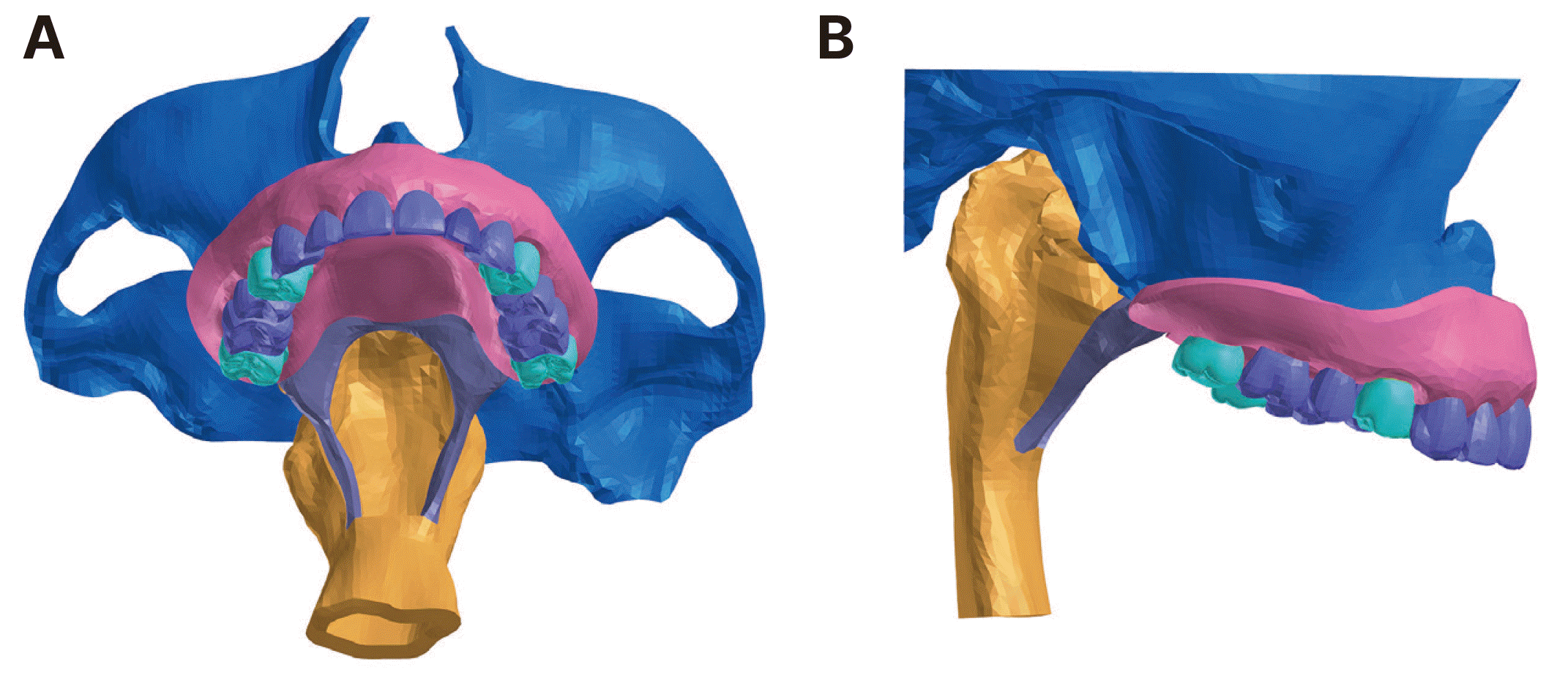
The right and left first premolars, as well as the second molars, were prepared for the fabrication of surveyed crowns following 3D printing using the 3D printer (3D printer MAX, Asiga, Sydney, Australia). After that, four prepared abutment teeth were scanned for the fabrication of their respective surveyed crowns using the dental lab scanner (E 4 scanner, 3 shape, Copenhagen, Denmark). Next, two types of surveyed crowns were virtually constructed based on different occlusal rest seat positions: one with mesial occlusal rest seats and the other with distal occlusal rest seats. All rest seats were prepared to the same dimensions, specifically 3 mm in width, 3 mm in length, and 1 mm in depth. Lingual ledges were also inserted in all surveyed crowns.
To simulate a congenital palatal defect, a portion of soft palate was resected (Fig. 1A). Two velopharyngeal obturators were virtually fabricated using Aker’s clasps for all abutment teeth, with different occlusal rest positions for each. The posterior extended portion of the obturator prosthesis was positioned at the level of the hard palate to simulate its effective position during function.15 Two experimental models were subsequently generated based on two different velopharyngeal obturator designs: one with “mesial occlusal rests” (Model 1) (Fig. 2A), and the other with “distal occlusal rests” (Model 2) (Fig. 2B).
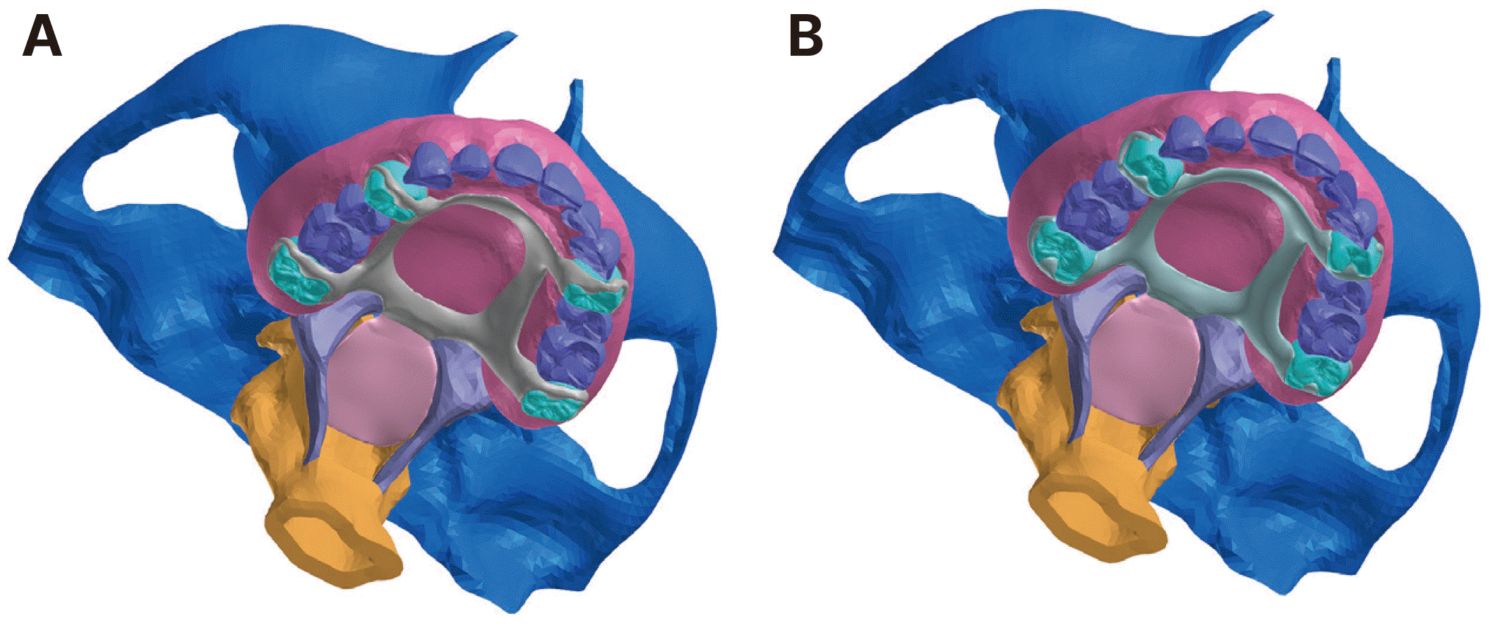
A meshing software (Visual-Mesh 18.0, ESI Group, Paris, France) was used to create tetrahedral meshes of all components in each experimental model. Model 1 generated a finite element mesh consisting of 632,847 nodes and 3,444,786 elements. Model 2 produced a mesh with 626,417 nodes and 3,433,595 elements. The material properties for this FEA study were assumed to be homogenous, isotropic, and linearly elastic. Table 1 provides a summary of the material properties of all the components used in this study.16-28
The friction coefficients (µ) of 0.01 and 0.10 were used for the contact behavior between the prosthesis and mucosa and between the occlusal rest and surveyed crown, respectively.29 All nodes on the cutting surface of the maxillary bone were constrained in all directions. To simulate swallowing pressure produced by the base of the tongue, 25 kPa was applied at the surface of the bulb of the obturator prosthesis opposite the base of the tongue.30 The analysis was processed using the Visual-Crash software (Visual-Crash for PAM 18.0, ESI Group, Paris, France), and the solver program (Launch VPS Solver 2020.0, ESI Group) was used to perform the analysis. The maximum von-Mises stress and displacement values of the velopharyngeal obturator prosthesis were analyzed and compared for each experimental model.

Results

Table 2 showed the maximum von-Mises stress and displacement values of the velopharyngeal obturator prosthesis and the abutment teeth for each model.
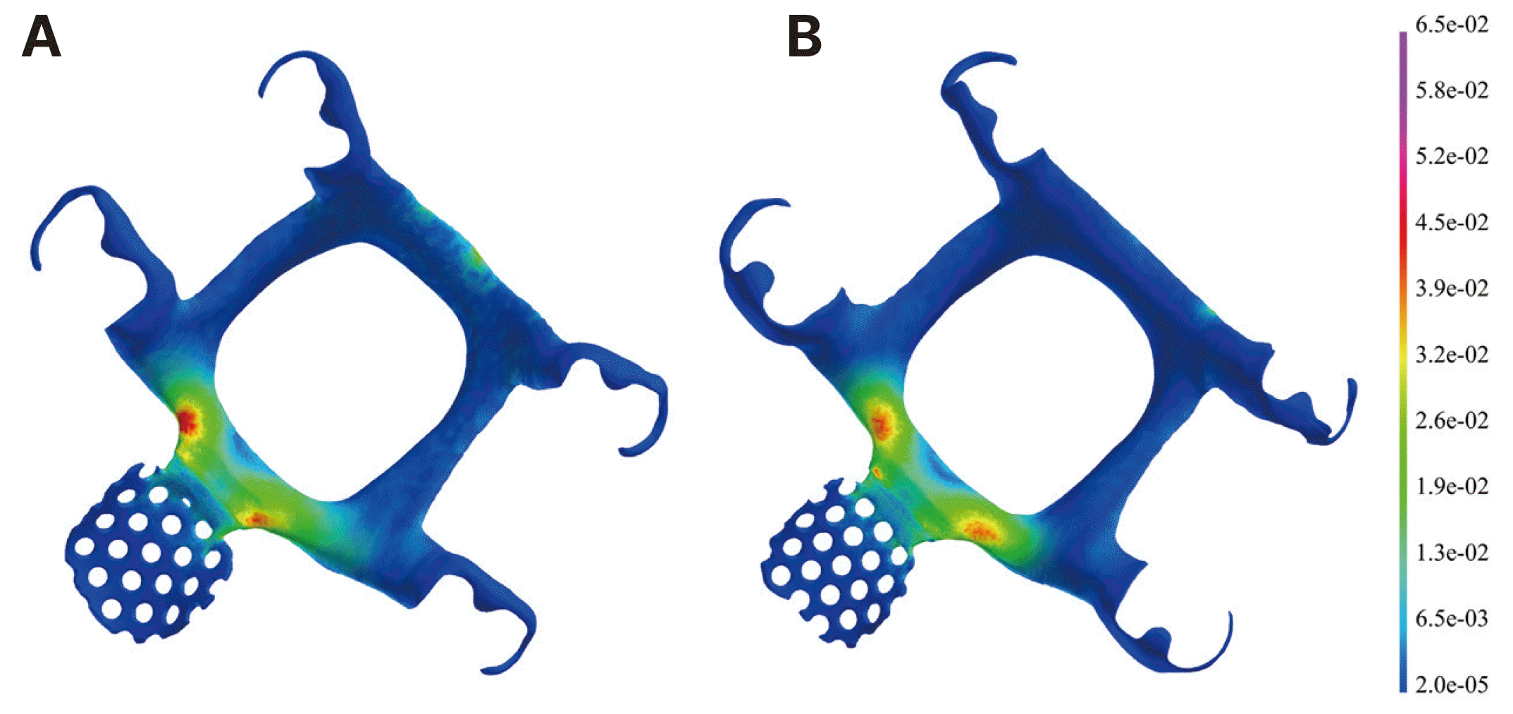
Concerning biomechanical stress distribution on the obturator prosthesis, the maximum von-Mises stress in the metal framework was slightly higher in model 1 (64.9 MPa) compared to model 2 (54.2 MPa), with both models having the same maximum stress concentration area at the posterior palatal strap adjacent to the junction of the metal framework and obturator bulb (Fig. 3). However, the acrylic resin obturator bulb showed the same maximum von-Mises stress distribution for both models, with a value of 4.3 MPa.
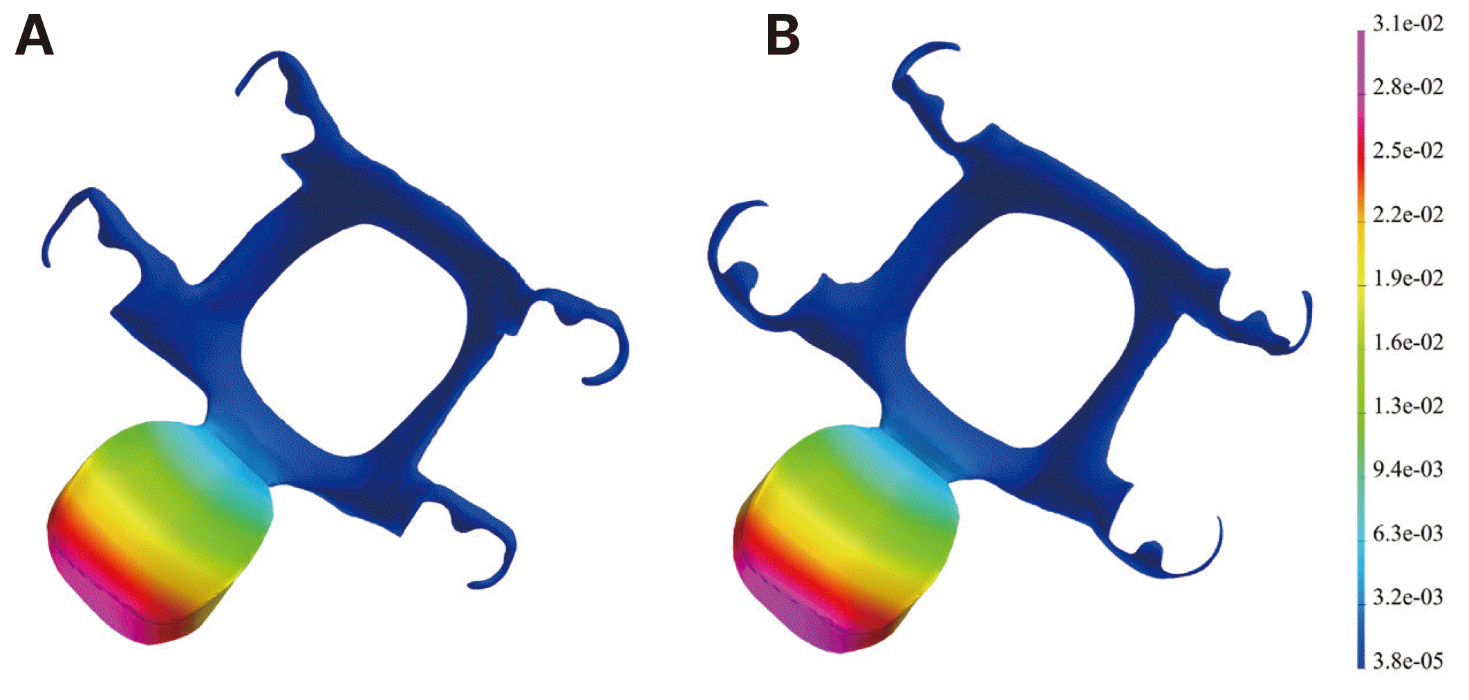
With regard to the displacement distribution of the obturator prosthesis and abutment teeth, model 1 and model 2 exhibited similar displacement values, with 31.3 µm and 33.6 µm for the prosthesis, and 0.42 µm and 0.31 µm for the abutment teeth, respectively. Fig. 4 illustrates the displacement distribution patterns of the velopharyngeal obturator prosthesis for both models.

Discussion

The dislodgement of the velopharyngeal obturator prosthesis can be potentially life-threatening for patients with soft palate defects who have been rehabilitated with this type of prosthesis. To ensure its efficient and safe function during speech and swallowing, it is crucial for the velopharyngeal obturator to remain stable in its position. This study investigated the effect of swallowing tongue pressure on the biomechanics of the velopharyngeal obturator prosthesis and determined the appropriate occlusal rest position for this type of prosthesis by comparing displacement values between the mesial and distal occlusal rest positions.
During swallowing mechanism, the pressure exerted can vary depending on the size and viscosity of the bolus, types of swallowing, and anatomical locations in the pharynx.30,31 In this study, a pressure of 25 kPa was used to simulate the pressure produced by the base of the tongue in the oropharynx during swallowing, based on data from the study conducted by Qazi et al.30
Previous literature indicates that most studies on occlusal rest positions have primarily focused on distal extension removable partial dentures. Considering the similarity of the posterior extension of the obturator bulb in the velopharyngeal obturator prosthesis to the distal extension partial denture scenario, it is reasonable to compare the findings of this study to those of previous investigations.
The present study found that the von-Mises stress in the metal framework was slightly higher for the mesial occlusal rests compared to the distal occlusal rests. Similarly, Shahmiri et al. suggested that distal occlusal rests should be utilized in distal extension partial dentures to improve stress distribution in the metal framework and acrylic resin denture base structures.29 In contrast, Zarrati et al. proposed using both mesial and distal occlusal rests in distal extension removable partial dentures.32
Basically, moving the occlusal rests from the mesial to the distal side of the abutment teeth could increase the rotation of the denture base. However, in this study, both mesial and distal occlusal rests showed almost the same prosthesis displacement values. The reason for this is the placement of the indirect retainer, which was identical in both models and had the same distance from the fulcrum of the obturator prosthesis.
In this FEA study, the simulation analysis of the biomechanics of the velopharyngeal obturator prosthesis was restricted to the fully dentulous scenario and only focused on the occlusal rest position. Further research is needed to investigate the biomechanical analysis of the velopharyngeal obturator prosthesis in a fully edentulous condition or with different retainer designs in partially edentulous conditions.

Conclusion

Within the limitations of this study, it can be concluded that swallowing tongue pressure had a minor impact on the biomechanics of the velopharyngeal obturator prosthesis and that distal occlusal rests showed a slightly better biomechanical response compared to mesial occlusal rests. The results of this study could probably assist in retainer design considerations during prosthetic rehabilitation for patients with velopharyngeal insufficiency.

Acknowledgements

This study was supported by grant no 04-2018-0097 from the SNUDH Research Fund.

References

1. Gaziano JE. 2002; Evaluation and management of oropharyngeal dysphagia in head and neck cancer. Cancer Control. 9:400–9. DOI: 10.1177/107327480200900505. PMID: 12410179.
crossref
2. Boseley ME, Hartnick CJ. 2004; Assessing the outcome of surgery to correct velopharyngeal insufficiency with the pediatric voice outcomes survey. Int J Pediatr Otorhinolaryngol. 68:1429–33. DOI: 10.1016/j.ijporl.2004.06.002. PMID: 15488976.
crossref
3. Thomas L, Jones TM, Tandon S, Katre C, Lowe D, Rogers SN. 2008; An evaluation of the University of Washington quality of life swallowing domain following oropharyngeal cancer. Eur Arch Otorhinolaryngol. 265 Suppl 1:S29–37. DOI: 10.1007/s00405-007-0470-2. PMID: 17952450.
crossref
4. Barata LF, de Carvalho GB, Carrara-de Angelis E, de Faria JCM, Kowalski LP. 2013; Swallowing, speech and quality of life in patients undergoing resection of soft palate. Eur Arch Otorhinolaryngol. 270:305–12. DOI: 10.1007/s00405-012-2006-7. PMID: 22526573.
crossref
5. Aramany MA, Myers EN. 1978; Prosthetic reconstruction following resection of the hard and soft palate. J Prosthet Dent. 40:174–8. DOI: 10.1016/0022-3913(78)90013-6. PMID: 278835.
crossref
6. Shetty NB, Shetty S, Nagraj E, D'Souza R, Shetty O. 2014; Management of velopharyngeal defects: A review. J Clin Diagn Res. 8:283–7. DOI: 10.7860/JCDR/2014/6220.4188. PMID: 24783161. PMCID: PMC4003670. PMID: 29b86e91cedb4dc9938604da638d2d39.
crossref
7. Saunders TR, Oliver NA. 1993; A speech-aid prosthesis for anterior maxillary implant-supported prostheses. J Prosthet Dent. 70:546–7. DOI: 10.1016/0022-3913(93)90271-O. PMID: 8277446.
crossref
8. Abreu A, Levy D, Rodriguez E, Rivera I. 2007; Oral rehabilitation of a patient with complete unilateral cleft lip and palate using an implant-retained speech-aid prosthesis: Clinical report. Cleft Palate Craniofac J. 44:673–7. DOI: 10.1597/06-169.1. PMID: 18177197.
crossref
9. Raol N, Hartnick CJ. 2015; Anatomy and physiology of velopharyngeal closure and insufficiency. Adv Otorhinolaryngol. 76:1–6. DOI: 10.1159/000368003. PMID: 25733226.
crossref
10. McConnel FM. 1988; Analysis of pressure generation and bolus transit during pharyngeal swallowing. Laryngoscope. 98:71–8. DOI: 10.1288/00005537-198801000-00015. PMID: 3336265.
crossref
11. Jacob RF, King G. 1990; Indirect retainers in soft palate obturator design. J Prosthet Dent. 63:311–5. DOI: 10.1016/0022-3913(90)90203-O. PMID: 2407829.
crossref
12. Tuna SH, Pekkan G, Gumus HO, Aktas A. 2010; Prosthetic rehabilitation of velopharyngeal insufficiency: pharyngeal obturator prostheses with different retention mechanisms. Eur J Dent. 4:81–7. DOI: 10.1055/s-0039-1697813. PMID: 20046485. PMCID: PMC2798795.
crossref
13. Collin JD, Main BGJ, Barber AJ, Thomas SJ. 2014; Airway compromise by dislodged obturator in a patient with severe trismus. J Prosthet Dent. 112:83–5. DOI: 10.1016/j.prosdent.2013.08.021. PMID: 24423458.
crossref
14. Hohmann A, Kober C, Young P, Dorow C, Geiger M, Boryor A, Sander FM, Sander C, Sander FG. 2011; Influence of different modeling strategies for the periodontal ligament on finite element simulation results. Am J Orthod Dentofacial Orthop. 139:775–83. DOI: 10.1016/j.ajodo.2009.11.014. PMID: 21640884.
crossref
15. Tachimura T, Nohara K, Wada T. 2000; Effect of placement of a speech appliance on levator veli palatini muscle activity during speech. Cleft Palate Craniofac J. 37:478–82. DOI: 10.1597/1545-1569_2000_037_0478_eopoas_2.0.co_2. PMID: 11034030.
crossref
16. de Sousa AA, Mattos BSC. 2014; Finite element analysis of stability and functional stress with implant-supported maxillary obturator prostheses. J Prosthet Dent. 112:1578–84. DOI: 10.1016/j.prosdent.2014.06.020. PMID: 25277029.
crossref
17. Rubin C, Krishnamurthy N, Capilouto E, Yi H. 1983; Stress analysis of the human tooth using a three-dimensional finite element model. J Dent Res. 62:82–6. DOI: 10.1177/00220345830620021701. PMID: 6571871.
18. Reinhardt RA, Pao YC, Krejci RF. 1984; Periodontal ligament stresses in the initiation of occlusal traumatism. J Periodontal Res. 19:238–46. DOI: 10.1111/j.1600-0765.1984.tb00815.x. PMID: 6235340.
crossref
19. Rees JS, Jacobsen PH. 1997; Elastic modulus of the periodontal ligament. Biomaterials. 18:995–9. DOI: 10.1016/S0142-9612(97)00021-5. PMID: 9212195.
crossref
20. Miyashita ER, Mattos BSC, Noritomi PY, Navarro H. 2012; Finite element analysis of maxillary bone stress caused by Aramany class IV obturator prostheses. J Prosthet Dent. 107:336–42. DOI: 10.1016/S0022-3913(12)60086-9. PMID: 22546312.
crossref
21. Haddad SMH, Dhaliwal SS, Rotenberg BW, Samani A, Ladak HM. 2018; Estimation of the Young's moduli of fresh human oropharyngeal soft tissues using indentation testing. J Mech Behav Biomed Mater. 86:352–8. DOI: 10.1016/j.jmbbm.2018.07.004. PMID: 30007184.
crossref
22. Birch MJ, odon PD Sr. 2009; Biomechanical properties of the human soft palate. Cleft Palate Craniofac J. 46:268–74. DOI: 10.1597/08-012.1. PMID: 19642755.
crossref
23. Carrigy NB, Carey JP, Martin AR, Remmers JE, Zareian A, Topor Z, Grosse J, Noga M, Finlay WH. 2016; Simulation of muscle and adipose tissue deformation in the passive human pharynx. Comput Methods Biomech Biomed Engin. 19:780–8. DOI: 10.1080/10255842.2015.1062477. PMID: 26213804.
crossref
24. Kim SM, Kim SJ, Bae HS, Choi BC, Mun JH. 2004; A nondestructive diagnostic modeling for muscular dysfunction of human pharynx using finite element method. Key Eng Mater. 270-273:2061–6. DOI: 10.4028/www.scientific.net/KEM.270-273.2061.
crossref
25. Ha SR, Kim SH, Lee JB, Han JS, Yeo IS, Yoo SH, Kim HK. 2016; Biomechanical three-dimensional finite element analysis of monolithic zirconia crown with different cement thickness. Ceram Int. 42:14928–36. DOI: 10.1016/j.ceramint.2016.06.133.
crossref
26. Heo KH, Lim YJ, Kim MJ, Kwon HB. 2018; Three-dimensional finite element analysis of the splinted implant prosthesis in a reconstructed mandible. J Adv Prosthodont. 10:138–46. DOI: 10.4047/jap.2018.10.2.138. PMID: 29713435. PMCID: PMC5917106.
crossref
27. Eom JW, Lim YJ, Kim MJ, Kwon HB. 2017; Three-dimensional finite element analysis of implant-assisted removable partial dentures. J Prosthet Dent. 117:735–42. DOI: 10.1016/j.prosdent.2016.09.021. PMID: 27914668.
crossref
28. Wang M, Qu X, Cao M, Wang D, Zhang C. 2013; Biomechanical three-dimensional finite element analysis of prostheses retained with/without zygoma implants in maxillectomy patients. J Biomech. 46:1155–61. DOI: 10.1016/j.jbiomech.2013.01.004. PMID: 23395509.
crossref
29. Shahmiri R, Das R, Aarts JM, Bennani V. 2014; Finite element analysis of an implant-assisted removable partial denture during bilateral loading: Occlusal rests position. J Prosthet Dent. 112:1126–33. DOI: 10.1016/j.prosdent.2014.04.023. PMID: 24951387.
30. Qazi WM, Ekberg O, Wiklund J, Kotze R, Stading M. 2019; Assessment of the food-swallowing process using bolus visualisation and manometry simultaneously in a device that models human swallowing. Dysphagia. 34:821–33. DOI: 10.1007/s00455-019-09995-8. PMID: 30840137. PMCID: PMC6825639.
crossref
31. Al-Toubi AK, Doeltgen SH, Daniels SK, Corey DM, Huckabee ML. 2015; Pharyngeal pressure differences between four types of swallowing in healthy participants. Physiol Behav. 140:132–8. DOI: 10.1016/j.physbeh.2014.12.029. PMID: 25527200.
crossref
32. Zarrati S, Bahrami M, Heidari F, Kashani J. 2015; Three dimensional finite element analysis of distal abutment stresses of removable partial dentures with different retainer designs. J Dent (Tehran). 12:389–97. PMID: 26884772. PMCID: PMC4754564.

Fig. 1
3D FEA soft palate defect model: (A) occlusal view, (B) lateral view.
jdras-39-2-61-f1.tif
Fig. 2
Velopharyngeal obturator models: (A) model 1 with mesial occlusal rests, (B) model 2 with distal occlusal rests.
jdras-39-2-61-f2.tif
Fig. 3
von-Mises stress distributions in the velopharyngeal obturator prostheses: (A) with mesial occlusal rests, (B) with distal occlusal rests.
jdras-39-2-61-f3.tif
Fig. 4
Displacement distribution pattern of the velopharyngeal obturator prostheses: (A) with mesial occlusal rests, (B) with distal occlusal rests.
jdras-39-2-61-f4.tif
Table 1
Elastic modulus and Poisson’s ratio for anatomical structures and materials
Materials Elastic modulus (GPa) Poisson’s ratio (v)
Cortical bone 13.7 0.30
Enamel 41 0.30
Dentin 18.6 0.31
PDL 0.05 0.49
Palatal mucosa 0.68 0.45
Soft palate 6E-6 0.45
Pharyngeal tissue 7E-6 0.45
Zirconia 210 0.27
Cement layer 8 0.33
Co-Cr alloy 211 0.30
Acrylic resin 2.7 0.35

GPa, gigapascal; PDL, periodontal ligament; Co-Cr, cobalt-chromium.

Table 2
Maximum von-Mises stress and displacement values for obturator prosthesis and abutment teeth
Model Maximum von-Mises stress (MPa) Maximum displacement (μm)
Metal frame Acrylic resin Prosthesis Abutment teeth
Model 1 (mesial occlusal rests) 64.9 4.3 31.3 0.42
Model 2 (distal occlusal rests) 54.2 4.3 33.6 0.31

MPa, megapascal; μm, micrometre.

TOOLS
Similar articles