

 PDF
PDF Citation
 Print
Print


INTRODUCTION
The coronavirus disease 2019 (COVID-19) pandemic has plagued humanity since 2019. Nonpharmaceutical interventions (NPIs) such as social distancing, respiratory etiquette, hand hygiene, and environmental cleaning, have been implemented worldwide to prevent the spread of COVID-19 [1]. The Korean Government mandated wearing facemasks in public places in November 2020. Other NPIs including, restrictions on private gatherings, curfew for entertainment facilities, and delay of school openings, were also implemented [2, 3]. Recent evidence suggests that decreased incidence of infectious diseases, especially respiratory infections [4-6], are associated with the NPIs. However, data on the relationship between incidence of central nervous system (CNS) infections and NPIs are limited.
The FilmArray Meningitis/Encephalitis (ME) panel (BioFire Diagnostics, Salt Lake City, UT, USA) is a novel multiplex polymerase chain reaction (PCR) test designed to rapidly and simultaneously identify 14 common pathogens that cause CNS infections: Escherichia coli K1, Haemophilus influenzae, Listeria monocytogenes, Neisseria meningitides (encapsulated), Streptococcus pneumoniae, Streptococcus agalactiae, cytomegalovirus, enterovirus, herpes simplex virus 1 and 2, human herpesvirus 6 (HHV-6), human parechovirus, varicella zoster virus, and Cryptococcus neoformans/Cryptococcus gattii [7]. The ME panel was the first nucleic acid-based cerebrospinal fluid (CSF) test approved in October 2015 by the U.S. Food and Drug Administration for use in CNS infections.
In Korea, the ME panel was approved in September 2017 by the National Healthcare Insurance Act as a new medical technology and introduced as a non-reimbursement test in November 2017. The ME panel was introduced in our institution on January 17, 2018, but was suspended on September 1, 2018, due to a cost issue regarding being reimbursed as per the National Healthcare Insurance Act. The ME panel was resumed in April 2021 after appropriate government cost adjustments, giving us a unique opportunity to compare the ME panel results before and during the pandemic.
Since then, HHV-6 has been the most frequently detected pathogen. Primary HHV-6 infections commonly occur in early childhood (<2 years of age). After primary infection, it remains latent in white blood cells (WBCs) but can reactivate in immunosuppressed hosts such as hematopoietic stem cell transplant (HSCT) recipients. In approximately 1% of the population, HHV-6 exists as chromosomally inherited HHV-6 (ciHHV-6), a condition in which the viral genome is integrated into a chromosome and inherited vertically [8, 9]. However, the interpretation of HHV-6 detection in ME panels is challenging. Previous reports from the U.S. and Botswana have suggested that HHV-6 detection in ME panels is inconsistent with CNS infection, ruling it out as the cause in most cases [10, 11]. However, another report has shown that 20% of all HHV-6 positive patients had true HHV-6 CNS infection and that HHV-6 detection in the ME panel led to faster administration of optimal antiviral treatment [12]. Thus, it is necessary to discuss whe-ther the detection of HHV-6 in the ME panel can be considered a true HHV-6 CNS infection.
This study aimed to determine the impact of the COVID-19 pandemic on CNS infections by comparing ME panel data before and during the pandemic at Pusan National University Yangsan Hospital. Furthermore, quantitative PCR (qPCR) and clinical evaluation of patients tested positive for HHV-6 in the ME panel were performed to determine the significance of detecting HHV-6 in the ME panel.
Go to : 
MATERIALS AND METHODS
1. Study population
This study included the data from the laboratory information system which were tested using the ME panel at Pusan National University Yangsan Hospital, a tertiary care center, from January 17, 2018 to August 31, 2018, and from April 9, 2021 to December 21, 2021, which represent the pre-pandemic (P1) and pandemic (P2) periods, respectively. In addition, we used the biospecimens showing positive HHV-6 results and clinical information from the biobank of the Pusan National University Yangsan Hospital. This study was approved by the Institutional Review Board (PNUYH IRB number 05-2022-088) and used biospecimens and clinical data from the Institutional Biobank Project according to the individual research protocol (OF-2022-10).
The ME panel is provided as a 24/7 stat service at the laboratory. It is mainly performed on emergency department patients suspected of having CNS infections. When a patient presents with clinical symptoms and signs of CNS infection such as, fever, headache, or seizure, CSF specimens are collected via lumbar puncture and sent to the laboratory. The ME panel is performed on the CSF with other laboratory workups to rule out bacterial meningitis. The ME panel results are reported within a maximum of 4 hrs after the request is made by the physician.
2. ME panel test results before and during the pandemic
ME panel test results for P1 and P2 periods were collected from the biobank data. The positive rates and distribution of pathogens in each period were calculated. The number of detections was derived by counting the detections of each pathogen.
3. Clinical significance of HHV-6
To assess the clinical significance of detecting HHV-6 in the ME panel, we collected specimens and clinical and laboratory data for patients tested positive for HHV-6 in the ME panel obtained in the hospital’s biobank. The clinical data included patients’ sex, age, presenting signs and symptoms, comorbidities, brain magnetic resonance imaging, antiviral treatment, clinical outcome at discharge, and diagnosis based on the treating physician’s documentation. The laboratory data included CSF analysis and other microbiological or serological tests performed during the hospital stay. CSF parameters included WBC count and differential, glucose, and protein levels. HHV-6 DNA quantification was assessed by qPCR of preserved CSF, serum, or plasma samples provided by the biobank. RealStar HHV-6 PCR Kit 1.0 (Altona Diagnostics, Hamburg, Germany) assay was used to differentiate and quantify human herpesvirus 6A (HHV-6A) and human herpesvirus 6B (HHV-6B) DNA. The limit of detection for the assay was set at 140 copies/mL.
Finally, clinical assessments were conducted to determine the clinical significance of HHV-6 as a true causative agent of CNS infection. Three of the study’s authors, including a pediatric infectious diseases specialist (S. E. Park), independently reviewed each patient’s clinical and laboratory data, including the HHV-6 qPCR test results. Each author individually classified the cases into one of the four groups: (1) primary infection; or one of the following regarding HHV-6 CNS infection: (2) likely (>90% probability), (3) possible (10–90% probability), or (4) unlikely (<10% probability) (modified from the criteria developed by Green et al. [10] and Slenker et al. [13]). Primary HHV-6 infections occur mostly in early childhood and cause acute febrile illness and rash, known as exanthema subitum. The symptoms are usually mild and typically self-limiting within a few days. CNS infection in primary exposure is less common. After primary infection, HHV-6 can enter a state of latency; many studies suggest that its reactivation later in life might result in neurologic symptoms [9, 14, 15]. Any inconsistencies between the authors regarding the differentials were discussed and reached to a consensus.
4. Statistical analyses
All statistical analyses were performed using R (version 4.1.2; R Foundation for Statistical Computing, Vienna, Austria) and RStudio (Desktop version 2022.07.2+548; POSIT, Boston, MA, USA). A chi-square test was conducted to compare the positive rates. Results with P<0.05 were considered statistically significant. Bar graphs were generated using RStudio.
Go to :
RESULTS
1. ME panel results before and during the pandemic
During P1, 432 patients were tested using the ME panel. The panel identified 125 pathogens in 116 patients (nine co-detections) (Table 1). During P2, 205 patients were tested, among which the ME panel identified 15 pathogens in 15 patients. The total positive rate markedly decreased from 28.9% (125/432) in P1 to 7.3% (15/205) in P2, showcasing a considerable statistical significance between the two periods (P<0.05). The positive rate of enterovirus decreased significantly from 15.3% (66/432) to 0.0% (P<0.05). The positive rate for human parechovirus also showed a significant reduction from 5.3% (23/432) to 0.0% (P<0.05). Those of the remaining 12 pathogens did not differ between P1 and P2.
Table 1
Positive rates of the ME panel before and during COVID-19 pandemic
| Pre-pandemic (N=432) | Pandemic (N=205) | P | |
|---|---|---|---|
| Virus | |||
| Enterovirus | 66 (15.3%) | 0 (0.0%) | < 0.001 |
| Herpes simplex virus 1 | 1 (0.2%) | 0 (0.0%) | 1 |
| Herpes simplex virus 2 | 4 (0.9%) | 1 (0.5%) | 0.916 |
| Varicella zoster virus | 6 (1.4%) | 2 (1.0%) | 0.955 |
| Cytomegalovirus | 2 (0.5%) | 1 (0.5%) | 1 |
| Human herpesvirus 6 | 11 (2.5%) | 7 (3.4%) | 0.717 |
| Human parechovirus | 23 (5.3%) | 0 (0.0%) | 0.002 |
| Bacteria | |||
| Escherichia coli K1 | 4 (0.9%) | 1 (0.5%) | 0.916 |
| Haemophilus influenzae | 0 | 0 | NA |
| Listeria monocytogenes | 0 | 0 | NA |
| Neisseria meningitidis | 0 | 0 | NA |
| Streptococcus agalactiae | 3 (0.7%) | 3 (1.5%) | 0.617 |
| Streptococcus pneumoniae | 4 (0.9%) | 0 (0.0%) | 0.398 |
| Yeast | |||
| Cryptococcus sp. | 1 (0.2%) | 0 (0.0%) | 1 |
| Total | 125 (28.9%)* | 15 (7.3%) | < 0.001 |
![]()
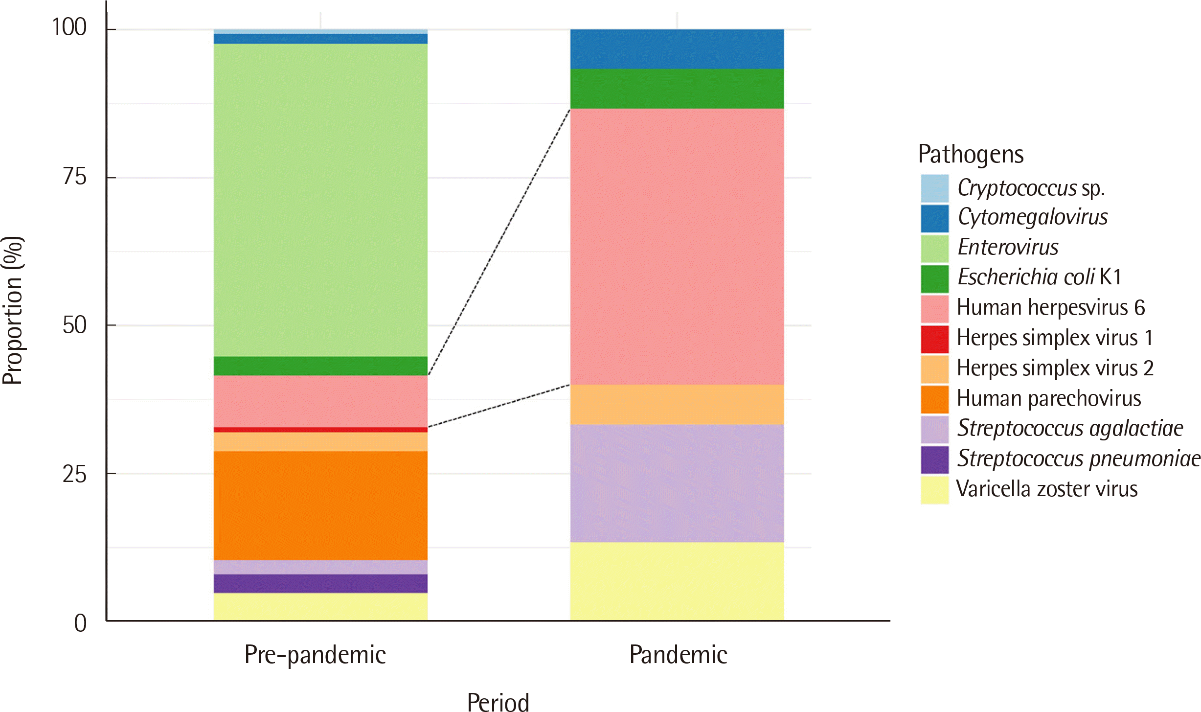
For each period, the proportion of each pathogen, with respect to the total number of detected pathogens, were analyzed. Enterovirus was the most commonly detected pathogen during P1, being discovered in 66/125 (52.8%) cases, followed by human parechovirus 23/125 (18.4%), and HHV-6 11/125 (8.8%). During P2, HHV-6 was the most commonly found pathogen, detected in 7/15 (46.7%) cases, followed by Streptococcus agalactiae 3/15 (20.0%). Enterovirus and human parechovirus, the top two most common pathogens in P1, were however not detected in P2 (Fig. 1).
Nine co-detections were identified during P1, all of which were positive for two pathogens. The most common pathogens in the co-detection were enterovirus and HHV-6: seven co-detections of enterovirus and HHV-6, one of Escherichia coli K1 and herpes simplex virus 2, and one of Streptococcus agalactiae and herpes simplex virus 1. Regarding HHV-6 detection in P1, 4/11 (36.4%) detection were solely of HHV-6, while the remaining 7/11 (63.6%) were co-detected with enterovirus. In contrast, there were no cases of co-detection during P2.
2. Clinical significance of HHV-6
Clinical evaluation was conducted on 18 patients positive for HHV-6 in the ME panel, and qPCR was conducted on those with available samples. Tables 2 and 3 present the data of 18 patients positive for HHV-6 in P1 and P2, respectively.
Table 2
Characteristics of patients tested positive for HHV-6 on ME panel before COVID-19 pandemic
![]()
Table 3
Characteristics of patients tested positive for HHV-6 on ME panel during COVID-19 pandemic
| Patient | Sex/Age | Immunosuppressed | Presenting signs and symptoms | Pathogens detected in ME panel other than HHV-6 | CSF | MRI consistent with CNS infection | |||
|
|
|||||||||
| WBC count (WBCs/μL) | Lymphocytes (%) | Glucose (mg/dL) | Protein (mg/dL) | ||||||
| 12 | M/51D | No | Fever, irritability, vomiting | None | 0 | 0 | 57 | 31.7 | NT |
| 13 | M/4Y | No | Fever, seizure | None | 53 | 75 | 48 | 38.1 | Yes |
| 14 | F/3M | No | Fever, whole body rash | None | 0 | 0 | 70 | 21.2 | NT |
| 15 | F/9Y | No | Headache, vomiting, neck stiffness, Brudzinski’s sign | None | 21.2* | 51.9 | 51 | 220 | No† |
| 16 | M/8Y | Yes (HSCT recipient) | Fever, disorientation | None | 62 | 88.7 | 67 | 61.9 | No |
| 17 | F/10Y | No | Fever, dizziness, headache, vomiting | None | 26 | 84 | 89 | 25.9 | Yes |
| 18 | M/82D | No | Fever | None | 383 | 7.3 | 65 | 62.1 | No |
| Patient | HHV-6 CSF viral load (copies/mL)‡ | HHV-6 Serum viral load (copies/mL)‡ | Antiviral treatment | Additional clinically relevant lab results | Outcome at discharge | Diagnosis | HHV-6 assessment | ||
| 12 | 20,380 | NT | None | Mildly increased WBCs in urine | Alive | Non-specific febrile illness | Primary infection | ||
| 13 | < 140 | < 140 | Ganciclovir | Blood positive for MOG antibody | Alive | Acute disseminated encephalomyelitis | Unlikely | ||
| 14 | 425 | NT | None | Urine gram stain positive for gram-negative bacilli | Alive | Acute pyelonephritis, exanthema subitum | Primary infection | ||
| 15 | NT | < 140 | None | None | Alive | Intraventricular hemorrhage | Unlikely | ||
| 16 | 2,770 | 2,006§ | Ganciclovir | None | Died during admission | HHV-6 encephalitis | Likely | ||
| 17 | < 140 | < 140 | Ganciclovir | Stool positive for norovirus; Blood positive for anti-Ro60 and anti-Ro52 antibodies | Alive | Brainstem encephalitis due to Sjögren’s syndrome | Unlikely | ||
| 18 | NT | NT | None | None | Alive | Bacterial meningitis | Primary infection | ||
*Corrected for blood contaminated CSF; †Intraventricular hemorrhage was seen; ‡All detected HHV-6 were HHV-6B; §Tested in plasma.
Abbreviations: ciHHV-6, chromosomally inherited HHV-6; CSF, cerebrospinal fluid; D, day; HHV-6, human herpesvirus 6; HSCT, hematopoietic stem cell transplant; M, month; ME panel, Meningitis/Encephalitis panel; MOG, myelin oligodendrocyte glycoprotein; MRI, magnetic resonance imaging; NT, not tested; WBC, white blood cell; Y, year.
![]()
Of the 11 HHV-6-positive patients (patient #1–11) from P1, seven were co-detected with enterovirus, while the remaining four were detected with HHV-6 alone (Table 2). Ten of these patients were children, while one was an adult, with ages ranging from 72 days to 74 years. All patients were immunocompetent. Fever was the most common presenting symptom, five had vomiting, four experienced headache, other four had cough, and two presented with a rash. Nine patients showed CSF pleocytosis. Among the 11 patients, none were considered likely to have HHV-6 CNS infections. Nine patients had clear alternative diagnoses and were classified as unlikely. The remaining two patients were assigned to the primary infection group. They lacked a definite alternative diagnosis and had clinical findings consistent with HHV-6 primary infections. HHV-6 qPCR could not be performed for patients in P1 due to unavailability of preserved samples.
All seven HHV-6 positive patients (patient #12–18) of P2 were children with ages ranging from 51 days to 10 years (Table 3). Six were immunocompetent hosts without a remarkable history, while one, who was an HSCT recipient, was an immunosuppressed host. Fever was the most common presenting symptom, three had vomiting, one experienced seizures, and one had altered mental status. Five patients had CSF pleocytosis. HHV-6 qPCR was conducted in six of those seven HHV-6 positive patients using their CSF and/or blood. HHV-6 qPCR was performed on CSF of five patients and HHV-6 DNA was detected in three patients. HHV-6 qPCR was performed on blood of four patients, and HHV-6 DNA was detected in one of them. All detected HHV-6 were HHV-6B. Among the seven patients, only the HSCT recipient (patient #16) was considered likely to have HHV-6 CNS infection. He had received HSCT three weeks prior to the onset of his symptoms. He had neurological symptoms consistent with a CNS infection, and his CSF analysis showed lymphocyte-dominant pleocytosis. Antiviral treatment improved the patient’s clinical and laboratory status. Unfortunately, he also developed graft-versus-host disease and died five months after the HSCT. Three patients had a clear alternative diagnosis and were considered unlikely to be consistent with HHV-6 CNS infection. The remaining three were classified as primary infection. They had no clear alternative diagnosis, were aged <2 years, and had symptoms and clinical courses consistent with HHV-6 primary infection. Patient #18 was diagnosed with bacterial meningitis during admission, due to CSF neutrophilic pleocytosis. However, upon our review, his symptoms, other laboratory findings, and clinical course were deemed inconsistent with those of bacterial meningitis. The contributing role of HHV-6 in his pathology seemed more likely, and he was thus classified as a primary infection.
Go to :
DISCUSSION
This study retrospectively analyzed ME panel results from the laboratory information system of a tertiary care center. The results showed that the positive rate of the ME panel markedly decreased by over 70% between the P1 and P2 periods. The significant reduction was mainly due to enterovirus, which constituted 52.8% of detected pathogens in P1, and was undetected in P2. These results are consistent with previous studies that reported a decreased incidence of common respiratory and gastrointestinal infections during the COVID-19 pandemic [4-6, 16-18]. In particular, studies in Korea, Taiwan, and Australia have reported a dramatic reduction in enterovirus infections during the pandemic [4, 17, 19]. These studies demonstrated that NPIs against COVID-19 have reduced the incidence of other common viral infections. Results obtained from this study are consistent with the implementation of NPIs.
The number of ME panel tests decreased from 54/month to 23.6/month. This decrease may be attributed to the implementation of NPIs, which resulted in fewer infectious diseases; thus, compared to P1, fewer patients presented with fever during P2. An Italian institute [16] also reported a 46% decrease in the number of gastrointestinal viruses PCR, from 2,547 to 1,368, during the pandemic, which were apparently attributed to the implementation of NPIs. They also suggested that some patients may have avoided visiting the hospital due to fear of contracting COVID-19. Korea had implemented restrictions on emergency department visitation during P2 if the patient presented fever, due to the risk of transmitting SARS-CoV-2 around the hospital. Moreover, all patients had to undergo nasopharyngeal swabs for COVID-19 testing before emergency department admission. All of these factors potentially prevented individuals from visiting the emergency department immediately after onset of symptom, thus giving time for the viral meningitis to self-limit [20].
HHV-6 was the most commonly detected pathogen in the ME panel during P2. The HHV-6 positive rate was similar between the two periods, but gained predominance in P2 due to the significant decrease in enterovirus and human parechovirus. Enterovirus and human parechovirus mainly spread through direct or indirect oral contact, with the virus being transmitted via feces and upper respiratory tract [21, 22]. The implementation of NPIs potentially prevented the transmission of these viruses and contributed to the decline in the positive rate. On the other hand, HHV-6 is most commonly transmitted person-to-person via saliva, infecting most children below the age of two. After a primary infection, it remains latent in WBCs [23-25]. The results of this study reflect the different transmission routes of each virus and the varying effects of NPIs on their prevalence.
However, the detection of HHV-6 in the ME panel was deemed inconsistent with HHV-6 CNS infection in most cases. Only one HSCT recipient was considered likely to have HHV-6 CNS infection. HHV-6 is known to reactivate in immunosuppressed hosts, such as this patient, to cause encephalitis [26, 27]. Twelve patients were assigned as unlikely. They were all immunocompetent and had a clear alternative diagnosis. The detection of HHV-6 in the ME panel of these patients was considered likely due to the asymptomatic reactivation of the latent virus or ciHHV-6 [10]. Notably, seven of the 12 unlikely patients had co-detection of enteroviruses. The co-detection of HHV-6 with other pathogens in the ME panel may be interpreted as a bystander, not a cause of CNS infection. Finally, five patients, aged <2 years, were classified as primary infection. They lacked clinical findings of CNS infection or a clear alternative diagnosis and had evidence suggesting HHV-6 primary infection. Our findings support the previous studies [10, 13] that the diagnosis of HHV-6 CNS infection based on the ME panel should be made on a comprehensive evaluation of the patient’s history, immune status, and clinical and laboratory findings.
Results obtained from the qPCR testing of CSF and whole blood further validate the obtained results. Patient #16, the only likely HHV-6 CNS infection case, was positive for HHV-6 qPCR in both, CSF and plasma. In contrast, three unlikely patients were negative for HHV-6 qPCR in the CSF and serum. If qPCR had been performed during their initial evaluation, patients #13 and #17 might not have unnecessarily received ganciclovir. Additional qPCR testing of whole blood in patients who test positive for HHV-6 in the ME panel will also help distinguish those with ciHHV-6. In individuals with ciHHV-6, all nucleated cells in the body contain HHV-6 DNA. They have consistently high levels of HHV-6 in their blood, which can lead to misdiagnosis of active infection and unnecessary treatment. Thus, exclusion of ciHHV-6 is needed to confirm HHV-6 as the cause of CNS infection [9]. Identification of ciHHV-6 can be performed by PCR of whole blood, hair follicles, fingernails, or by testing the patient’s parents or siblings. Detection of ≥106 copies/mL in whole blood is indicative of ciHHV-6 [9, 28]. Notably, a study has suggested that viral loads of thousands of copies in the CSF and millions of copies in the blood should prompt the suspicion of ciHHV-6 [29]. Although we did not test for ciHHV-6 and could not accurately assess the ciHHV-6 status of the patients; patient #12, who was assigned as primary infection, could have had ciHHV-6 based on his CSF viral load of 20,380 copies/mL. Additional qPCR testing on whole blood will thus help assess his ciHHV-6 status. Therefore, we propose the need for additional testing of HHV-6 qPCR on CSF and blood when HHV-6 is detected in CSF using qualitative tests, such as the ME panel, which will allow accurate diagnosis and optimal treatment.
The distinction between HHV-6A and HHV-6B may prove helpful in other countries. All four patients with HHV-6, detected by qPCR in our study, had HHV-6B. Our results are consistent with previous findings that HHV-6B causes 97–100% of HHV-6 primary infections [30]. HHV-6B is associated with exanthema subitum and HHV-6 reactivation in HSCT recipients. Although there have been reports of primary infections caused by HHV-6A in African populations, there are limited studies in this regard. A review article [9] demonstrated that among 34 ciHHV-6 reported cases, nine were HHV-6A and 25 were HHV-6B, while 10 cases from Japan were all HHV-6B. Therefore, when HHV-6 is detected in a routine diagnostic laboratory setting in Korea, it is most likely HHV-6B, whether it is a primary infection or ciHHV-6.
This study, however, had some limitations. First, we could not perform HHV-6 qPCR on all HHV-6 positive patients due to unavailability of the samples, given the retrospective nature of the study. Only six of the 18 patient samples were available for this study, as some had previously been used in other studies or lacked sufficient volumes. Second, our study did not test for ciHHV-6 due to the lack of whole blood samples available. Finally, the presented outcomes were limited by the small sample size, as this study was conducted in a single tertiary center. Further multicenter studies with larger sample sizes are required to confirm the impact of the COVID-19 pandemic and NPIs on the prevalence and distribution of CNS infections.
In conclusion, we report a significant decrease in the positive rates of the ME panel during the COVID-19 pandemic at our institution. This reduction can be explained by NPIs implemented worldwide to prevent the spread of COVID-19. As NPIs are still in effect during the ongoing pandemic, HHV-6 can continue to be frequently detected in the ME panel. The detection of HHV-6 in the ME panel should be interpreted cautiously with comprehensive clinical information and supplemented with additional qPCR on CSF and blood to ensure accurate diagnosis and optimal treatment.
Go to :
 XML Download