

 PDF
PDF Citation
 Print
Print


INTRODUCTION
An infectious disease involves an external agent, a host, and the environment. Therefore, hosts infected with the same pathogen (agent) in the same environment may show variations in disease progression and prognosis depending on the host characteristics [1, 2]. The novel coronavirus disease 2019 (COVID-19) is caused by severe acute respiratory syndrome coronavirus 2 (SARS-CoV-2) [3, 4]. The Centers for Disease Control and Prevention estimated that over 57% of the US population and 75% US children have been infected by SARS-CoV-2 until February 2022 [3]. Further, the World Health Organization has announced that COVID-19 has resulted in 616,427,419 confirmed cases and 6,528,557 deaths as of October 5, 2022 [4]. The clinical manifestations of SARS-CoV-2 infection exhibit a wide range of severity, from asymptomatic and mild to severe pneumonia, myocarditis, acute kidney injury, respiratory distress syndrome, multiorgan failure, and death. Various studies have reported that the severity of COVID-19 symptoms is related to the host epidemiological characteristics, hematological and immunological profile, similar to that in other infectious diseases [5-13]. In previous studies, the disease severity and mortality rates have been reported to differ depending on the study period. The laboratory profiles of infected patients showed increased white blood cell counts, neutrophil-lymphocyte ratios, troponin, D-dimer, and c-reactive protein levels, and decreased platelet counts and hemoglobin [5-8]. Unfortunately, early studies in the pandemic were limited in terms of complete investigation, including varying severities, and laboratory test parameters, while the latest studies had analytical bias owing to the influence of vaccine inoculation and various SARS-CoV-2 variants.
The composition and function of the human immune response vary among individuals and are influenced by several factors [13-15], including specific antibodies (immunoglobulins) to the pathogen. Antibodies represent humoral immunity and are generated in response to external stimuli such as pathogens. Therefore, the presence or absence of antibodies can be used to evaluate whether an individual has been previously infected with a pathogen [16, 17]. Further, antibodies have protective and therapeutic effects and are commonly used as therapeutic agents. COVID-19 mRNA vaccines, the most widely used COVID-19 vaccine type, use a specific mechanism to activate antibody production [18-20]. Monoclonal antibody therapies, such as casirivimab/imdevimab (REGEN-COV™, Regeneron Pharmaceuticals, Inc., Tarrytown, NY, USA), regdanvimab (Regkirona™, Celltrion, Inc., Incheon, Korea), sotrovimab (Xevudy™, GlaxoSmithKline LLC, Zebulon, NC, USA), and tixagevimab/cilgavimab (Evusheld™, AstraZeneca, Cambridge, UK) have also been actively administered to high-risk patients with COVID-19 worldwide [21-23].
Cytokines are important components of the human immune system and are secreted by various immune cells such as macrophages, lymphocytes, and mast cells to send signals to other cells. They are classified into several families according to their structures. The major cytokine families include the interleukins (ILs), members of the tumor necrosis factor (TNF) superfamily, interferons, and chemokines family. Each family has dozens to hundreds of cytokines, each of which promotes or suppresses inflammation and regulates cellular functions [24, 25]. Cytokine kinetics change dynamically over the course of disease and play important roles in the response to common viral infections [26]. Therefore, IL-6, IL-8, IL-1β, TNF-α, and interferon-γ which show potential as anti-inflammatory therapies, are being actively studied in patients with COVID-19 [11-13, 27, 28].
In this study, we recruited and collected samples from patients diagnosed with COVID-19 (strongly presumed to be the wild-type, Wuhan strain) in the early pandemic period, before the emergence of major SARS-CoV-2 variants and vaccine development, to exclude variant and vaccine effects. Furthermore, we performed a broader and more in-depth investigation than that in previous studies on the relationship between patient baseline characteristics, serial antibody titers, serial cytokine (IL-6 and TNF-α) levels, and COVID-19 severity for up to three months after SARS-CoV-2 infection.
MATERIALS AND METHODS
1. Study population
Patients diagnosed with COVID-19 (based on a reverse-transcription polymerase chain reaction test of a nasopharyngeal or oropharyngeal sample) and hospitalized at Boramae Medical Center, a designated Korean COVID-19 treatment center from February to August 2020, were included in the study. None of the patients had been vaccinated against COVID-19, and all participants were thought to have been infected with wild-type SARS-CoV-2. The time (days) from infection was calculated based on the date of symptom onset in symptomatic patients and the date of diagnosis in asymptomatic patients. The days from infection were divided into five intervals: 0–7 days (1 week), 8–14 days (2 weeks), 15–21 days (3 weeks), 22–42 days (4–6 weeks), and 43–84 days (7–12 weeks).
The study was approved by the Institutional Review Board of Boramae Medical Center (IRB 21-2021-38); consent was waived because the study involved no more than minimal risk. Moreover, the waiver did not adversely affect the rights and welfare of the participants, and the research could not have been feasibly conducted without the waiver being granted. Whenever appropriate, the participants would be provided with additional pertinent information after participation.
2. Clinical severity and outcomes
The clinical severity of SARS-CoV-2 infection was classified from asymptomatic infection to critical illness according to the clinical features and treatment required based on the established guidelines [29-31]. Patients without symptoms were classified as having “asymptomatic infection”; those with mild symptoms, which could be controlled through only supportive care, and without dyspnea were classified as having “mild illness”; patients with clinical and radiographic evidence of lower respiratory tract disease, who were administered oxygen via nasal prongs or a face mask were classified as having “moderate illness”; those with oxygen saturation <94%, representing severe pneumonia, or treated with specific therapies such as dexamethasone, were classified as having “severe illness”; and patients with respiratory failure, shock, or multiorgan dysfunction or failure were classified as having “critical illness”.
The main bases of outcomes were requiring oxygen treatment (“asymptomatic and mild” group or “moderate, severe, and critical” group) and requiring intensive care unit care (“asymptomatic, mild and moderate” group or “severe and critical” group).
3. Clinical and routine laboratory characteristics
In addition to collection of history, the patient underwent physical examination, chest X-ray, and initial blood sampling within two days of hospital admission. The blood tests included the complete blood counts (hemoglobin, white blood cells, and platelets), a chemistry panel (blood urea nitrogen, creatinine, protein, albumin, bilirubin, alkaline phosphatase, aspartate transaminase, and alanine aminotransferase), a coagulation panel (prothrombin time, activated partial thromboplastin time, fibrinogen, and D-dimer), cardiac markers (creatine kinase, creatine kinase-myocardial band, troponin I, and pro-B-type natriuretic peptide), and inflammatory markers (C-reactive protein, procalcitonin, lactate dehydrogenase, and ferritin).
4. Serologic testing for immunologic profiles
Blood collection was performed using a serum separator tube followed by serum separation via centrifugation at 3,000 rpm for 10 minutes. The samples were then stored at -80°C until use. All tests were performed within one month of blood collection.
Anti-nucleocapsid (N) antibody: An automated electrochemiluminescence immunoassay (ECLIA)-based Elecsys Anti-SARS-CoV-2 assay (Elecsys Anti-N; Roche Diagnostics, Mannheim, Germany) was performed using a Cobas 800/e 801 analyzer (Roche Diagnostics). The Elecsys Anti-N assay uses a recombinant protein representing the nucleocapsid (N) antigen to qualitatively detect anti-SARS-CoV-2 antibodies. The results were recorded as reactive (positive) or non-reactive (negative) depending on whether the cutoff index was ≥1.0 or <1.0, according to the manufacturer’s directions.
Anti-spike/receptor-binding domain (S/RBD) antibody: An automated ECLIA-based quantitative Elecsys Anti-SARS-CoV-2 S assay (Elecsys Anti-S/RBD; Roche Diagnostics) was performed using a Cobas 800/e 801 analyzer. The Elecsys Anti-S/RBD assay uses a recombinant protein representing the receptor binding domain (RBD) of the spike (S) antigen, which enables quantitative detection of high-affinity anti-SARS-CoV-2 antibodies. Titers ≥0.8 U/mL were reported as reactive (positive), whereas those <0.8 U/mL were reported as non-reactive (negative) according to the manufacturer’s instructions.
Neutralizing antibody: To identify neutralizing antibodies, we used the cPass SARS-CoV-2 Neutralization Antibody Detection kit ver. RUO 3.0, (cPass Neutralizing Antibody; GenScript Biotech, Piscataway, NJ, USA), an enzyme-linked immunosorbent assay (ELISA)-based surrogate virus neutralization test (sVNT). The results are based on the inhibition rate. According to the manufacturer’s instructions, the cut-off value is 30%; therefore, if neutralizing antibodies were detected to be ≥30%, the result was considered as positive, whereas values <30% were considered as negative. This cut-off value is based on validation of sera from patients with COVID-19 and healthy controls, and is reported to have 100.0% (95% CI: 87.1–100.0%) and 100.0% (95% CI: 95.8–100.0%) positive percent agreement with the plaque reduction neutralization tests, PRNT50 and PRNT90, respectively.
IL-6: An automated ECLIA-based Elecsys IL-6 (Roche Diagnostics) was performed using a Cobas 800/e 801 analyzer. According to the manufacturer’s instructions, the reference value is ≤7 pg/mL.
TNF-α: The Quantikine® HS ELISA (R&D Systems, Minneapolis, MN, USA) is a 4 h solid phase ELISA designed to measure human TNF-α in serum. This assay employs the quantitative sandwich enzyme immunoassay technique. According to the manufacturer’s instructions, the reference value is ≤10 pg/mL.
5. Statistical analysis
Data were compared using the independent samples t-test or Mann-Whitney U-test for continuous variables and the chi-square test for categorical variables. Pearson correlation analysis was used to evaluate the relationship between the Elecsys Anti-S/RBD and cPass Neutralizing Antibody results. Two-sided P values of P<0.05 were considered statistically significant. Multivariate analysis (logistic regression analysis) was performed to eliminate the influence of confounding variables and explain the degree of association among various risk factors. Selected correlates with P<0.2 in the univariable analysis were included in the multivariable logistic regression model. All statistical analyses were performed using SPSS version 27.0 (SPSS, Inc., Chicago, IL, USA) and Excel software (Microsoft Corporation, Redmond, WA, USA).
RESULTS
In total, 195 patients were included in the study, from whom 274 serum samples were collected within 84 days of infection. The number of samples obtained in each time interval was as follows: 96 in days 0–7, 72 in days 8–14, 34 in days 15–21, 53 in days 22–42, and 19 in days 43–84. The samples were obtained in only one interval from 137 patients, two intervals from 42 patients, three intervals from 12 patients, four intervals from three patients, and all five intervals from only one patient.
1. Demographic and clinical characteristics of the study participants
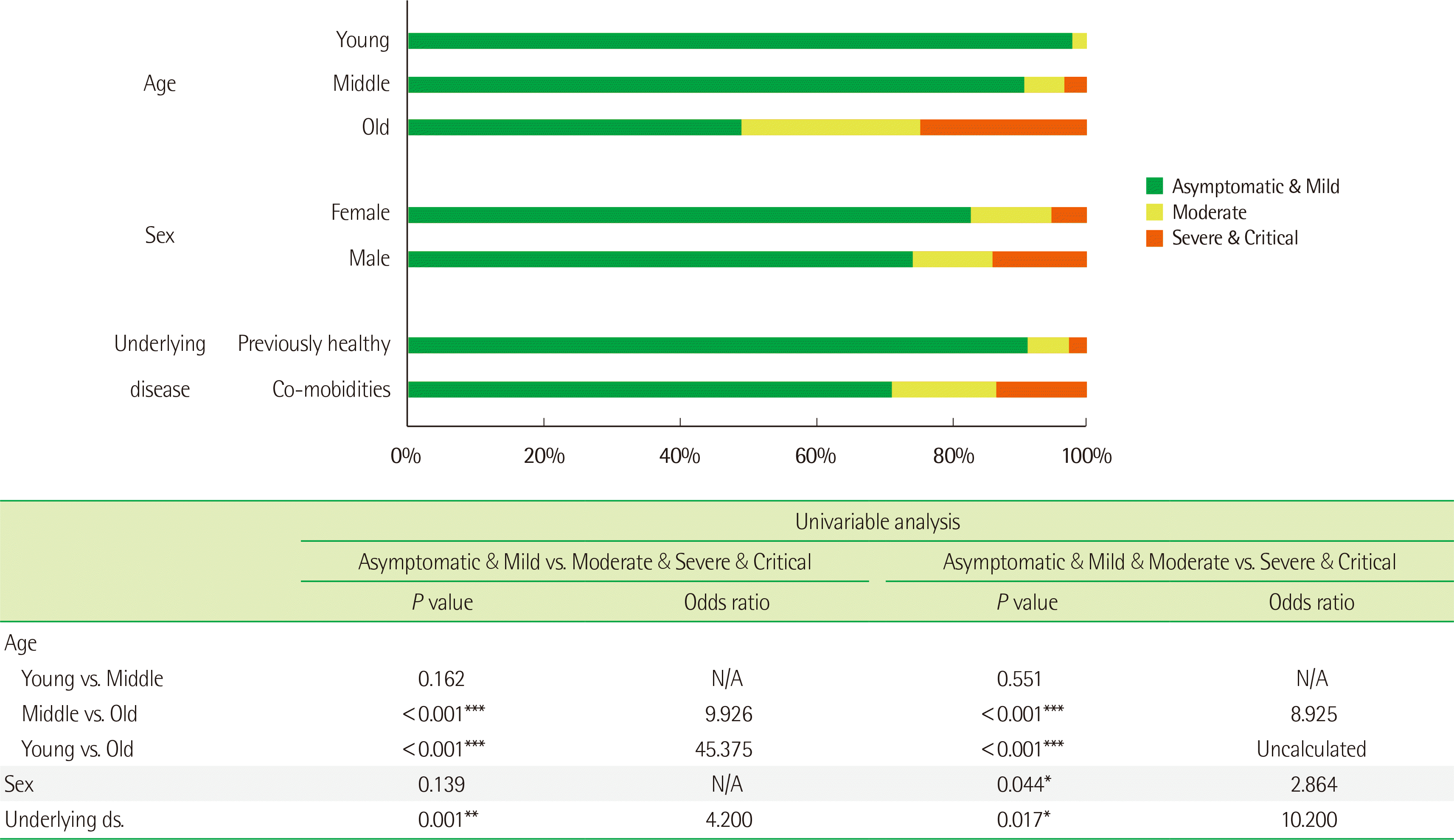
The demographic and clinical characteristics of the 195 participants are shown in Table 1. Forty-five patients (23.1%) were less than 40 years of age, 85 (43.6%) were between 40–64 years of age, and 65 (33.3%) were aged ≥65 years. There were 94 female patients (48.2%) and 101 male patients (51.8%). Sixty-nine patients (35.4%) were previously healthy, and 126 (64.6%) had underlying conditions such as hypertension, diabetes mellitus, coronary artery disease, chronic lung disease, or cancer. Most participants (96.4%) were Koreans. Of the patients, 139 (71.3%) reported general symptoms, 125 (64.1%) reported respiratory symptoms, 22 (11.3%) reported loss of taste and smell, and 18 (9.2%) reported gastrointestinal symptoms. Further, 153 patients (78.5%) had asymptomatic or mild illness, 23 (11.8%) had moderate illness, and 19 (9.7%) had severe or critical illness.
Of the 156 patients who could specify the date of symptom onset, an average of 3.6, 4.3, and 5.4 days passed from symptom onset to diagnosis, hospital admission, and radiography and first blood sampling, respectively. Further, during the analysis period, the quarantine release criteria of South Korean domestic COVID-19 guidelines were eased (as of June 25, 2020), after which the average hospital stay of patients who were asymptomatic and had mild illness was significantly reduced from 23.7 to 17.4 days (P=0.002).
2. Relationship between baseline participant characteristics and COVID-19 severity
The disease severity and outcome according to patient age, sex, and underlying disease status are shown in Fig. 1. The probability of an unfavorable outcome increased significantly with increasing age and the presence of comorbidities; further, the probability of requiring ventilator/intensive care unit care was significantly higher in males than in females.
The relation between laboratory data at the initial blood sampling and COVID-19 severity is shown in Table 2. In the severe and critical (high severity) group who later required intensive care, the initial hemoglobin, platelet, total protein, and albumin levels were significantly lower and the neutrophil-to-lymphocyte ratio, blood urea nitrogen, and creatinine levels were significantly higher than those in the asymptomatic, mild, and moderate (low severity) group. In addition, coagulation, inflammatory, and cardiac markers were significantly higher in the high severity group.
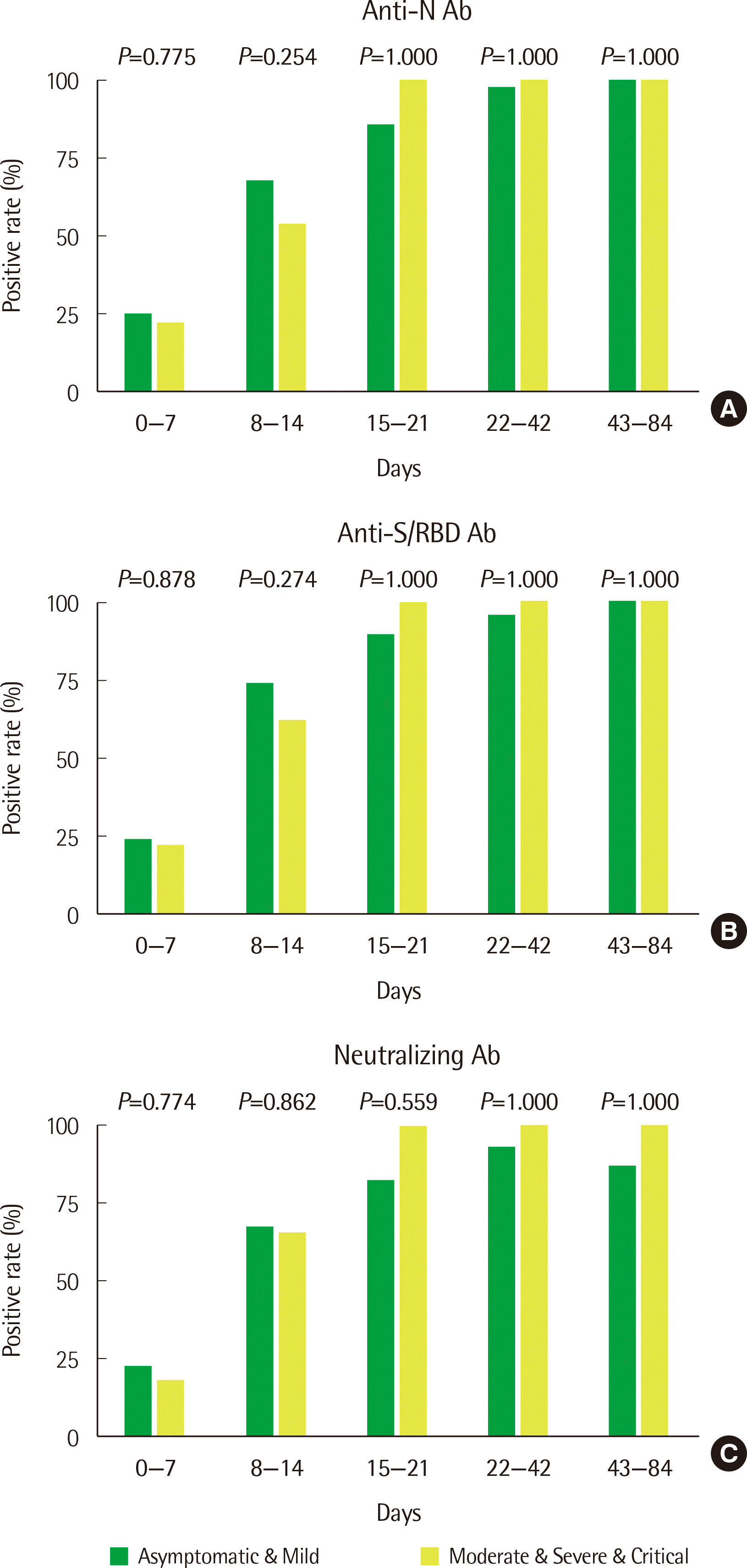
3. Qualitative changes in anti-N, anti-S/RBD, and neutralizing antibody levels in the early stage of infection
Most patients had anti-SARs-CoV-2 antibodies starting at two weeks (15 days) from the date of infection and maintained these antibodies for up to three months. The antibody-positive rates were somewhat higher in patients with low severity illness before two weeks and were somewhat higher in patients with high severity illness after two weeks, but these differences were not significant (Fig. 2).
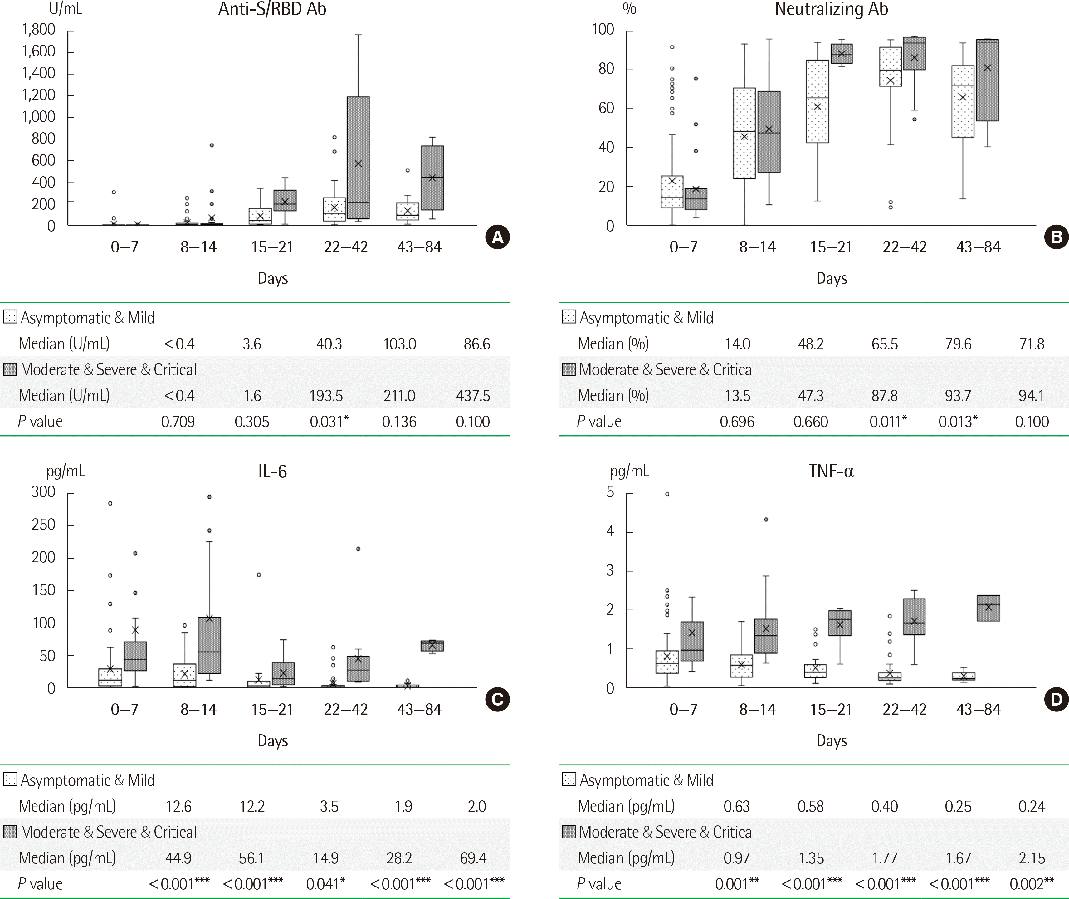
4. Quantitative changes in anti-S/RBD antibody, neutralizing antibody, IL-6, and TNF-α levels in the early stage of infection
The anti-S/RBD and neutralizing antibody titers were significantly higher in patients with high severity disease than in patients with low severity disease at the time of initial antibody production at approximately three weeks after infection (Fig. 3A, B). Cytokine (IL-6 and TNF-α) levels were consistently higher in patients with high severity disease than in patients with low severity disease from immediately after infection until at least three months (Fig. 3C, D).
5. Relationship between anti-S/RBD antibody and neutralizing antibody levels
The neutralizing antibodies and the anti-S/RBD antibodies in the Elecsys Anti-SARS-CoV-2 S assay and cPass SARS-CoV-2 Neutralization Antibody Detection Kit (ELISA-based sVNT method) showed a significantly positive correlation (r=0.540, P<0.001). The cPass Neutralizing Antibody Kit cut-off value of 30.0%, suggested by GenScript, was equivalent to approximately 340 U/mL using the Elecsys Anti-SARS-CoV-2 S assay (data not shown).
6. Factors associated with outcomes
The parameters included in the multivariable logistic regression analysis were age, sex, underlying disease, and initial X-ray findings. The results of logistic regression analysis are shown in Table 3.
DISCUSSION
During the initial period of the SARS-CoV-2 epidemic, South Korea responded with very strict quarantine restrictions and detailed investigations and disclosures regarding the movement of all people known to be infected. According to this national policy, all people with respiratory symptoms, close contacts with infected patients, and foreign visitors entering the country were required to undergo a PCR test, and those with confirmed infection were required to be admitted to a medical facility as soon as infection was confirmed. Because this study was performed at the beginning of the epidemic, all patients were hospitalized regardless of symptoms; thus, patients were not selected according to disease severity. Additionally, all study participants were interviewed about their medical history and underwent a timely SARS-CoV-2 PCR test, blood collection, and radiographic imaging. After October 2020, the national policy was changed, and home quarantine and non-face-to-face treatment was implemented; thus, detailed data on patients with asymptomatic and mild illness could not be collected. Therefore, research on the early stages of the pandemic is important for analyzing the full spectrum of COVID-19 clinical manifestations.
Several previous studies have shown that cancer, cerebrovascular disease, chronic kidney disease, liver cirrhosis, diabetes mellitus, disabilities, and cardiovascular disease are high risk factors for severe outcomes in COVID-19 [8, 32, 33]; however, we found no significant associations between underlying disease and COVID-19 severity. This may be because the underlying disease was identified based on the patient questionnaire, and the comorbidities were well-controlled at the time of admission. To reduce the potential for bias related to underlying disease, it is necessary to collect objective data and obtain accurate data on the patient condition at the time of hospitalization.
As in previous studies [9-11], serum antibodies against SARS-CoV-2 were detected by the third week (15 days) of infection in all study participants, and the patients showing high severity illness had high titers of S/RBD antibodies and neutralizing antibodies. Furthermore, there was no significant difference in the antibody titer in the first two weeks of infection (before 14 days), but this difference was significant in the third week.
In agreement with previous studies [11-13, 34-38], the pro-inflammatory cytokines, IL-6 and TNF-α, showed higher levels in patients with more severe disease than in those with less severe disease. Therefore, inhibitors of pro-inflammatory cytokines may be useful as therapies for reducing disease severity. Although, differences in infectivity and cytokine secretion were observed among variants such as alpha (B.1.1.7), beta (B.1.351), delta (B.1.617.2), and omicron (B.1.1.529) [39], a cytokine pattern similar to that of the wild-type strain was observed as indicated by several studies during the specific variant outbreak period showing similar results [34-38]. Based on these cytokine dynamics, several new drugs targeting IL-6 and TNF-α have been developed to prevent cytokine storms [12]. These drugs have various mechanisms of action, including blocking cytokine receptors, promoting anti-cytokine antibody production, and reducing cytokine levels to suppress their function, and have shown promising results [12, 40-42].
Overall, this study clearly highlights the immunological characteristics of unvaccinated patients with confirmed SARS-CoV-2 infections in South Korea. However, it does have some limitations. First, it has a retrospective, single-center design with a small sample size. As the center is located in Seoul, most patients were residents of Seoul, and the possibility of regional bias could not be ruled out. Second, serial samples through consecutive three-month intervals were only obtained from a few patients, which makes it difficult to examine individual antibody titers and cytokine level dynamics.
We showed that the levels of anti-S/RBD and neutralizing antibodies as well as of pro-inflammatory cytokines, IL-6 and TNF-α, varied significantly according to disease severity during the early stage of SARS-CoV-2 infection. Although we examined the early stages of the COVID-19 pandemic, individuals with more recent SARS-CoV-2 infection and those with other respiratory viruses may exhibit similar trends. Therefore, the immunological characteristics of patients confirmed through blood sampling at the appropriate time may be useful for predicting clinical outcomes to provide effective treatments for patients with respiratory viral infections. Further, we focused on infections with the early wild-type SARS-CoV-2, providing comparative data for studying the differences among variant types in further studies. COVID-19 has greatly impacted society as well as led to research and clinical data collection, which can advance medical technology.
 XML Download